. 2018 Feb 16;46(3):1102-1112.
doi: 10.1093/nar/gkx1262.

A coarse-grained model for DNA origami

Affiliations

A coarse-grained model for DNA origami

Roman V Reshetnikov et al. Nucleic Acids Res. .

Abstract

Modeling tools provide a valuable support for DNA origami design. However, current solutions have limited application for conformational analysis of the designs. In this work we present a tool for a thorough study of DNA origami structure and dynamics. The tool is based on a novel coarse-grained model dedicated to geometry optimization and conformational analysis of DNA origami. We explored the ability of the model to predict dynamic behavior, global shapes, and fine details of two single-layer systems designed in hexagonal and square lattices using atomic force microscopy, Förster resonance energy transfer spectroscopy, and all-atom molecular dynamic simulations for validation of the results. We also examined the performance of the model for multilayer systems by simulation of DNA origami with published cryo-electron microscopy and atomic force microscopy structures. A good agreement between the simulated and experimental data makes the model suitable for conformational analysis of DNA origami objects. The tool is available at http://vsb.fbb.msu.ru/cosm as a web-service and as a standalone version.


Figures

Figure 1.
Figure 1.
The DNA origami fundamentals: lattice and crossovers. (A) The helices (black numbered circles) are located in the nodes of the honeycomb lattice (gray). Colored lines between the circles represent crossovers between the helices and their colors correspond to individual staples from the panel B. The colored sectors within the circles refer to the twist of the staples on their path between the helices shown with a dashed line in the panel B; (B) a side view of the design from the panel A, numbers of the helices correspond between the panels. The helices are formed by a scaffold chain (black) and a number of staple chains (colored). The dashed line shows the staple-formed path from helix 1 to helix 5 corresponding to the colored sectors and crossovers from the panel A; (C) a full-atom view of the area highlighted in gray on the panel B, colors of the DNA chains correspond between the panels.
Figure 2.
Figure 2.
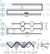
Transition from initial 2D scheme to the COSM model. Blue-encircled numbers specify the index numbers of the corresponding strands. (A) 2D-plot of the DNA origami. Black: scaffold chain; gray-blue: staple chains. Each cell corresponds to one base pair. Sites of insertions are denoted with a tear-shaped sign with a plus symbol; (B) the design from the panel A schematically shown in its COSM representation. The scaffold chain is shown by a black line. Crossover-forming particles are outlined by gray; (C) correspondence between the COSM model for the strand 1 and a B-DNA geometry. COSM particles are in gray, staple crossover site is shown with arrows; (D) an actual geometry of the encircled area from the panel B.
Figure 3.
Figure 3.
Designs of the benchmark structures. (A) scheme of the Hc-system; (B) scheme of the Sq-system with putative arrangement of the arms.
Figure 4.
Figure 4.
Analysis of experimental and simulated shapes of the Hc-system. (A) AFM images of individual structures; (B) conformations of the Hc-system predicted by our model in side and front views; (C) scheme of the characteristic movements of the Hc-system and corresponding torsional angles: γ for swinging and θ for twisting motions of the legs; (D) probability distribution of the γ angle values according to our model (red) and AFM imaging (blue); (E) Dynamics of the θ angle according to coarse-grained (red) and all-atom (black) models; (F) mobility of atoms/particles of all-atom (black; only phosphorus atoms of the scaffold chain were considered for the RMSF calculation) and coarse-grained (red) models during MD simulation.
Figure 5.
Figure 5.
Analysis of experimental and simulated shapes of the Sq-system. (A) AFM images of individual structures; (B) scheme of the Sq-system showing the parameters used for characterization of the system’s geometry: angles α, β and a distance between the ends of the Sq-system; (C) conformations of the Sq-system predicted by our computational model; (D, E) probability distribution of the α and β angle values, correspondingly. Red: the values according to our model, blue: obtained from AFM imaging; (F) mobility of atoms/particles of all-atom (black; only phosphorus atoms of the scaffold chain were considered for the RMSF calculation) and coarse-grained (red) models during MD simulation.
Figure 6.
Figure 6.
Verification of the model using simulations of previously published DNA origami structures. (A) Different views of the cryo-EM ‘pointer’ object, PDB entry 4v5x (37); (B) idealized COSM model of the pointer object used as a starting structure for the simulation; (C–E) different views of the pointer shape after simulation (black framework), superposed with the cryo-EM coordinates (blue and pink); F: predicted structure of an ‘S’-shaped object (10). Left: side view; right: snapshots of a front view at different timeframes of the trajectory, showing out-of-bending-plane flexibility of the object; (G) a hexagonal nanocontainer opening (38); (H) an icosahedron assembly from initial system to a final shape (39). Crossover sites are not shown for clarity.

References

    1. Ke Y. Designer three-dimensional DNA architectures. Curr. Opin. Struct. Biol. 2014; 27:122–128. - PubMed
    1. Rothemund P.W.K. Folding DNA to create nanoscale shapes and patterns. Nature. 2006; 440:297–302. - PubMed
    1. Holliday R. A mechanism for gene conversion in fungi. Genet Res. 1964; 5:282–304. - PubMed
    1. Andersen E.S., Dong M., Nielsen M.M., Jahn K., Lind-Thomsen A., Mamdouh W., Gothelf K.V., Besenbacher F., Kjems J.. DNA origami design of dolphin-shaped structures with flexible tails. ACS Nano. 2008; 2:1213–1218. - PubMed
    1. Zhu J., Wei B., Yuan Y., Mi Y.. UNIQUIMER 3D, a software system for structural DNA nanotechnology design, analysis and evaluation. Nucleic Acids Res. 2009; 37:2164–2175. - PMC - PubMed

Publication types

Substances

LinkOut - more resources