. 2018 Feb:515:52-65.
doi: 10.1016/j.virol.2017.12.005. Epub 2017 Dec 18.

Localization to detergent-resistant membranes and HIV-1 core entry inhibition correlate with HIV-1 restriction by SERINC5

Affiliations

Localization to detergent-resistant membranes and HIV-1 core entry inhibition correlate with HIV-1 restriction by SERINC5

Bianca Schulte et al. Virology. 2018 Feb.

Abstract

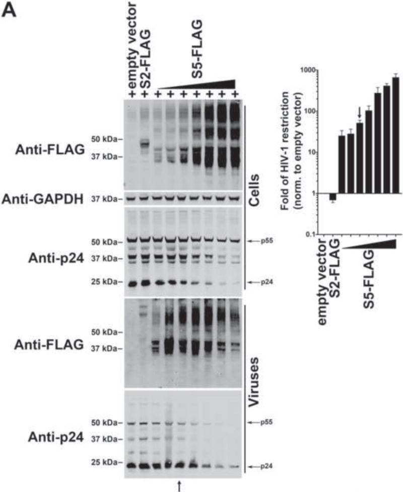
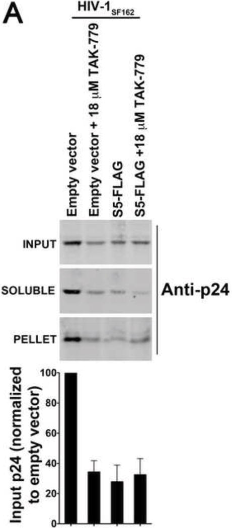
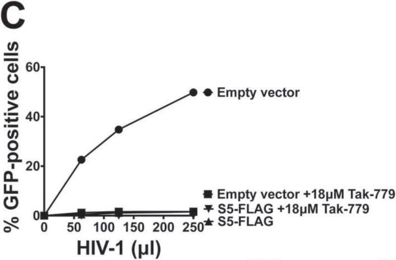
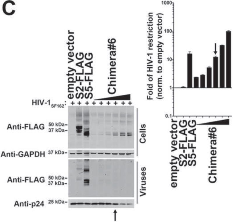
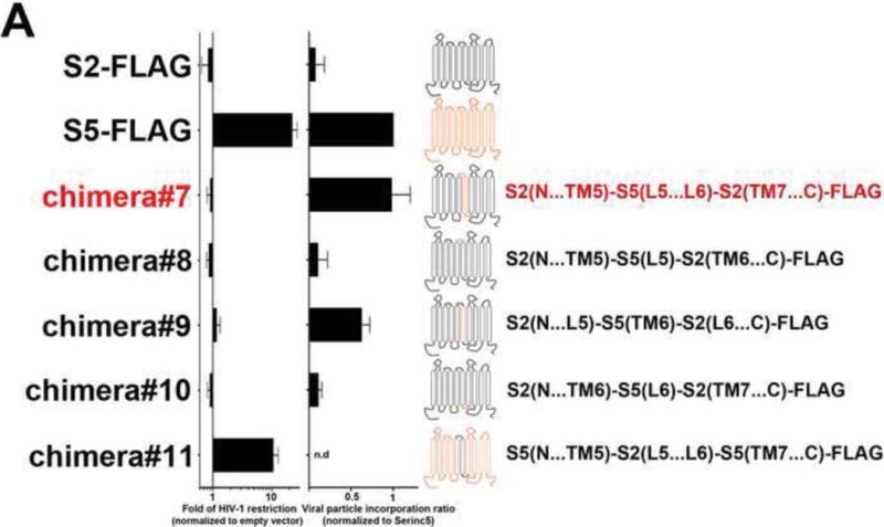
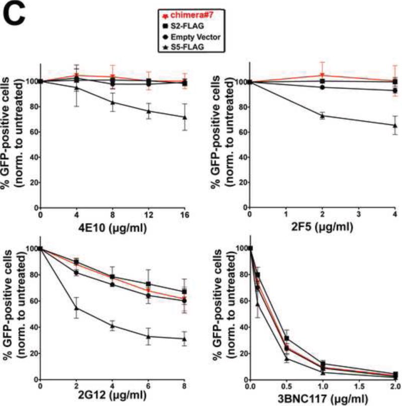
SERINC5(S5) is a multi-span transmembrane protein that potently blocks the infectivity of HIV-1 produced by human T-cells. The ability of S5 to restrict infectivity correlates with its presence in the virion, but the exact mechanism by which S5 restricts HIV-1 is unknown. Here we tested whether the core from HIV-1 virions containing S5 is delivered to the cytoplasm. Using the "fate of the capsid" assay, we demonstrated that the viral core of S5-restricted HIV-1 does not reach the cytoplasm of target cells, suggesting a block in the delivery of the core to the cytoplasm. In agreement with evidence suggesting that the viral determinants for S5 restriction map to the envelope of HIV-1, we observed that S5 induces conformational changes to the HIV-1 envelope. Further, we demonstrated that S5 localizes to detergent-resistant membranes (DRMs), as has been shown previously for the HIV-1 envelope in producer cells. In order to identify the determinants of S5 restriction, we explored the ability of all human SERINC proteins to restrict HIV-1. In contrast to human S5, we observed that human SERINC2(S2) did not restrict HIV-1, and was inefficiently incorporated into HIV-1 virions when compared to S5. Experiments using S5-S2 chimeric proteins revealed two functional domains for restriction: one necessary for S5 incorporation into virions, which does not seem to be necessary for restriction, and a second one necessary to change the HIV-1 envelope conformation, localize to DRMs, and block infection.

Keywords: Core; DRMs; Envelope; HIV-1; Restriction; SERINC2; SERINC5.


Figures

Figure 1
Figure 1. Ability of human SERINC proteins to restrict HIV-1
To test the ability of S5 (A), S2 (B), S4 (C), S3 (C), and S1 (C) to restrict HIV-1, HIV-1ΔNef particles expressing the SF162 envelope (HIV-1SF162) in the presence of increasing amounts of the indicated SERINC protein were produced. Viruses and producer cells were harvested 48 hours post-transfection. Producer cells (Cells) were lysed and analyzed for expression of the indicated SERINC protein, p24 and GAPDH by Western blotting using anti-FLAG, anti-p24 and anti-GAPDH antibodies (left panels), respectively. Produced HIV-1SF162 viruses (Viruses) were partially purified using a 20% sucrose cushion and analyzed for expression of the indicated SERINC protein and p24 using anti-FLAG and anti-p24 antibodies (left panels), respectively. At the same time, TZM-bl GFP-reporter cells were challenged with the different HIV-1SF162 viruses. At 24 h post-challenge, infection was determined by measuring the percentage of GFP-positive cells (right panel). Fold-restriction is defined as the ratio of %infection by viruses produced in the presence of empty vector to %infection by viruses produced in the presence of the indicated SERINC protein (right panel). The fold of HIV-1 restriction shown is the average of 3 independent experiments. Black arrows point to the experiments where the levels of SERINC expression did not affect virus production as measured by p24. Experiments were repeated at least three times and the Western blot from a representative example is shown. (C) The fold of HIV-1 restriction at SERINC levels that do not affect particle release (black arrow) for three independent experiments with standard deviation is shown. In addition, accession number, molecular weight and number of amino acids for each human SERINC protein is illustrated.
Figure 2
Figure 2. S5 topology and contribution of the different loops to HIV-1 restriction
(A) The theoretical membrane topology for human S5 (top left panel) predicted by the software TOPCONS is shown. N and C indicates the N- and C-terminus, respectively. The protein loops (L) are numbered L1-L9. Similarly, the transmembrane (TM) regions are numbered TM1-TM10. The residues spanning the TM regions are shown. Surface (fixed) and total expression (fixed & permeabilized) of the indicated S5 variants containing a FLAG epitope peptide (red circle) inserted into each of the nine loops (L1-L9) of S5 were analyzed by flow cytometry using anti-FLAG antibodies (red line). As a control, flow cytometric analysis was performed using an Isotype-matched control (blue line). The percentage of FLAG-positive cells for the illustrated experiment is presented. The standard deviation of three independent experiments is shown (right panels). (B) HeLa cells expressing the indicated S5 variants containing a FLAG epitope tag were imaged by fluorescence microscopy using anti-FLAG antibodies (red). Nuclei were stained with DAPI (blue). (C) The ability of the indicated S5 variants to restrict HIV-1 was measured. The fold of HIV-1 restriction at SERINC levels that do not affect particle release for three independent experiments with standard deviation is shown.
Figure 3
Figure 3. S5 prevents the delivery of HIV-1 cores to the cytosol of target cells
Human TZM-bl GFP-reporter cells were challenged with normalized amounts of HIV-1SF162 that do or do not contain S5. Eight hours post-infection cells were harvested and processed as described in Methods. Briefly, cells were incubated in a hypotonic buffer and homogenized using a Dounce homogenizer. A post-nuclear supernatant (INPUT) was obtained by pelleting the nuclear fraction. The INPUT represents the total capsid delivered into the cytoplasm of the target cell. The post-nuclear supernatant was then spun on 50% sucrose cushion to separate the pelletable capsid (PELLET) from the soluble capsid (SOLUBLE), as described in Methods. The PELLET represents the capsid that is forming HIV-1 cores. The SOLUBLE fraction represents the soluble capsid. INPUT, SOLUBLE and PELLET fractions were analyzed by Western blot using anti-p24 antibodies (A upper panel). As a control, similar infections were also performed in the presence of the inhibitor TAK-779, which is a CCR5 antagonist that prevents the fusion of viral membrane with the cellular membrane. The standard deviation of the INPUT for three independent experiments is shown (A lower panel). For these experiments viruses that do or do not contain S5 were normalized using p24 levels (B). Infectivity of viruses used in the fractionation assay is shown (C).
Figure 4
Figure 4. S5 affects the conformation of the HIV-1 envelope
(A) The ability of different HIV-1 neutralizing antibodies to block HIV-1SF162 viruses produced in the presence S5 was measured. HIV-1SF162 viruses produced in human HEK293T cells in the presence of S5, S2, or empty vector were incubated with increasing amounts of the indicated neutralizing antibody for 1 h at 37 °C. The virus- antibody mixture was then used to infect TZM-bl GFP-reporter cells. Infectivity was determined by measuring the percentage of GFP-positive cells twenty-four hours post-infection. Infection values were normalized to the infection of viruses that were not incubated with neutralizing antibodies. (B) Similar neutralization experiments were performed using HIV-1SF162 viruses with anti-gp41 and anti-gp120 antibodies, which either don´t neutralize SF162 viruses or with anti-gp120 antibodies, which strongly neutralize SF162 viruses. (C) Neutralization experiments were also performed using HIV-1SF162 viruses produced in human Jurkat TAg cells that do or do not express S3 and S5 (S3/S5 KO). (D) HIV-1SF162 viruses produced in HEK293T (top) or Jurkat TAg S3/S5 KO cells (bottom) in the presence of S5, S2, or empty vector were used to challenge TZM-bl GFP-reporter cells in the presence of increasing concentrations of the small molecule inhibitor 484. Infectivity was determined by measuring the % of GFP-positive cells 24 h post-infection. Infection values were normalized to the infection of viruses that were not incubated with the drug. Infections were performed in triplicates and standard deviations are shown.
Figure 5
Figure 5. S5 localizes to detergent-resistant membranes (DRMs)
(A) HEK293T cells expressing S5-FLAG or S2-FLAG were lysed and fractionated using a flotation gradient as described in Methods. Cells were homogenized in a Triton X-100 detergent solution and fractionated by discontinuous sucrose gradient centrifugation (5–40%). Gradient fractions 1–12 were analyzed by Western blotting using anti-FLAG antibodies. As positive controls, similar analysis was performed using HEK293T cells separately transfected with Caveolin-1 fused to GFP (Caveolin-1-GFP), which localizes to DRMs. As a negative control, we studied the membrane localization of the receptor TVBS3 fused to GFP (TVBS3-GFP), which does not localize to DRMs. Caveolin-1-GFP and TVBS3-GFP expression was detected by Western blotting using anti-GFP antibodies. Fractions for S2 and S5 from three independent experiments were quantified using a Li-Cor instrument, and they are shown as % of total FLAG protein with standard deviations (lower panel). (B) Similar fractionation experiments were performed in human HEK293T cells producing HIV-1SF162 viruses in the presence of S5-FLAG or S2-FLAG. The DNA mix for each transfection included 8 μg NL4–3ΔNef, 0.5 μg SF162, plus either empty vector, S2-FLAG, S5-FLAG, caveolin-1-GFP, or TVB-1. Forty-eight hours post-transfection cells were lyzed and fractionated as described in Methods. Fractions were analyzed by Western blotting using anti-FLAG, anti-GFP, or anti-Env antibodies. Fractions for S2 and S5 from three independent experiments were quantified using a Li-Cor instrument, and they are shown as % of total FLAG protein with standard deviations (lower panel).
Figure 6
Figure 6. S5 residues 203–411 containing regions TM5-L5-TM6-L6-TM7-L7-TM8-L8-TM9 are important for HIV-1 restriction
(A) The ability of the indicated S5-S2 chimeric proteins to restrict HIV-1 is shown as fold-restriction. The amino acid region of each chimera is shown (right panel). The fold-restriction is defined as the ratio of % infection by viruses produced in the presence of empty vector to % infection by viruses produced in the presence of the indicated S5-S2 chimera. The HIV-1 fold-restriction where expression of the chimera did not affect viral production is shown with a standard deviation from six independent experiments. Schematic representations of the chimeras are shown, with protein regions of S5 and S2 colored in orange and black, respectively. The ability of S5-S2 chimera#5 (B) and chimera#6 (C) to restrict HIV-1 is shown. Black arrows point to the experiments where the levels of chimera expression did not affect virus production as measured by p24. Experiments were repeated at least three times and the Western blot from a representative example is shown.
Figure 7
Figure 7. The S5 region L5-TM6-L6 is required for incorporation into HIV-1 virions
(A) Ability of S2 proteins containing S5 regions to restrict HIV-1 infection and be incorporated into virions. S2 proteins containing individual elements (L5, TM6 or L6), or the entire L5-TM6-L6 region of S5, were analyzed for the ability to restrict HIV-1 (fold-restriction) and be incorporated into virions (viral particle incorporation ratio). Fold-restriction is defined as the ratio of %infection by viruses produced in the presence of empty vector to %infection by viruses produced in the presence of the indicated S5-S2 chimera. The HIV-1 fold-restriction where expression of the chimera did not affect viral production is shown with a standard deviation from six independent experiments. Incorporation of the chimera into viral particles is defined as the ratio of protein incorporated into virions to the protein expressed in producer cells. Viral particle incorporation ratio where expression of the chimera did not affect viral production is shown with a standard deviation from three independent experiments. Schematic representations of S2 proteins containing S5 regions are shown with protein regions of S2 and S5 colored in black and orange, respectively. (B) The ability of chimera#7 and chimera#9 to restrict HIV-1 is shown. Black arrows point to the experiments where the levels of chimera expression did not affect virus production as measured by p24. Experiments were repeated at least three times and the Western blot from a representative example is shown. (C) The ability of the indicated HIV-1 neutralizing antibodies to block HIV-1SF162 viruses produced in the presence of chimera#7 was measured. Viruses produced in the presence of the indicated SERINC proteins were incubated with increasing amounts of the indicated neutralizing antibody for 1 hr at 37 °C. The virus-antibody mixture was then used to infect TZM-bl GFP-reporter cells. Infectivity was determined by measuring the percentage of GFP-positive cells at 24 h post-infection. Infection values were normalized to the infection of virions that were not incubated with neutralizing antibodies. Infections were performed in triplicates and standard deviations are shown. (D) Human 293T cells expressing the indicated proteins were lysed in Triton X-100 and fractionated on a discontinuous sucrose gradient (5–40%) as described in Methods. As controls, human HEK293T cells expressing caveoilin-1 fused to GFP (Caveolin-1-GFP) and tumor virus B receptor (TVBS3-GFP) were analyzed as probes for proteins that did or did not localize to DRMs, respectively. Twelve fractions (1–12) were analyzed per sample using anti-FLAG or anti-GFP antibodies. Fractions for the indicated proteins from three independent experiments were quantified using a Li-Cor instrument, and they are shown as % of total FLAG protein with standard deviations (lower panel). (E) The ability of chimera#11 to block HIV-1 infection was measured, as described above.
Figure 8
Figure 8. The S5 region L5-TM6-L6-TM7-L7 is required for it’s the ability to restrict the infectivity of HIV-1
(A) HIV-1 restriction ability of S2 proteins containing S5 regions. S2 proteins containing the L5-TM6-L6 region of S5 were extended using S5 elements toward the N- and C-terminus of the protein. The new variants were then tested for the ability to block HIV-1 infection. The HIV-1 fold-restriction where expression of the chimera did not affect viral production is shown with a standard deviation from six independent experiments. Schematic representations of S2 proteins containing S5 regions are shown, and regions of S2 and S5 colored in black and orange, respectively. (B) The ability of S2 containing the S5 region L5-TM6-L6-TM7-L7 (chimera#15) to block HIV-1 infection is shown. The fold of HIV-1 restriction shown is the average of 3 independent experiments. Black arrows point to the experiments where the levels of chimera expression did not affect virus production as measured by p24. Experiments were repeated at least three times and the Western blot from a representative example is shown. (C) The ability of the indicated HIV-1 neutralizing antibodies to block HIV-1SF162 viruses produced in the presence of the S2 protein containing the S5 region L5-TM6-L6-TM7-L7 (chimera#15) was measured. HIV-1SF162 viruses produced in the presence of the indicated SERINC proteins were incubated with increasing amounts of the indicated neutralizing antibody for 1 hr at 37 °C. The virus-antibody mixture was then used to infect TZM-bl GFP-reporter cells. Infectivity was determined by measuring the percentage of GFP-positive cells at 24 h post-infection. Infection values were normalized to the infection of viruses that were not incubated with neutralizing antibodies. (D) S2 protein containing the S5 region L5-TM6-L6-TM7-L7 (chimera#15) localized to DRMs in a manner similar to wild-type S5. (E) S2 protein containing the S5 region L5-TM6-L6-TM7-L7 (chimera#15) prevented the delivery of HIV-1 cores to the cytosol as measured by the fate of the capsid. The standard deviation of the INPUT for three independent fate of the capsid experiments is shown (lower panel).

Similar articles

Cited by

References

    1. Aiken C, Trono D. Nef stimulates human immunodeficiency virus type 1 proviral DNA synthesis. J Virol. 1995;69:5048–5056. - PMC - PubMed
    1. Baba M, Nishimura O, Kanzaki N, Okamoto M, Sawada H, Iizawa Y, Shiraishi M, Aramaki Y, Okonogi K, Ogawa Y, Meguro K, Fujino M. A small-molecule, nonpeptide CCR5 antagonist with highly potent and selective anti-HIV-1 activity. Proc Natl Acad Sci U S A. 1999;96:5698–5703. - PMC - PubMed
    1. Beitari S, Ding S, Pan Q, Finzi A, Liang C. Effect of HIV-1 Env on SERINC5 Antagonism. J Virol. 2017;91 - PMC - PubMed
    1. Bhattacharya J, Peters PJ, Clapham PR. Human immunodeficiency virus type 1 envelope glycoproteins that lack cytoplasmic domain cysteines: impact on association with membrane lipid rafts and incorporation onto budding virus particles. J Virol. 2004;78:5500–5506. - PMC - PubMed
    1. Bhattacharya J, Repik A, Clapham PR. Gag regulates association of human immunodeficiency virus type 1 envelope with detergent-resistant membranes. J Virol. 2006;80:5292–5300. - PMC - PubMed

Publication types