

Preliminary study on the diagnostic value of single-source dual-energy CT in diagnosing cervical lymph node metastasis of thyroid carcinoma
- PMID: 29268547
- PMCID: PMC5721007
- DOI: 10.21037/jtd.2017.09.151
Preliminary study on the diagnostic value of single-source dual-energy CT in diagnosing cervical lymph node metastasis of thyroid carcinoma
Abstract
Background: To investigate the value of single-source dual-energy spectral CT imaging in improving the accuracy of preoperative diagnosis of lymph node metastasis of thyroid carcinoma.
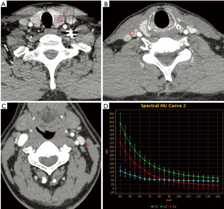
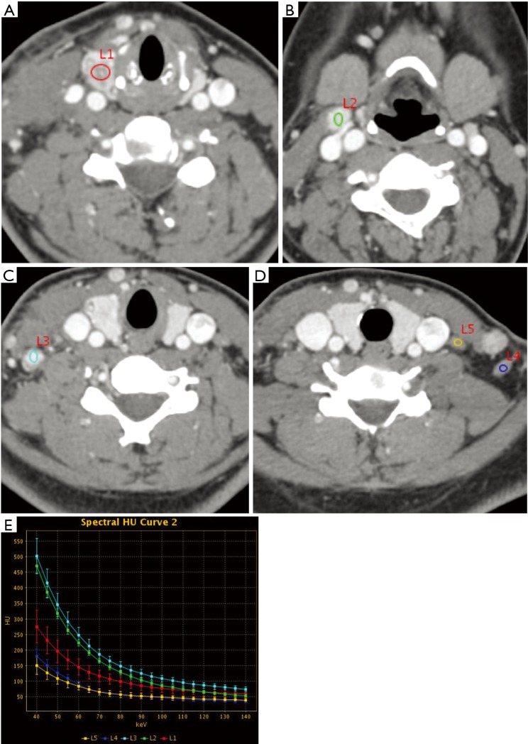
Methods: Thirty-four thyroid carcinoma patients were enrolled and received spectral CT scanning before thyroidectomy and cervical lymph node dissection surgery. Iodine-based material decomposition (MD) images and 101 sets of monochromatic images from 40 to 140 keV were reconstructed after CT scans. The iodine concentrations (IC) of lymph nodes were measured on the MD images and was normalized to that of common carotid artery to obtain the normalized iodine concentration (NIC). The CT number of lymph nodes as function of photon energy was measured on the 101 sets of images to generate a spectral HU curve and to calculate its slope λHU. The measurements between the metastatic and non-metastatic lymph nodes were statistically compared and receiver operating characteristic (ROC) curves were used to determine the optimal thresholds of these measurements for diagnosing lymph nodes metastasis.
Results: There were 136 lymph nodes that were pathologically confirmed. Among them, 102 (75%) were metastatic and 34 (25%) were non-metastatic. The IC, NIC and the slope λHU of the metastatic lymph nodes were 3.93±1.58 mg/mL, 0.70±0.55 and 4.63±1.91, respectively. These values were statistically higher than the respective values of 1.77±0.71 mg/mL, 0.29±0.16 and 2.19±0.91 for the non-metastatic lymph nodes (all P<0.001). ROC analysis determined the optimal diagnostic threshold for IC as 2.56 mg/mL, with the sensitivity, specificity and accuracy of 83.3%, 91.2% and 85.3%, respectively. The optimal threshold for NIC was 0.289, with the sensitivity, specificity and accuracy of 96.1%, 76.5% and 91.2%, respectively. The optimal threshold for the spectral curve slope λHU was 2.692, with the sensitivity, specificity and accuracy of 88.2%, 82.4% and 86.8%, respectively.
Conclusions: The measurements obtained in dual-energy spectral CT improve the sensitivity and accuracy for preoperatively diagnosing lymph node metastasis in thyroid carcinoma.
Keywords: Thyroid carcinoma; lymph node; metastasis; spectral CT.
Conflict of interest statement
Conflicts of Interest: The authors have no conflicts of interest to declare.
Figures
Similar articles
-
Diagnostic accuracy of single-source dual-energy computed tomography and ultrasonography for detection of lateral cervical lymph node metastases of papillary thyroid carcinoma.J Thorac Dis. 2019 Dec;11(12):5032-5041. doi: 10.21037/jtd.2019.12.45. J Thorac Dis. 2019. PMID: 32030219 Free PMC article.
-
Diagnosing cervical lymph node metastasis in oral squamous cell carcinoma based on third-generation dual-source, dual-energy computed tomography.Eur Radiol. 2023 Jan;33(1):162-171. doi: 10.1007/s00330-022-09033-6. Epub 2022 Sep 7. Eur Radiol. 2023. PMID: 36070090 Free PMC article.
-
Papillary thyroid cancer: dual-energy spectral CT quantitative parameters for preoperative diagnosis of metastasis to the cervical lymph nodes.Radiology. 2015 Apr;275(1):167-76. doi: 10.1148/radiol.14140481. Epub 2014 Dec 17. Radiology. 2015. PMID: 25521777
-
The accuracy of ultrasonography in the preoperative diagnosis of cervical lymph node metastasis in patients with papillary thyroid carcinoma: A meta-analysis.Eur J Radiol. 2012 Aug;81(8):1798-805. doi: 10.1016/j.ejrad.2011.04.028. Epub 2011 May 4. Eur J Radiol. 2012. PMID: 21536396 Review.
-
Diagnostic potential of ultrasonography and computed tomography in differentiating cervical lymph node metastasis of thyroid cancer: a systematic review and meta-analysis.Arch Med Sci. 2020 May 11;19(4):965-975. doi: 10.5114/aoms.2020.95104. eCollection 2023. Arch Med Sci. 2020. PMID: 37560722 Free PMC article. Review.
Cited by
-
Quantitative parameter analysis of pretreatment dual-energy computed tomography in nasopharyngeal carcinoma cervical lymph node characteristics and prediction of radiotherapy sensitivity.Radiat Oncol. 2024 Jun 26;19(1):81. doi: 10.1186/s13014-024-02468-9. Radiat Oncol. 2024. PMID: 38918834 Free PMC article.
-
Integration of dual-source dual-energy CT quantitative parameters and ultrasound image features: A diagnostic method for extraglandular invasion of papillary thyroid carcinoma.Oncol Lett. 2025 May 21;30(1):356. doi: 10.3892/ol.2025.15102. eCollection 2025 Jul. Oncol Lett. 2025. PMID: 40454243 Free PMC article.
-
Diagnostic accuracy of ultrasound, CT and their combination in detecting cervical lymph node metastasis in patients with papillary thyroid cancer: a systematic review and meta-analysis.BMJ Open. 2022 Jul 4;12(7):e051568. doi: 10.1136/bmjopen-2021-051568. BMJ Open. 2022. PMID: 35788082 Free PMC article.
-
Prediction of lymph node metastasis in advanced gastric adenocarcinoma based on dual-energy CT radiomics: focus on the features of lymph nodes with a short axis diameter ≥6 mm.Front Oncol. 2024 Mar 1;14:1369051. doi: 10.3389/fonc.2024.1369051. eCollection 2024. Front Oncol. 2024. PMID: 38496754 Free PMC article.
-
Utility of non-contrast Dual Energy Computed Tomography in diagnosis of differentiated thyroid cancer - two case study.Cancer Imaging. 2023 Apr 18;23(1):39. doi: 10.1186/s40644-023-00555-w. Cancer Imaging. 2023. PMID: 37072868 Free PMC article.
References
LinkOut - more resources
Full Text Sources
Other Literature Sources