. 2018 Feb 15;434(2):278-291.
doi: 10.1016/j.ydbio.2017.12.011. Epub 2017 Dec 19.

Temporal and SUMO-specific SUMOylation contribute to the dynamics of Polo-like kinase 1 (PLK1) and spindle integrity during mouse oocyte meiosis

Affiliations

Temporal and SUMO-specific SUMOylation contribute to the dynamics of Polo-like kinase 1 (PLK1) and spindle integrity during mouse oocyte meiosis

Weber Beringui Feitosa et al. Dev Biol. .

Abstract

During mammalian meiosis, Polo-like kinase 1 (PLK1) is essential during cell cycle progression. In oocyte maturation, PLK1 expression is well characterized but timing of posttranslational modifications regulating its activity and subcellular localization are less clear. Small ubiquitin-related modifier (SUMO) posttranslational modifier proteins have been detected in mammalian gametes but their precise function during gametogenesis is largely unknown. In the present paper we report for mouse oocytes that both PLK1 and phosphorylated PLK1 undergo SUMOylation in meiosis II (MII) oocytes using immunocytochemistry, immunoprecipitation and in vitro SUMOylation assays. At MII, PLK1 is phosphorylated at threonine-210 and serine-137. MII oocyte PLK1 and phosphorylated PLK1 undergo SUMOylation by SUMO-1, -2 and -3 as shown by individual in vitro assays. Using these assays, forms of phosphorylated PLK1 normalized to PLK1 increased significantly and correlated with SUMOylated PLK1 levels. During meiotic progression and maturation, SUMO-1-SUMOylation of PLK1 is involved in spindle formation whereas SUMO-2/3-SUMOylation may regulate PLK1 activity at kinetochore-spindle attachment sites. Microtubule integrity is required for PLK1 localization with SUMO-1 but not with SUMO-2/3. Inhibition of SUMOylation disrupts proper meiotic bipolar spindle organization and spindle-kinetochore attachment. The data show that both temporal and SUMO-specific-SUMOylation play important roles in orchestrating functional dynamics of PLK1 during mouse oocyte meiosis, including subcellular compartmentalization.

Keywords: Cell cycle; Centrosome; Kinetochore; MTOC; Meiosis; SUMOylation; Spindle.


Figures

Figure 1
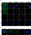
Figure 1. The pattern of subcellular PLK1 is dynamic during oocyte maturation
Mouse oocytes were matured in vitro and the following stages harvested: 0h (germinal vesicle, GV), 4h (GV breakdown, GVBD), 8h (metaphase-I) and 12h (telophase and metaphase-II). PLK1 (b, f, j, n, r) and alpha-tubulin (c, g, k, o, s) were localized in mouse oocytes using confocal microscopy following immune-fluorescence labeling with mouse anti-PLK1 and rabbit anti-alpha-tubulin antibodies. DNA was counterstained with DAPI (d, h, l, p, t); merged images shown (a, e, i, m, q). Large white squares shown in “m” and “q” are enlargements of each corresponding small box. At GV (a–d), PLK1 is mainly observed in nuclei with a punctate staining pattern. After GVBD, PLK1 is seen as foci on condensed chromosomes (e–h) and is coincident with alpha-tubulin at organizing spindle poles (see arrows, e–g). At metaphase I, PLK1 concentrates at centromeres and is coincident with alpha-tubulin in the spindle poles (i–l; arrows, i–k). At telophase, PLK1 is observed at the spindle mid-zone, a change from its localization at the poles (m–p); however, PLK1 still localizes with chromatids (large box, m). After telophase, PLK1 localizes to the centromeric region and spindle poles in metaphase II (q–t). Note, PLK1 is coincident with the microtubules at the centromeric region (box, q). Bar, 10μM.
Figure 2
Figure 2. PLK1 and SUMO-1 are coincident during meiosis
Oocytes were staged and harvested as in Figure 1. Subcellular localization for SUMO-1 (b, f, j, n, r) and PLK1 (c, g, k, o, s) were visualized by bioimaging using confocal microscopy, following immunolabeling with mouse anti-PLK1 and rabbit anti-SUMO-1 antibodies. DNA was counterstained with DAPI (d, h, l, p, t); merged images are shown (a, e, i, m, q). White large square boxes shown in “a” and “m” are enlargements of each corresponding smaller box. At GV, SUMO-1 is in the nucleus, predominantly localized at the nuclear membrane and non-nucleolar heterochromatin (a–d). Non-nucleolar SUMOylated heterochromatin closely associate with PLK1 (boxes, a–d). After GVBD (e–h), SUMO-1 is detected in the organizing spindle poles and is coincident with PLK1 (see arrows in e–g). At MI, SUMO-1 localizes over the spindle with a marked intensity at the spindle poles (i–l; arrows, i–k), where SUMO-1 is coincident with PLK1. As oocyte progress to telophase (m–p), SUMO-1, similar to that observation with PLK1, now locates from the spindle poles to the mid-zone where it overlaps with PLK1 (arrows, m–o); however, PLK1 still localizes with chromatids (large box, m). At MII, SUMO-1 localizes on the spindle coincident with PLK1, most prominently at the poles (q–t). Bar, 10μM.
Figure 3
Figure 3. SUMO-2/3 and PLK1 dynamics during oocyte maturation
(A) GV, GVBD, MI, telophase and MII stages of matured oocytes were collected as above. SUMO-2/3 (b, f, j, n, r) and PLK1 (c, g, k, o, s) were immuno-fluorescently labeled with mouse anti-PLK1 and rabbit anti-SUMO-2/3 antibodies and visualized by confocal microscopy. DNA was counterstained with DAPI (d, h, l, p, t), and merged images are shown (a, e, i, m, q). The white larger square boxes show enlargements of each corresponding small dotted box. At GV (a–d), SUMO-2/3 is diffusely distributed throughout the nuclei. Prominent perinucleolar chromatin rim and non-nucleolar heterochromatin staining is observed (arrows, b, d) where PLK1 overlaps with SUMO (white box, a). After GVBD (e–h), SUMO-2/3 is coincident with PLK1 in foci on condensed chromosomes (box, e–h). At MI, SUMO-2/3 is coincident with PLK1 adjacent to centromeres (i–l; see box, i–l). At telophase (m–p), SUMO-2/3 is coincident with PLK1 at chromatids (box, m, o) and spindle mid-zone (arrows, m–o). In MII (q–t), SUMO-2/3 is again detected with PLK1 at the centromeres (boxes, q–t). Bar, 10μM. (B) SUMO-2/3 is observed between chromatin and PLK1. Chromatin enlargement of mouse oocyte in metaphase I showing in detail SUMO-2/3 coincident with PLK1 at kinetochore.
Figure 4
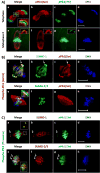
Figure 4. Shared meiotic patterns of phosphorylated-PLK1 kinases and SUMO proteins
Oocytes matured in vivo were employed to delineate temporal cellular localizations for phosphorylated (p) PLK1 proteins p-threonine-PLK1(T210) and p-serine-PLK1(S137), SUMO-1 and SUMO-2/3. Subcellular localization was determined using high-resolution bioimaging following immunodetection using phosphorylation-specific antibodies. DAPI was used to counterstain DNA. (A) Oocytes in telophase (a- d) and metaphase II (e- h) were labeled with anti-p-serine-PLK1 (b, f), and anti-p-threonine-PLK1 (c, g); DNA (d, h). Merged images are shown (a, e). (B) MII mouse oocytes were labeled with anti-SUMO-1 (b), anti-SUMO-2/3 (f) and anti-p-serine-PLK1 (c, g) antibodies; DNA (d, h). Merged images are shown (a, e). p-serine-PLK1 markedly is coincident with SUMO-1 at the spindle poles (arrows, a–c) and appears coincident over the spindle apparatus. At the centromere, some overlap with SUMO-2/3 is observed (e; inset, arrow). (C) Patterns for SUMO-1 (b), SUMO-2/3 (f) and p-threonine-PLK1 (c, g) were evaluated in MII oocytes. DNA (d, h) and the merged images are shown (a, e). Only a modest p-threonine-PLK1 signal is detected with SUMO-1 at the spindle pole (arrows, a–c). In contrast, a pronounced signal with SUMO-2/3 is observed at centromeres (arrows, e–g and box, e). Bar, 10μM.
Figure 5
Figure 5. PLK1 and phosphorylated PLK1 kinases are posttranslationally modified by SUMOylation
(A) Proteins extracted from freshly harvested MII oocytes (MII OC) and those subsequently subjected to immunoprecipitation (IP) with anti-PLK1 antibody were simultaneously separated using gel electrophoresis under denaturing conditions and transferred to the same membrane as described. As a control, IP was conducted with the antibody but without MII proteins added (Ø). Sequential immunodetection analyses were performed to determine specific PLK1 posttranslational modifications and associations (see left-sided labels). An illustrative single membrane is shown with sequential hybridizations with particular antibodies. Although a linear range of film exposures were made for each protein, representative signal intensities determined the exposure used for illustration purposes. (B) In vitro sumoylation assays specific for SUMO-1, SUMO-2 and SUMO-3 were each performed using the protein extract equivalent from 55-freshly harvested MII oocytes incubated with components of the sumoylation cascade for 2h incubation at 37°C as indicated in Methods. For each SUMO-specific assay, Western analyses are shown for PLK1 forms for the matched “starting material” (Lane 1, substrate, time0), PLK1 forms following a 2h assay with substrate and indicated reaction conditions (Lanes 2, 3) in comparison to the reaction materials without MII OC (Lane 4, non-substrate control). Immunodetection of proteins on the same membranes using sequential hybridizations with the specific anti-PLK1 and phosphorylated (p) PLK1 proteins p-threonine-PLK1(T210) and p-serine-PLK1(S137) antisera for Western analyses is shown on a representative membrane for each. The order of hybridizations for each duplicate experiment was randomized. Immunoblotting with the anti-α-tubulin antisera was negative for modification in the assay (data not shown). (C) Densitometry analyses of serine- or threonine-phosphorylated PLK1 levels following normalization with those of pan-PLK1. Results in the presence or absence of ATP for 2h are illustrated, representing densitometric measurements from either triplicate or duplicate membranes.
Figure 6
Figure 6. SUMO-1-PLK1 localization depends on spindle integrity and is involved in MTOC organization
MII oocytes matured in vivo were treated with microtubule-disturbing drugs followed by immunofluorescence bioimaging using confocal microscopy. DNA was detected using DAPI. (A) Mouse oocytes were cultured with Nocodazole (0.1 or 20μM; 10min), or without (DMSO-matched vehicle controls). Nocodazole-treated or vehicle-matched control MII oocytes were either processed immediately for immuno-bioimaging or rinsed and cultured for an additional 1h to allow microtubule re-assembly (rescue). Oocytes were immunostained using rabbit anti-SUMO-1 (b, f, j, n, r), mouse anti-PLK1 (c, g, k, o, s) and rat anti-α-tubulin (white boxes, a, e, i, m, q) antibodies; DNA (d, h, l, p, t). The merged image is shown (a, e, i, m, q); tubulin signals are not shown to better visualize SUMO-1 localization with PLK1. In vehicle-matched control oocytes, intact spindles are observed (box, a). PLK1 localizes at centromeres and is coincident with SUMO-1 at the spindle poles (a–d). Treatment with 0.1μM Nocodazole partially disassembled microtubules (box, e) and PLK1 localizes both at centromeres and coincident with SUMO-1 at the remaining spindle poles (arrows, e–g). Treatment with 20μM Nocodazole completely disorganized oocyte microtubules and no intact spindles are observed (see absence of any spindle structure, box, i). PLK1 remains at the centromeres but no longer coincident with SUMO-1 (i–l). A 1h rescue after Nocodazole treatment with 0.1μM (m–p) or 20μM (q–t) Nocodazole allowed spindle reorganization and SUMO-1-PLK1 localization at the poles. Bar, 10μM. (B) MII oocytes were treated with Taxol (10μM, 30min) to initiate microtubule organizing centers (MTOC) formation. Oocytes were labeled with specific antibodies for SUMO-1 (a), PLK1 (b), and α-tubulin (c); DNA (d). Treatment induced spindle relaxation (arrow, c) and formation of cytoplasmic microtubule asters (ǂ, asterisk; a, b, c). (C) Representative cytoplasmic aster (merged images, d) shows PLK1 localized with SUMO-1 (a) and each with tubulin (b, c).
Figure 7
Figure 7. Independent of spindle attachment or tension, centromeric SUMO-2/3 and PLK1 remain coincident
Spindle dependency was evaluated using pharmacological disruptors and immuno-bioimaging. (A) MII mouse oocytes were treated (10min) with Nocodazole (0.1 or 20μM) or without (DMSO-matched vehicle controls) and either processed immediately for immuno-bioimaging or rinsed and cultured for an additional 1h to allow microtubule re-assembly (rescue). Oocytes were analyzed by confocal microscopy after immunolabeling using rabbit anti-SUMO-2/3 (b, f, j, n, r), mouse anti-PLK1 (c, g, k, o, s) and rat anti-α-tubulin (white squares, a, e, i, m, q) antibodies; DNA counterstained with DAPI shown (d, h, l, p, t). The merge for DNA, SUMO-2/3 and PLK1 is shown without tubulin for better visualization of SUMO with PLK1 (a, e, i, m, q). In MII vehicle-matched control oocytes, intact spindles are observed (box, a); PLK1 is coincident with SUMO-2/3 at the centromeres (a–d). Treatment with 0.1μM Nocodazole partially disrupts microtubules (box, e) but at the centromeres PLK1is still coincident with SUMO-2/3 (e–h). 20μM Nocodazole completely disorganizes oocyte microtubules and no intact spindles are observed (box, i). PLK1 remains centromeric with SUMO-1 (i–l). Spindle reorganization was observed following 1h rescue after either 0.1μM (m–p) or 20μM (q–t) Nocodazole. Bar, 10μM. (B) MII oocytes matured in vivo were treated with Nocodazole to disrupt the spindle or Taxol to reduce spindle tension. In matched-vehicle treated control oocytes, SUMO-2/3 (b) and PLK1 (c) are observed with overlapping signals at centromeres (a). Treatment with 20μM Nocodazole (10min) does not alter SUMO-2/3 (f) and PLK1 (g) coincident pattern (e). Similarly, Taxol-treatment of oocytes (10μM, 30min) does not affect SUMO-2/3 (j), PLK1 (k) or their coincident pattern (i) at the centromeric region. (C) Illustration represents a control (a, c) or Taxol-treated (b, d) oocyte and the localization of tubulin with DNA and SUMO-2/3 (a, b, b’) and SUMO-2/3 with PLK1 (c, d, d’). White boxes (b, d) enlarged respectively (b’ and d’). SUMO-2/3 localization with PLK1 is unaffected by Taxol-induced spindle relaxation.
Figure 8
Figure 8. SUMOylation is required for proper spindle organization
MII in vivo-matured oocytes were incubated with 100μM Ginkgolic Acid (GA; 1h), then treated with 20μM Nocodazole (10min). Nocodazole was removed and oocytes rinsed and incubated again with 100μM GA (2h). After treatment with GA or matched-vehicle control, oocytes were analyzed by bioimaging analysis as described previously. Rabbit anti- SUMO-1 (A, a–h) SUMO-2/3 (B, a–h), rat anti-α-tubulin (A, B; a–d) and mouse anti-PLK1 (A, B; e–h) antibodies were used for immunodetection; DNA counterstained by DAPI. GA inhibition of SUMOylation prevents proper spindle organization after Nocodazole (A, B). In (A), very few oocytes show typical SUMO-1 at spindle poles (a, b), with most demonstrating no localization or scattered signals on the disorganized spindle (c, d). Following GA treatment, aberrant SUMO-1 localization with PLK1 is observed (e–h). In (B), most GA-treated oocytes are not able to reorganize their spindle properly and abnormal patterns for SUMO-2/3 are observed at the chromatin (a–d). In contrast, even with chromosomes spread in the cytoplasm after GA, SUMO-2/3 is coincident with PLK1 and still associated with DNA.

Similar articles

Cited by

References

    1. Abrieu A, Brassac T, Galas S, Fisher D, Labbé JC, Dorée M. The Polo-like kinase Plx1 is a component of the MPF amplification loop at the G2/M-phase transition of the cell cycle in Xenopus eggs. J Cell Sci. 1998:1751–7. - PubMed
    1. Arnaud L, Pines J, Nigg EA. GFP tagging reveals human Polo-like kinase 1 at the kinetochore/centromere region of mitotic chromosomes. Chromosoma. 1998;107:424–9. - PubMed
    1. Azuma Y, Arnaoutov A, Anan T, Dasso M. PIASy mediates SUMO-2 conjugation of Topoisomerase-II on mitotic chromosomes. EMBO J. 2005;24:2172–2182. - PMC - PubMed
    1. Ban R, Nishida T, Urano T. Mitotic kinase Aurora-B is regulated by SUMO-2/3 conjugation/deconjugation during mitosis. Genes to Cells. 2011;16:652–669. - PubMed
    1. Bernardi R, Pandolfi PP. Structure, dynamics and functions of promyelocytic leukaemia nuclear bodies. Nat Rev Mol Cell Biol. 2007;8:1006–1016. - PubMed

Publication types