. 2018 Feb 16;122(4):583-590.
doi: 10.1161/CIRCRESAHA.117.311490. Epub 2017 Dec 21.

Infarct Fibroblasts Do Not Derive From Bone Marrow Lineages

Affiliations

Infarct Fibroblasts Do Not Derive From Bone Marrow Lineages

Thomas Moore-Morris et al. Circ Res. .

Abstract

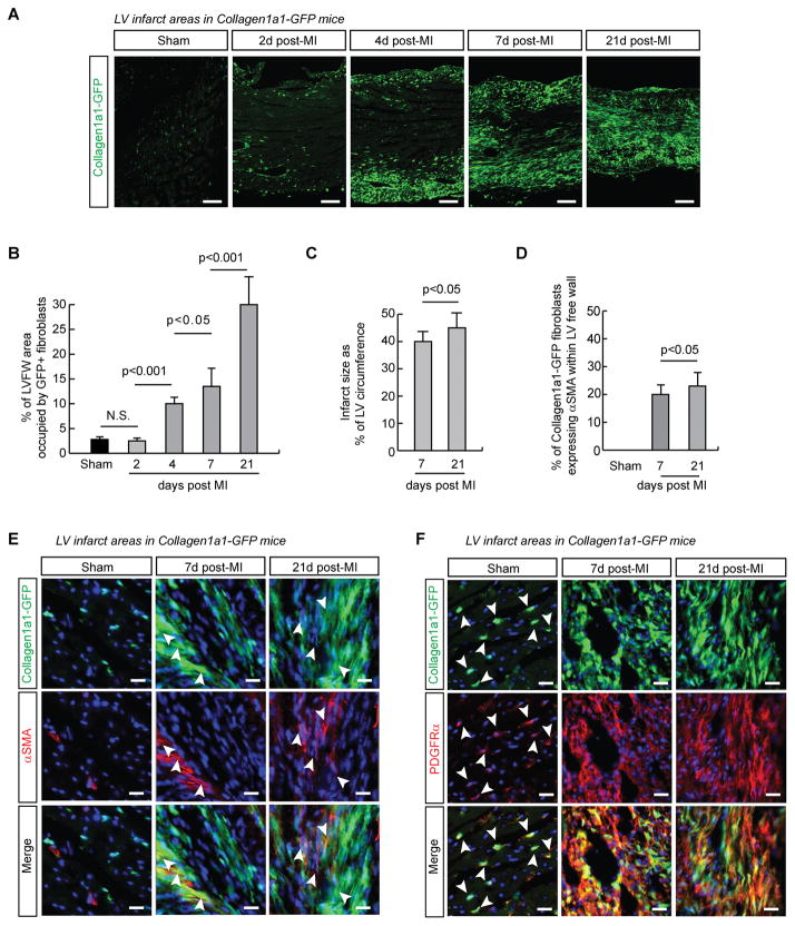
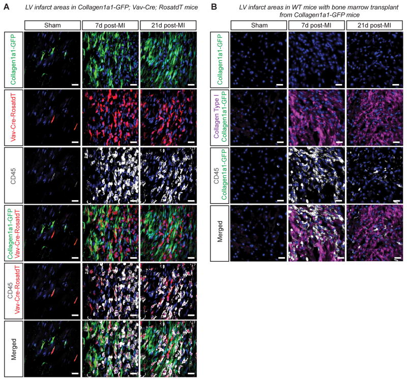
Rationale: Myocardial infarction is a major cause of adult mortality worldwide. The origin(s) of cardiac fibroblasts that constitute the postinfarct scar remain controversial, in particular the potential contribution of bone marrow lineages to activated fibroblasts within the scar.

Objective: The aim of this study was to establish the origin(s) of infarct fibroblasts using lineage tracing and bone marrow transplants and a robust marker for cardiac fibroblasts, the Collagen1a1-green fluorescent protein reporter.

Methods and results: Using genetic lineage tracing or bone marrow transplant, we found no evidence for collagen-producing fibroblasts derived from hematopoietic or bone marrow lineages in hearts subjected to permanent left anterior descending coronary artery ligation. In fact, fibroblasts within the infarcted area were largely of epicardial origin. Intriguingly, collagen-producing fibrocytes from hematopoietic lineages were observed attached to the epicardial surface of infarcted and sham-operated hearts in which a suture was placed around the left anterior descending coronary artery.

Conclusions: In this controversial field, our study demonstrated that the vast majority of infarct fibroblasts were of epicardial origin and not derived from bone marrow lineages, endothelial-to-mesenchymal transition, or blood. We also noted the presence of collagen-producing fibrocytes on the epicardial surface that resulted at least in part from the surgical procedure.

Keywords: bone marrow; fibroblasts; fibrosis; heart diseases; myocardial infarction.


Figures

Figure 1
Figure 1. Collagen1a1-GFP comprehensively labels infarct fibroblasts
A. Collagen1a1-GFP expression marks activated fibroblasts successively invading the infarcted area. The epicardial surface is in the lower part of the images. B. Quantification of the percentage of the left ventricular free wall (LVFW) area occupied by Collagen1a1-GFP+ fibroblasts in Sham operated (n =4) and infarcted hearts at 2d (n=3), 4d (n=3), 7d (n=4) and 21d (n=4) post infarction. ANOVA with Bonferroni’s post-hoc test. C. Quantification of infarct size as the percentage of left ventricle (LV) circumference from trichrome stained sections (n=5 hearts per time point). Un-paired, 2-tailed Student’s t-test. D. Quantification of αSMA+ fibroblasts in Sham LV, and infarcts 7d and 21d after surgery (n=3 hearts per time point). Un-paired, 2-tailed Student’s t-test. E. Immunofluorescence images showing a subset of myofibroblasts expresses Collagen1a1-GFP and αSMA (arrowheads). F. Fibroblasts co-express Collagen1a1-GFP and PDGFRα in Sham operated (arrowheads) and infarcted hearts. DAPI, blue. Scale bars are 20μm, except in A 100μm.
Figure 2
Figure 2. Bone marrow derived cells do not give rise to infarct fibroblasts
A. 7 and 21 days following infarction, Vav-Cre labelled cells in infarcted hearts were CD45+ hematopoietic cells and not Collagen1a1-GFP+ fibroblasts. Scale bars are 20μm. B. No Collagen1a1-GFP+ bone marrow derived cells were found in the infarct area 7 and 21 days following infarction in wild-type recipients of bone marrow from Collagen1a1-GFP mice. Collagen type I and CD45 staining indicate infarct area of left ventricle. DAPI, blue. Scale bars are 20μm.
Figure 3
Figure 3. Fibroblasts of hematopoeitic origin (fibrocytes) locate to the epicardial surface following infarct or sham operation with suture
A. In wild-type recipients of Collagen1a1-GFP bone marrow, Collagen1a1-GFP+ cells were spherical CD45+ fibrocytes found outside the infarcts 7d post-infarction. Collagen1a1-GFP+;CD45+ cells were also found outside infarcts of Collagen1a1-GFP mice 7d post-infarction. Similar results were obtained 21d post-infarction. B. Collagen1a1-GFP+;CD45+ cells were not found on the surface of hearts from Collagen1a1-GFP sham-operated mice 7d post-sham operation, when the sham was performed by opening the chest and pericardium of anesthetized mice without needle punch or suture around the LAD. In contrast, when the sham operation was performed in a more invasive manner, also including a needle punch and suture around the LAD, Collagen1a1-GFP+;CD45+ cells were found on the surface of hearts 7d post-operation. Similar results were obtained 21d post-sham or post-sham-with-suture surgery. Scale bars represent 50μm. C. Collagen1a1-GFP+;CD45+ cells on the surface of hearts from infarcted Collagen1a1-GFP+/−;Vav-Cre+/−;RosatdT/+ mice co-expressed Cxcr4 and were labelled by Vav-Cre. The dashed lines represent the epicardium. DAPI, blue. Scale bars are 150μm and 50μm (insets).
Figure 4
Figure 4. The great majority of fibroblasts within infarcted areas display an epicardial signature
A. Tie2-Cre labelled fibroblasts were relatively scarce at 7d or 21d post-infarction, representing a similar fraction of fibroblasts to that observed in sham-operated hearts. B. Wt1-Cre labeled fibroblasts represent the vast majority of infarct fibroblasts at 7d or 21d post-infarction. C. Quantification of Tie2-Cre and Wt1-Cre lineage traced fibroblasts in Sham-operated and infarcted hearts. Data are presented as absolute counts and percentages. D. A negative binomial regression analysis was run to statistically assess the main effect of the lineage (Tie2-Cre, WT1-Cre), intervention (sham, 7 days post MI, 21 day post MI) and sampling area (scar vs remote) on numbers of the lineage traced fibroblasts. Only the Cre-lineage, but not intervention or area of sampling had a significant effect on number of labelled fibroblasts. DAPI, blue. Scale bars represent 20μm.

Comment in

References

    1. Camelliti P, Borg TK, Kohl P. Structural and functional characterisation of cardiac fibroblasts. Cardiovascular research. 2005;65:40–51. - PubMed
    1. Moore-Morris T, Guimaraes-Camboa N, Banerjee I, Zambon AC, Kisseleva T, Velayoudon A, Stallcup WB, Gu Y, Dalton ND, Cedenilla M, Gomez-Amaro R, Zhou B, Brenner DA, Peterson KL, Chen J, Evans SM. Resident fibroblast lineages mediate pressure overload-induced cardiac fibrosis. The Journal of clinical investigation. 2014;124:2921–2934. - PMC - PubMed
    1. Ali SR, Ranjbarvaziri S, Talkhabi M, Zhao P, Subat A, Hojjat A, Kamran P, Muller AM, Volz KS, Tang Z, Red-Horse K, Ardehali R. Developmental heterogeneity of cardiac fibroblasts does not predict pathological proliferation and activation. Circulation research. 2014 - PubMed
    1. Sun Y, Weber KT. Infarct scar: A dynamic tissue. Cardiovascular research. 2000;46:250–256. - PubMed
    1. Zhou B, Honor LB, He H, Ma Q, Oh JH, Butterfield C, Lin RZ, Melero-Martin JM, Dolmatova E, Duffy HS, Gise A, Zhou P, Hu YW, Wang G, Zhang B, Wang L, Hall JL, Moses MA, McGowan FX, Pu WT. Adult mouse epicardium modulates myocardial injury by secreting paracrine factors. The Journal of clinical investigation. 2011;121:1894–1904. - PMC - PubMed

Publication types

Substances