. 2018 Jan 19;359(6373):339-343.
doi: 10.1126/science.aar2781. Epub 2017 Dec 21.

Structural mechanisms of centromeric nucleosome recognition by the kinetochore protein CENP-N

Affiliations

Structural mechanisms of centromeric nucleosome recognition by the kinetochore protein CENP-N

Sagar Chittori et al. Science. .

Abstract

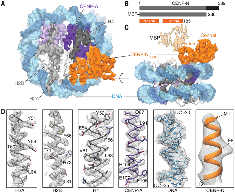
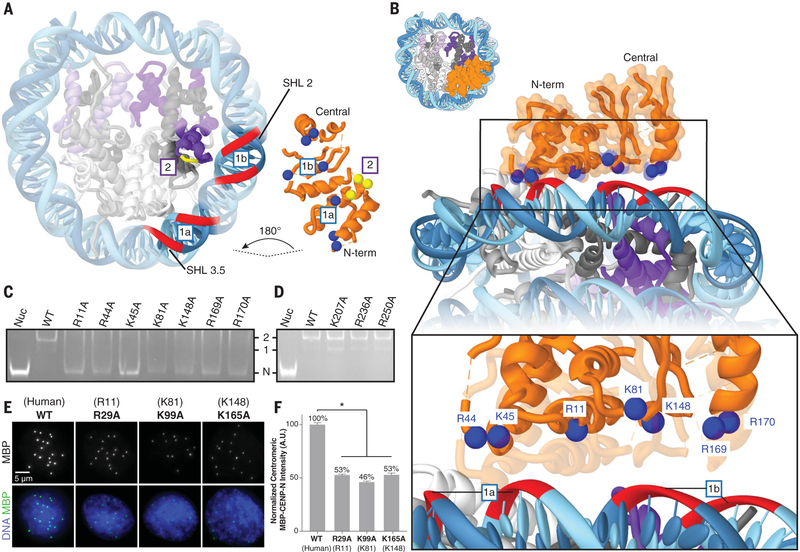
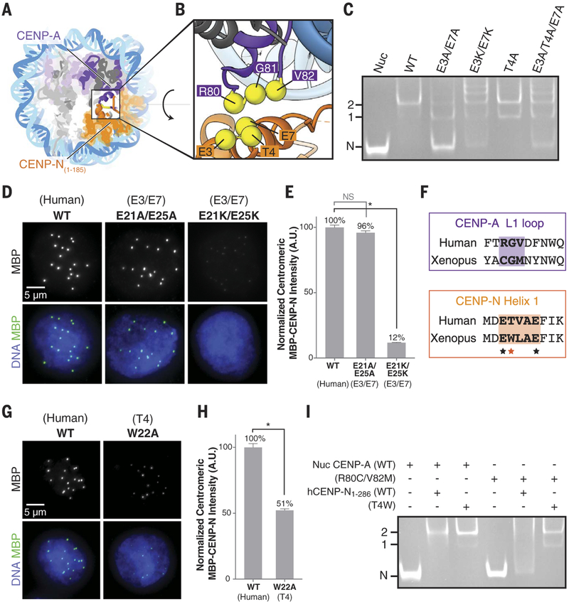
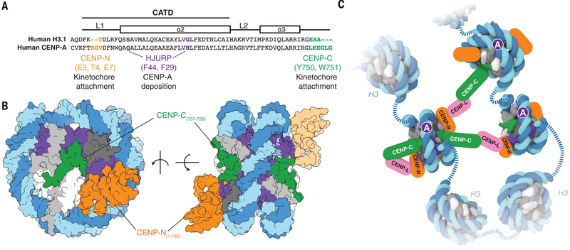
Accurate chromosome segregation requires the proper assembly of kinetochore proteins. A key step in this process is the recognition of the histone H3 variant CENP-A in the centromeric nucleosome by the kinetochore protein CENP-N. We report cryo-electron microscopy (cryo-EM), biophysical, biochemical, and cell biological studies of the interaction between the CENP-A nucleosome and CENP-N. We show that human CENP-N confers binding specificity through interactions with the L1 loop of CENP-A, stabilized by electrostatic interactions with the nucleosomal DNA. Mutational analyses demonstrate analogous interactions in Xenopus, which are further supported by residue-swapping experiments involving the L1 loop of CENP-A. Our results are consistent with the coevolution of CENP-N and CENP-A and establish the structural basis for recognition of the CENP-A nucleosome to enable kinetochore assembly and centromeric chromatin organization.


Figures

Fig. 1.
Fig. 1.. Structure of the human CENP-N/CENP-A nucleosome complex.
(A) Cryo-EM density map of the hCENP-N1–286/CENP-A nucleosome complex viewed down theaxis of the DNA supercoil. (B) Schematicof the functional domains of CENP-N known to bind the CENP-A nucleosome (gray) and CENP-L (black) (top panel). The CENP-N construct used for the present structural analysis (hCENP-N1–286) and the regions of the sequence whose structure we report here [N-terminal domain: residues 1 to 81, and central domain: residues 101 to 185; hCENP-N(1–185)] are shown in the middle and bottom panels, respectively. (C) Cryo-EM density mapof the hCENP-N1–286/CENP-A nucleosome complex as viewed from the side, at an orientation 90° to the view shownin (A). This view also depicts the extra density connected to the N-terminal domain that we assign to MBP, shown with lighter shading. (D) Representative regions of the cryo-EM density mapto illustrate map quality (from left to right) for canonical histones H2A, H2B, and H4, centromere-specific H3 variant CENP-A, nucleosomal DNA, and CENP-N.
Fig. 2.
Fig. 2.. Interaction of CENP-N with nucleosomal DNA.
(A) Cut-away view of the hCENP-N1–286/CENP-A nucleosome model to highlight interfaces involved in complex formation (see also fig. S11, A and B). For the CENP-N/DNA interface (labeled “1a” and “1b”), nucleosomal DNAis shown as a red ribbon, whereas positively charged residues of CENP-N that are proposed to interact with it are shown as blue spheres. Forthe CENP-N/CENP-A interface (labeled “2”), CENP-A residues (R80, G81, and V82) are marked by the short yellow ribbon, whereas interacting CENP-N residues (E3, T4, and E7) are shown as yellow spheres. (B) View of the CENP-N/DNA interface at different magnifications to highlight details of interactions between the nucleosomal DNA and positively charged residues of CENP-N. (C) Gel mobility shift experiment to examine the effects of CENP-N mutations (indicated atop the gel) on binding to the CENP-A nucleosome. Impaired binding is reflected by increased intensity of the free nucleosome (Nuc) band, concomitant with the disappearance of defined 1:1 and 2:1 bands. “N” indicates the migration position of the free CENP-A nucleosome; “1” and “2” denote the migration positions of CENP-A nucleosomes bound with either one or two molecules of CENP-N, respectively. WT, wild type. (D) Similar analysis to that in (C), carried out with a set of CENP-N mutations involving residues distal from the binding interface. (E) Images of interphase nuclei in Xenopus egg extracts with exogenous MBP-xCENP-N and xCENP-L proteins containing the indicated mutations (with analogous human mutations in parentheses), stained with an antibody forMBP (green) and Hoechst (blue). (F) Centromeric MBP fluorescence intensity normalized as a percentage of that observed for wild-type MBP-xCENP-N. Error bars represent SEM (n > 200 centromeres). A.U., arbitrary units.
Fig. 3.
Fig. 3.. Interaction between theL1 loop of CENP-A and helix 1 of CENP-N.
(A and B) Overall (A) and close-up (B) view of the hCENP-N1–286/CENP-A interface formed betweenR80, G81, and V82 on the L1 loop of CENP-A and E3, T4, and E7 on helix 1of CENP-N. (C) Gel mobility shift experiment to examine the effects of CENP-N mutations (indicated atopthe gel) on binding to the CENP-A nucleosome. (D) Images of interphase nuclei in Xenopus egg extracts with exogenous MBP-xCENP-L andxCENP-L proteins containing the indicated mutations of xCENP-N residues E21 and E25 (correspondingto residues E3 and E7 in hCENP-N), stained with an antibody forMBP (green) and Hoechst (blue).(E) Centromeric MBP fluorescence intensity normalized as a percentageof that observed for wild-type MBP-xCENP-N. Error bars representSEM (n > 200 centromeres). (F) Alignment of human and Xenopus laevis sequences corresponding to the L1 loop of CENP-A and helix 1 of CENP-N. Closely interacting segments of the L1 loop of CENP-A and helix 1 of CENP-N are highlighted bythe shaded areas. The asterisks indicate conserved glutamic acid residues(black asterisks) and variability in the hydrophobic residue correspondingto position T4 (red asterisk) of human CENP-N. (G) Images of interphase nuclei in Xenopus egg extracts with exogenous MBP-xCENP-N and xCENP-L proteins containing the indicated mutations of xCENP-N, as in (D).(H) Centromeric MBP fluorescence intensity, determined as in (E). (I) Gel mobility shift experiment to examine the effects of correlated amino acid substitutions between the L1 loop of CENP-A and helix 1 of CENP-N.
Fig. 4.
Fig. 4.. Structural determinants of kinetochore assembly on the CENP-A nucleosome.
(A) Sequence alignment between humanH3.1 and CENP-A to highlight distinct CENP-A motifs involvedin deposition and recognition of CENP-A at centromeric chromatin (see also fig. S11, C and D). Single-letter abbreviations for the amino acid residues are as follows: A, Ala; C, Cys; D, Asp; E, Glu; F, Phe; G, Gly; H, His; I, Ile; K, Lys; L, Leu; M, Met; N, Asn; P, Pro; Q, Gln; R, Arg; S, Ser; T, Thr; V, Val; W, Trp; and Y, Tyr. (B) Two different views of the CENP-A nucleosome bound to hCENP-N and a modeled CENP-C motif peptide (5) to highlight potential dual binding of full-length CENP-C and CENP-N proteins on the CENP-A nucleosome. The second CENP-N (shown with lighter shading) is modeled on the basis of the cryo-EM density map obtained in the presence of excess hCENP-N1–286 (fig. S4), whereas the CENP-C motif peptides (human numbering shown for clarity) on each face of the nucleosome are positioned according to the crystal structure of the nucleosome in complex with the rat CENP-C motif (5). (C) Schematic view to highlight recognition and possible enrichment of CENP-A nucleosomes by the CCAN proteins CENP-C, CENP-N, and CENP-L. Other kinetochore proteins and the dimerization of CENP-C have been omitted for clarity.

References

    1. Kops GJ, Weaver BA, Cleveland DW, Nat. Rev. Cancer 5, 773–785 (2005). - PubMed
    1. McKinley KL, Cheeseman IM, Nat. Rev. Mol. Cell Biol. 17, 16–29 (2016). - PMC - PubMed
    1. Verdaasdonk JS, Bloom K, Nat. Rev. Mol. Cell Biol. 12, 320–332 (2011). - PMC - PubMed
    1. Carroll CW, Milks KJ, Straight AF, Cell Biol J. 189, 1143–1155 (2010). - PMC - PubMed
    1. Kato H et al., Science 340, 1110–1113 (2013). - PMC - PubMed

Publication types

MeSH terms