. 2018 Feb;38(2):335-343.
doi: 10.1161/ATVBAHA.117.310491. Epub 2017 Dec 21.

Vascular Semaphorin 7A Upregulation by Disturbed Flow Promotes Atherosclerosis Through Endothelial β1 Integrin

Affiliations

Vascular Semaphorin 7A Upregulation by Disturbed Flow Promotes Atherosclerosis Through Endothelial β1 Integrin

Shuhong Hu et al. Arterioscler Thromb Vasc Biol. 2018 Feb.

Abstract

Objective: Accumulating evidence suggests a role of semaphorins in vascular homeostasis. Here, we investigate the role of Sema7A (semaphorin 7A) in atherosclerosis and its underlying mechanism.

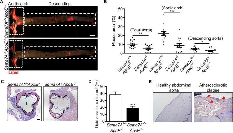
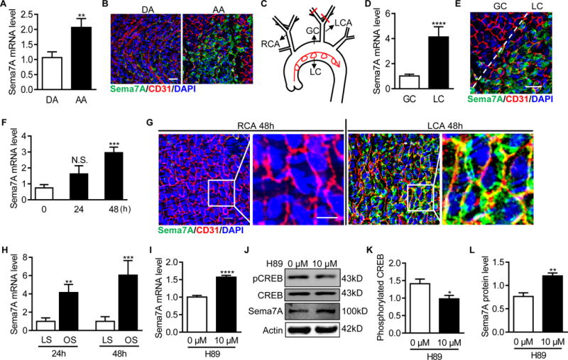
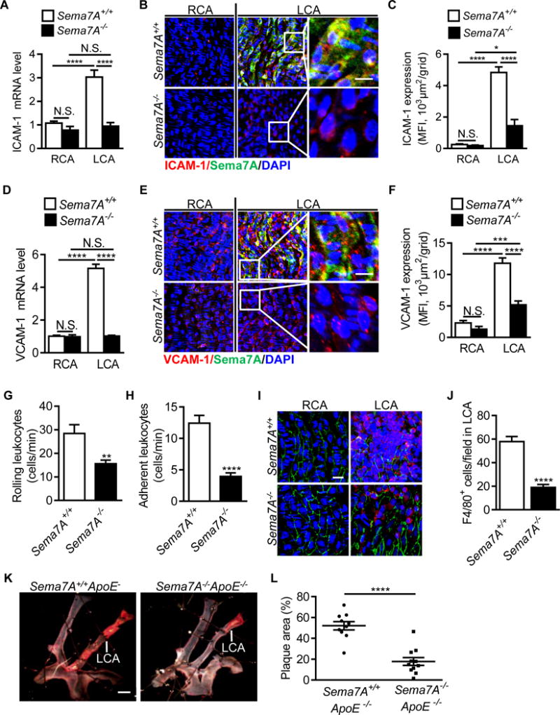
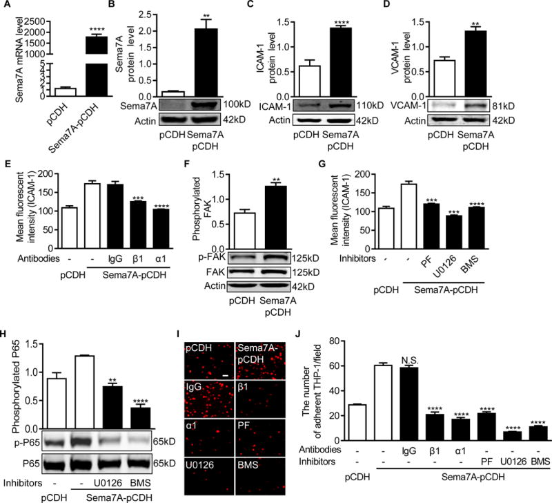
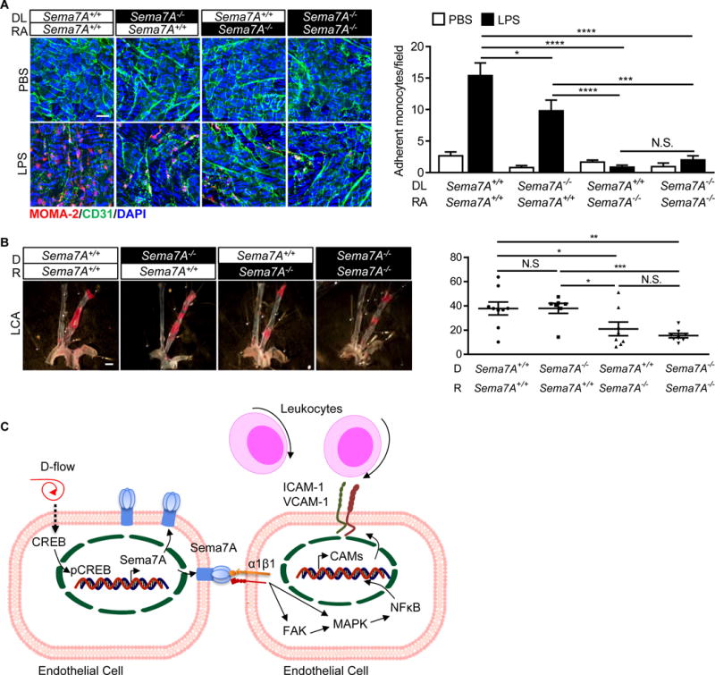
Approach and results: Using genetically engineered Sema7A-/-ApoE-/- mice, we showed that deletion of Sema7A attenuates atherosclerotic plaque formation primarily in the aorta of ApoE-/- mice on a high-fat diet. A higher level of Sema7A in the atheroprone lesser curvature suggests a correlation of Sema7A with disturbed flow. This notion is supported by elevated Sema7A expression in human umbilical venous endothelial cells either subjected to oscillatory shear stress or treated with the PKA (protein kinase A)/CREB (cAMP response element-binding protein) inhibitor H89 (N-[2-(p-bromocinnamylamino)ethyl]-5-isoquinolinesulfonamide·2HCl hydrate). Further studies using the partial carotid artery ligation model showed that disturbed flow in the left carotid artery of Sema7A+/+ApoE-/- mice promoted the expression of endothelial Sema7A and cell adhesion molecules, leukocyte adhesion, and plaque formation, whereas such changes were attenuated in Sema7A-/-ApoE-/- mice. Further studies showed that blockage of β1 integrin, a known Sema7A receptor, or inhibition of FAK (focal adhesion kinase), MEK1/2 (mitogen-activated protein kinase kinase 1/2), or NF-κB (nuclear factor-κB) significantly reduced the expression of cell adhesion molecules and THP-1 (human acute monocytic leukemia cell line) monocyte adhesion in Sema7A-overexpressing human umbilical venous endothelial cells. Studies using chimeric mice suggest that vascular, most likely endothelial, Sema7A plays a major role in atherogenesis.

Conclusions: Our findings indicate a significant role of Sema7A in atherosclerosis by mediating endothelial dysfunction in a β1 integrin-dependent manner.

Keywords: atherosclerosis; diet, high fat; monocytes; semaphorins; upregulation.


Figures

Figure 1
Figure 1
Sema7A deletion reduces lipid deposition in the aorta of ApoE−/− mice and vascular Sema7A is up-regulated during atherogenesis (A) Aortic plaque areas in Sema7A+/+ApoE−/− and Sema7A−/−ApoE−/− mice on HFD for 12 w were analyzed by en face Sudan IV-staining (Bar=2 mm). (B) Quantitative analysis for total aorta, aortic arch and descending aorta are shown. Data are mean ± SEM (n≥10 mice per group). *P<0.05; **P<0.01; ***P<0.001. (C, D) Aortic root sections from Sema7A+/+ApoE−/− and Sema7A−/−ApoE−/− mice on HFD for 12 w were stained with Oil Red O. Plaque area were demarcated by dotted lines (Bar=200 μm). (E) Sema7A expression in human atherosclerotic plaques was detected by immunostaining in brown (arrows). Nuclei were stained by hematoxylin (Bar=100 μm).
Figure 2
Figure 2
Disturbed blood flow up-regulates vascular endothelial Sema7A expression. (A, D) Total RNAs in the aortic arch (AA) or the descending aorta (DA) (A), and in the lesser (LC) or greater (GC) curvature regions of the aortic arch (D) from C57BL/6J mice were isolated. Sema7A mRNA expression was analyzed by qPCR normalized to GAPDH. Fold-changes are shown. Data are mean ± SEM. **P<0.01; ****P<0.0001. Results are representative of ≥3 independent experiments. (B, E) En face staining for CD31 (red) and Sema7A (green) on the aortic arch (AA) or the descending aorta (DA) (B) and the lesser (LC) or greater (GC) curvature (E) was shown. Nuclei were stained with DAPI (blue). Bar = 50 μm. The data are representative of ≥3 independent experiments. (C) The diagram of aortic arch and carotid arteries. (F) Total RNAs of LCA and the control RCA from C57BL/6J mice were isolated 24h and 48 h after PCL. The ratio of LCA to RCA was calculated (n≥5 mice per group). Data are mean ± SEM. ***P<0.001. The bar graphs represent the results from 3 independent experiments (one-way ANOVA with non-parametric multiple comparison test) (G) Carotid arteries from C57BL/6J 48 h after PCL were en face stained for CD31 (red) and Sema7A (green). Nuclei were stained with DAPI (blue). Bar = 10 μm. (H) Sema7A mRNA levels in HUVECs exposed to LS (15 dyn/cm2) or OS (±5 dyn/cm2, at 1 Hz) were analyzed by qPCR. Data are mean ± SEM, n=3, **P<0.01, ***P<0.001. (I-L) HUVECs treated with H89 for 24 h and Sema7A mRNA expression was analysed by qPCR (I) Sema7A protein expression (J, L) and CREB phosphorylation were analyzed by Western blotting (J, K). Data are mean ± SEM, n=3, *P<0.05; **P<0.01; ***P<0.001; ****P<0.0001. RCA: right common carotid artery; LCA: left common carotid artery; DA: descending aorta; AA: aortic arch; GC: greater curvature; LC: lesser curvature; LS: laminar shear stress; OS: oscillatory shear stress.
Figure 3
Figure 3
Sema7A deficiency reduces d-flow induced endothelial expression of ICAM-1 and VCAM-1, leukocyte adhesion and plaque formation. ICAM-1 and VCAM-1 mRNA (A, D) and protein (B, E) level in LCA and RCA in Sema7A+/+ and Sema7A−/− mice after PCL were analyzed by qPCR (n=9 mice per group) and en face immunostaining of aortic luminal surface (red: ICAM-1 or VCAM-1; green: Sema7A; blue: nuclei. Bar = 10 μm). Data are representative of ≥3 independent experiments. Mean fluorescent intensity for ICAM-1 (C) and VCAM-1 (F) was used for the statistical analysis of panels B and E, respectively. Each result includes 15-30 fields from 3 mice. Data are mean ± SEM. *P<0.05; ***P<0.001; ****P<0.0001. (G, H) Mice were injected with TNF-α around the cremaster muscle. Rolling and adhesion of leukocytes on cremaster venules (4-5 vessels per mouse) were analyzed under a real-time intravital microscope and counted visually as numbers of rolling (G) or adherent cells (H) on the vascular surface per min (n=6 mice per group). Data are mean ± SEM. **P<0.01; ****P<0.0001. Results are representative of ≥3 independent experiments. (I, J) Adhesion of F4/80+ leukocytes in partially ligated LCA was observed under a confocal microscope and recorded as numbers of adherent cells on the vascular surface per field (Bar = 20 μm). Data are mean ± SEM (n≥9 per group). ****P<0.0001. Results are representative of ≥3 independent experiments. (K) Sema7A+/+ApoE−/− and Sema7A−/−ApoE−/− mice were subjected to PCL. After 2 w on HFD, lipid deposition (in red) was analyzed by en face Sudan IV-staining. Bar=1 mm. The lesion surface areas were quantified and displayed as the percentage area of the LCA (L). Data are mean ± SEM (n≥9 mice per group). ****P<0.0001. RCA: right common carotid artery; LCA: left common carotid artery.
Figure 4
Figure 4
Sema7A overexpression enhances ICAM-1 and VCAM-1 expression in HUVECs and monocyte-endothelial cell interaction via β1 integrin and the downstream FAK/MEK1/2/NFκB pathway (A) Sema7A mRNA expression in the Lenti-pCDH-hSema7A-GFP-transduced HUVECs was analyzed by qPCR (n≥5 per group). Data are mean ± SEM. ***P<0.001. (B~D) Sema7A (B), ICAM-1 (C) and VCAM-1 (D) proteins in Lenti-pCDH-hSema7A-GFP-transduced HUVECs were analyzed by Western blotting normalized to β-actin and displayed as fold-changes relative to Lenti-pCDH-GFP-transduced control HUVECs. Data are mean ± SEM. Results are representative of ≥3 independent experiments. **P<0.01; ***P<0.001; ****P<0.0001. (E) Sema7A-overexpressing and control HUVECs were pre-treated with blocking antibodies against β1 (P5D2) or α1 (5E8D9) integrin subunits for 24 h and analyzed for ICAM-1 expression by flow cytometry. Data are mean ± SEM. ****P<0.0001. Results are representative of ≥3 independent experiments. (F) FAK phosphorylation in Sema7A-overexpressing HUVECs was analyzed by Western blotting normalized to total FAK, and displayed as fold-changes relative to the control HUVECs. Data are mean ± SEM. **P<0.01. Results are representative of ≥3 independent experiments. (G) Sema7A-overexpressing and control HUVECs were pre-treated with PF573228 (10μM), U0126 (10 μM) and BMS-345541 (20 μM) for 24 h. The cells were analyzed by flow cytometry for ICAM-1 expression. (H) Cells were treated with U0126 (10 μM) and BMS-345541 (20 μM), and the lysates were analyzed by Western blotting for NFκB (p65) phosphorylation. Data are mean ± SEM. **P<0.01; ****P<0.0001. Results are representative of ≥3 independent experiments. (I, J) THP-1 cells were stained by DiL and incubated with HUVECs for 30 min at 37°C. HUVECs were also pretreated with blocking antibodies against β1 or α1 integrin subunits or inhibitors for FAK, MAPK/MEK1/2 or NFκB for 24 h. Adherent THP-1 cells (red) on the monolayer of Sema7A-overexpressing or control HUVECs were visualized under a microscope (I) and statistically analyzed (J). Bar=20 μm. Data are mean ± SEM. ****P<0.0001. Results are representative of ≥3 independent experiments. PF: PF573228, FAK inhibitor; U0126: MAPK/MEK1/2 inhibitor; BMS: BMS-345541, NFκB inhibitor.
Figure 5
Figure 5
Endothelial Sema7A plays a major role in promoting leukocyte adhesion and plaque formation in ApoE−/− mice (A) Mouse thoracic aortas were isolated from Sema7A−/−ApoE−/− and Sema7A+/+ApoE−/− mice (8-w-old). The exposed endothelium was treated with LPS (5 μg in 2 mL medium per well) for 4 h and cultured with leukocytes from Sema7A−/−ApoE−/− or Sema7A+/+ApoE−/− mice for 30 min at 37°C. Adhered monocytes on the aortic endothelium were stained with a fluorescence-labeled anti-MOMA-2 antibody (red). Bar = 20 μm. Data are mean ± SEM. Results are representative of ≥3 independent experiments. *P<0.05; ***P<0.001; ****P<0.0001. (B) Sema7A+/+ApoE−/− and Sema7A−/−ApoE−/− mice were transplanted with Sema7A+/+ApoE−/− and Sema7A−/−ApoE−/− bone marrow cells and recovered for the following 4 w before subjected to PCL. After additional 2 w on HFD, atherosclerotic lipid deposition (in red) was analyzed by en face Sudan IV-staining. Bar=1 mm. The lesion surface areas were quantified and displayed as the percentage area of the LCA. Data are mean ± SEM (n≥7 mice per group). *P<0.05; **P<0.01; ***P<0.001. DL: donor leukocyte; RA: recipient aorta; D: donor; R: recipient; LCA: left common carotid artery. (C). A proposed model for the role of Sema7A in d-flow-induced endothelial phenotypic changes and leukocyte recruitment in atherogenesis. When exposed to d-flow, endothelial Sema7A expression is upregulated, potentially by the cAMP/CREB pathway. Endothelial Sema7A activates the transcription of ICAM-1/VCAM-1 genes through binding to its receptor integrin α1β1, resulting in FAK/MAPK/NFκB signaling pathway activation and the recruitment of leukocytes into atherosclerotic lesions. D-flow: disturbed flow.

References

    1. Davies PF, Zilberberg J, Helmke BP. Spatial microstimuli in endothelial mechanosignaling. Circ Res. 2003;92:359–70. doi: 10.1161/01.RES.0000060201.41923.88. - DOI - PubMed
    1. Feaver RE, Gelfand BD, Blackman BR. Human haemodynamic frequency harmonics regulate the inflammatory phenotype of vascular endothelial cells. Nat Commun. 2013;4:1525. doi: 10.1038/ncomms2530. - DOI - PMC - PubMed
    1. Giannotta M, Trani M, Dejana E. VE-cadherin and endothelial adherens junctions: active guardians of vascular integrity. Dev Cell. 2013;26:441–54. doi: 10.1016/j.devcel.2013.08.020. - DOI - PubMed
    1. Gimbrone MA. The Gordon Wilson lecture. Understanding vascular endothelium: a pilgrim’s progress. Endothelial dysfunction, biomechanical forces and the pathobiology of atherosclerosis. Trans Am Clin Climatol Assoc. 2010;121:115–27. discussion 127. - PMC - PubMed
    1. Garcia-Cardena G, Comander J, Anderson KR, Blackman BR, Gimbrone MA., Jr Biomechanical activation of vascular endothelium as a determinant of its functional phenotype. Proc Natl Acad Sci U S A. 2001;98:4478–85. doi: 10.1073/pnas.071052598. - DOI - PMC - PubMed

Publication types

MeSH terms