. 2017 Dec 21;7(1):18015.
doi: 10.1038/s41598-017-18237-6.

Ablation of peri-insult generated granule cells after epilepsy onset halts disease progression

Affiliations

Ablation of peri-insult generated granule cells after epilepsy onset halts disease progression

Bethany E Hosford et al. Sci Rep. .

Abstract

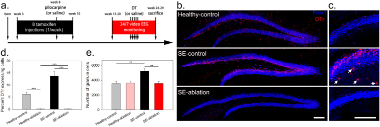
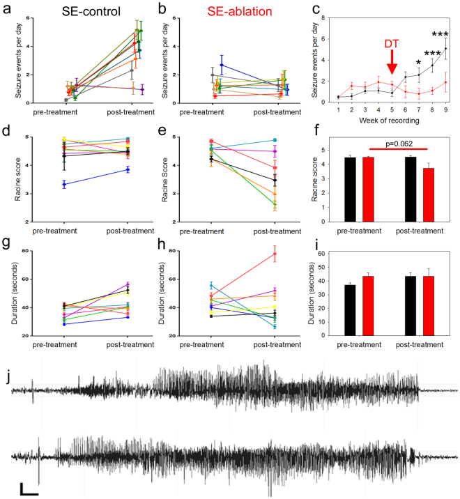
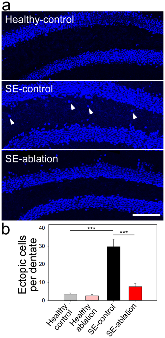
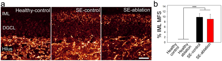
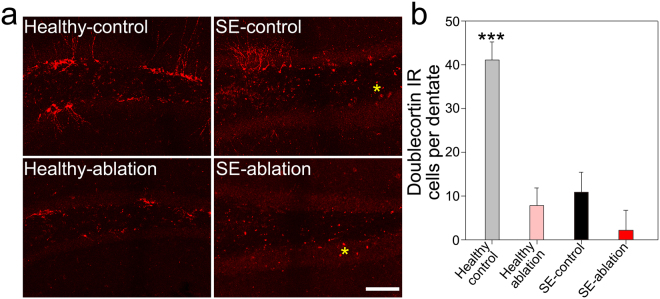
Aberrant integration of newborn hippocampal granule cells is hypothesized to contribute to the development of temporal lobe epilepsy. To test this hypothesis, we used a diphtheria toxin receptor expression system to selectively ablate these cells from the epileptic mouse brain. Epileptogenesis was initiated using the pilocarpine status epilepticus model in male and female mice. Continuous EEG monitoring was begun 2-3 months after pilocarpine treatment. Four weeks into the EEG recording period, at a time when spontaneous seizures were frequent, mice were treated with diphtheria toxin to ablate peri-insult generated newborn granule cells, which were born in the weeks just before and after pilocarpine treatment. EEG monitoring continued for another month after cell ablation. Ablation halted epilepsy progression relative to untreated epileptic mice; the latter showing a significant and dramatic 300% increase in seizure frequency. This increase was prevented in treated mice. Ablation did not, however, cause an immediate reduction in seizures, suggesting that peri-insult generated cells mediate epileptogenesis, but that seizures per se are initiated elsewhere in the circuit. These findings demonstrate that targeted ablation of newborn granule cells can produce a striking improvement in disease course, and that the treatment can be effective when applied months after disease onset.


Conflict of interest statement

The authors declare that they have no competing interests.

Figures

Figure 1
Figure 1
DT ablation effectively eliminates DTr expressing newborn dentate granule cells. (a) Timeline depicting the experimental treatment paradigm. (b) Images of Prox1 (blue) and DTr (red) immunostained tissue from healthy-control, SE-control and SE-ablation groups. (c) Higher resolution images of Prox1 and DTr immunostaining in the dentate gyrus showing DTr induction in a small number of reactive astrocytes in the dentate molecular layer (arrows) of a SE-control mouse. (d) Percentage of granule cells that were DTr-positive within each treatment group. (e) Number of Prox1-positive dentate granule cells per dentate section. **p < 0.01, ***p < 0.001, scale bars: 100 µm.
Figure 2
Figure 2
Cell ablation treatment blocks epilepsy progression. Pre-treatment and post-treatment seizure frequencies (a,b), severities (d,e), and durations (g,h) are shown for SE-control mice (left, black) and SE-ablation mice (middle, red). Each line shows the means ± SEM for one animal. (c) Average number of seizure events during each week of recording for SE-control (black) and SE-ablation (red) groups (DT was given during week 5, red arrow). (f) Average behavioral seizure scores and (i) durations. (j) Representative post-treatment electrographic seizures from SE-control (top) and SE-ablation (bottom) mice. *p < 0.05, **p < 0.01, ***p < 0.001, scale bars: 300 μV and 2 seconds.
Figure 3
Figure 3
Cell ablation reduces ectopic cell numbers. (a) Prox1-stained dentate gyri from healthy-control (top), SE-control (middle), and SE-ablation (bottom) mice. Arrowheads denote ectopic cells. (b) Quantification of the number of ectopic granule cells per dentate section in each of the four treatment groups. ***p < 0.001, scale bar: 100 µm.
Figure 4
Figure 4
Cell ablation does not reduce mossy fiber sprouting. (a) ZnT3-immunostaining of mossy fiber terminals in healthy-control (left), SE-control (middle), and SE-ablation (right) mice. (b) Graph shows the percentage of the inner molecular layer (IML) occupied by ZnT3-immunoreative terminals for each group. DGCL = dentate granule cell body layer, ***p < 0.001, scale bar scale bar: 25 µm.
Figure 5
Figure 5
Representative confocal images of doublecortin immunoreactivity in the dentate gyri of (a) Healthy-control, healthy-ablation, SE-control and SE-ablation mice. Yellow asterisks denote examples of autofluorescent cellular debris in epileptic mice, and not doublecortin immunoreactivity (b) Quantification of doublecortin-immunoreactive granule cells per dentate gyrus in the four groups. ***p < 0.001 vs. all other groups. Scale bar = 100 µm.
Figure 6
Figure 6
Confocal maximum projections of the dentate hilus showing immunostaining for microglial (Iba1, red) and astrocytic (GFAP, green) markers. (a) Healthy-control that received diphtheria toxin but lacked the receptor, so ablation did not occur (C + DT, −A). (b) Healthy-control that expressed the diphtheria toxin receptor, but received saline, so ablation did not occur (C + NaCl, −A). (c) Healthy-ablation, expressing the receptor and receiving toxin (C + DT, + A). (d) SE-control that received toxin but lacked the receptor (SE + DT, −A). (e) SE-control that expressed the diphtheria toxin receptor, but received saline (SE + NaCl, −A). (f) SE-ablation, expressing the receptor and receiving toxin (SE + DT, + A). Scale bar = 50 µm. (g) Graph showing the average soma area for GFAP immunopositive astrocytes. Status epilepticus was associated with increased astrocyte soma area. ***p < 0.001 vs. Group B. *p < 0.05 vs. Group B. ##p < 0.01 vs. Group A. (h) Graph detailing the average soma area for Iba1 positive microglia cells within each treatment group. Status epilepticus (SE) was associated with increased microglial soma area. **p < 0.01 vs. Group B. *p < 0.05 vs. Group B. #p < 0.05 vs Group C. See Table 1 for group details.

References

    1. Walter C, Murphy BL, Pun RY, Spieles-Engemann AL, Danzer SC. Pilocarpine-induced seizures cause selective time-dependent changes to adult-generated hippocampal dentate granule cells. J. Neurosci. 2007;27:7541–7552. doi: 10.1523/JNEUROSCI.0431-07.2007. - DOI - PMC - PubMed
    1. Scharfman HE, Sollas AE, Berger RE, Goodman JH, Pierce JP. Perforant path activation of ectopic granule cells that are born after pilocarpine-induced seizures. Neuroscience. 2003;121:1017–1029. doi: 10.1016/S0306-4522(03)00481-0. - DOI - PubMed
    1. Singh, S. P., LaSarge, C. L., An, A., McAuliffe, J. J. & Danzer, S. C. Clonal Analysis of Newborn Hippocampal Dentate Granule Cell Proliferation and Development in Temporal Lobe Epilepsy. eNeuro2, 10.1523/eneuro.0087-15.2015 (2015). - PMC - PubMed
    1. Althaus AL, Sagher O, Parent JM, Murphy GG. Intrinsic neurophysiological properties of hilar ectopic and normotopic dentate granule cells in human temporal lobe epilepsy and a rat model. J. Neurophysiol. 2015;113:1184–1194. doi: 10.1152/jn.00835.2014. - DOI - PMC - PubMed
    1. Zhan RZ, Timofeeva O, Nadler JV. High ratio of synaptic excitation to synaptic inhibition in hilar ectopic granule cells of pilocarpine-treated rats. J. Neurophysiol. 2010;104:3293–3304. doi: 10.1152/jn.00663.2010. - DOI - PMC - PubMed

Publication types

LinkOut - more resources