. 2017 Dec 21;8(1):2234.
doi: 10.1038/s41467-017-01604-2.

A molecular basis for water motion detection by the mechanosensory lateral line of zebrafish

Affiliations

A molecular basis for water motion detection by the mechanosensory lateral line of zebrafish

Shih-Wei Chou et al. Nat Commun. .

Abstract

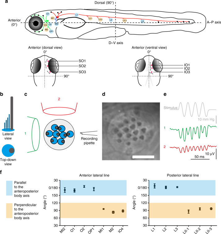
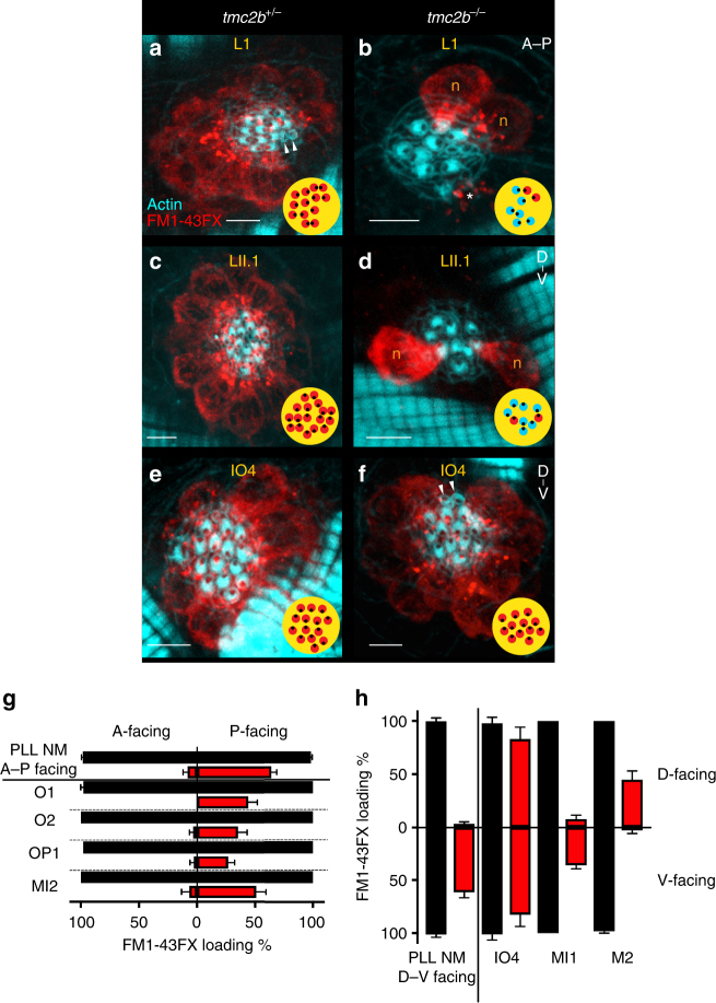
Detection of water motion by the lateral line relies on mechanotransduction complexes at stereocilia tips. This sensory system is comprised of neuromasts, patches of hair cells with stereociliary bundles arranged with morphological mirror symmetry that are mechanically responsive to two opposing directions. Here, we find that transmembrane channel-like 2b (Tmc2b) is differentially required for mechanotransduction in the zebrafish lateral line. Despite similarities in neuromast hair cell morphology, three classes of these cells can be distinguished by their Tmc2b reliance. We map mechanosensitivity along the lateral line using imaging and electrophysiology to determine that a hair cell's Tmc2b dependence is governed by neuromast topological position and hair bundle orientation. Overall, water flow is detected by molecular machinery that can vary between hair cells of different neuromasts. Moreover, hair cells within the same neuromast can break morphologic symmetry of the sensory organ at the stereocilia tips.


Conflict of interest statement

The authors declare no competing financial interests.

Figures

Fig. 1
Fig. 1
Axes of best sensitivity for anterior and posterior lateral line neuromasts. a Schematic of the zebrafish lateral line at ~6 dpf. (top) Side view. Neuromasts that have been scored are blue or tan and correspond to the graph below. ALL(green), PLL(red), and medial–lateral-line (blue) ganglia contact neuromasts. (bottom) Dorsal and ventral views of the head. Supraorbital (SO) neuromasts 1 to 3 and infraorbital (IO) neuromasts 1 to 3 are marked in red. The mean angle of the orientation of the axis of best sensitivity relative to the A–P body axis of each neuromast is presented as a double-headed arrow. be Evaluation of the axis of best sensitivity for a neuromast. b Schematics of hair bundles. Gray and blue protrusions are a kinocilium and stereocilia, respectively. c Experimental configuration: a top-down view of a neuromast with a fluid jet pipette that provides stimuli (1) parallel to the axis  along which the bevel-shaped hair bundles face opposite directions or (2) orthogonal to that axis. The pipettes in the diagram are not to scale. d DIC image of hair bundles of a posterior neuromast from which stimulus-evoked microphonic potentials were recorded. Scale bar = 5 μm. e Recordings from an A–P oriented L3 neuromast with fluid jet pipettes stimulating, serially, from two different directions depicted in c. f Graphs of mean angle of the orientation of the axis of best sensitivity relative to the A–P body axis of each anterior and posterior neuromast position ± SEM (n= 4–7). Each axis with a range from 60° to 120° from the A–P body axis is defined perpendicular (tan). Each axis with a range from 0° to 30° or 150° to 180° from the A–P body axis is defined as parallel (blue). ALL nomenclature: opercular (OP), mandibular (M), infraorbital (IO), supraorbital (SO), middle (MI) and otic (O). PLL nomenclature: L neuromasts are A–P oriented; LII neuromasts are D–V oriented. These different neuromast types are derived from different migrating primordia during development
Fig. 2
Fig. 2
Localization of Tmc2b and TALEN-mediated disruption of the tmc2b gene. a Schematic of the hair bundle. b Tmc2b localizes to the tips of stereocilia in a neuromast. Two lateral line hair cells expressing Tmc2b-GFP (green) demonstrate that the fusion protein localizes to the tips of β-actin-mCherry-labeled stereocilia (red). Scale bar = 0.5 μm. This pattern was observed in 130 of 381 hair cells of somatic transgenics. In transgenic hair cells with weak expression, localization to stereocilia was difficult to recognize. c Graphical representation of the tmc2b genomic locus in zebrafish. Putative exons and splice sites are displayed. Red arrow marks the targeted exon, exon 4. d Segment of exon 4 subjected to genome editing. Two differently engineered TALENs bind their corresponding half-sites to enable FokI dimerization and DNA cleavage. Mutagenesis deleted seven nucleotides. e Amino acid sequences of wild-type and mutant proteins. The alteration results in an opal mutation upstream of all putative transmembrane domains. f Sequencing results of mutagenized and control loci. Blue highlight and blue delta indicates deleted 7-nucleotide stretch absent in mutant. Red highlight denotes the opal mutation that was generated at the site of the TALEN targeting. g (left) Topographical representation of the Tmc2b protein. Red arrowhead indicates point of introduced mutation. Amino acids of putative transmembrane domains are labeled in blue and the TMC domain is in green. (right) Predicted truncated product of mutation
Fig. 3
Fig. 3
Variable dependence on Tmc2b for mechanotransduction in posterior neuromast hair cells. a Extracellular recordings of microphonic potentials measured from posterior neuromasts with A-P orientations of 6-dpf zebrafish larvae are displayed. The responses in tmc2b −/− are often absent, bottom trace (n = 9/15); however, several responses (n = 6/15) are highly asymmetric, with weakened amplitude for one direction of stimulus and no response for the other direction, penultimate trace. Deflection anteriorly (A) or posteriorly (P). b, c Qualitative confocal images of hair cells labeled with FM1-43FX (red), to assess mechanotransduction function, and parvalbumin 3 (cyan), as a counter label, to visualize mature hair cells. b A posterior neuromast from a tmc2b +/− animal demonstrates co-labeling of all hair cells. c In a tmc2b −/− animal, a posterior neuromast contains hair cells that label with FM1-43FX and a subset that do not (label:no label; 2:5). Orange n, hair cells labeled with parvalbumin 3 antiserum. Blue n, hair cells that load with FM1-43FX. Green n, hair cells that do not load. Scale bar = 5 μm. d, e Percentages of hair cells within PLL neuromasts that label with 4-Di-2-ASP. d In neuromasts of tmc2b +/+ and tmc2b +/− animals, most hair cells take up fluorophore. Approximately 35% of hair cells in each PLL neuromast of tmc2b −/− fish take up 4-Di-2-ASP. In contrast, in cdh23 aj64a/aj64a mutants, all hair cells take up dye, though at much lower quantities than wild-type animals (see f and Supplementary Fig. 4b). **** Kruskal–Wallis test P value < 0.0001. n values ≥ 20. e In mutants, A–P-facing and D–V-facing posterior neuromasts have similar percentages of hair cells that gather fluorophore, ~38 and 32%, respectively. ****Student’s t-test P value < 0.0001. n values ≥ 23. f Mean fluorescence intensity of 4-Di-2-ASP uptake of the brightest cell per neuromast. n values ≥ 17. Kruskal–Wallis test ****P < 0.0001, ***P = 0.001, **P = 0.0215
Fig. 4
Fig. 4
Spatial positioning of anterior neuromasts regulates diverse Tmc2b dependence. a Confocal micrographs of MI1 hair cells incubated with 4-Di-2-ASP from tmc2b +/− (left) or tmc2b −/− (right) animals at 6 dpf, viewed under low gain. b Hair cells from a, right, viewed under high gain. c Percentages of MI1 hair cells at different fluorescence intensities (n = 5). d Images and e percentages of hair cells at different fluorescence intensities from IO4 neuromasts (n = 5). f Percentages of hair cells of ALL neuromasts that take up 4-Di-2-ASP. ****One-way ANOVA with Holm-Sidak’s multiple comparisons test P< 0.0001. n values (het/homo): IO4 = 6/4, MI1 = 7/5, M2 = 5/3, O1 = 5/2, O2 = 5/4, OP1 = 5/3, MI2 = 5/5. g Mean whole-neuromasts normalized fluorescence intensity ratios, I tmc2b −/−/ (I tmc2b +/+, I tmc2b +/). n values ≥ 5. Scale bar = 6 µm
Fig. 5
Fig. 5
Hair cell PCP and neuromast position govern dependence of mechanotransduction channel function on Tmc2b. af Confocal images of hair cells from L1, LII.1, and IO4 neuromasts of tmc2b +/− or tmc2b −/− animals. FM1-43FX (red) uptake reveals functional channels, and phalloidin (cyan) shows hair bundle polarity. Qualitative maps (yellow) of micrographs from tmc2b −/− mutants show that subpopulations of posterior- and ventral-facing hair bundles preferentially function (red) in L1 and LII.1, respectively; in contrast, hair cell uptake in IO4 is non-biased. g In PLL neuromasts with A–P orientations of tmc2b −/− mutants, the vast majority (n = 21) of FM1-43FX uptake is by hair bundles that are posterior facing (P-facing). Similarly, in the A–P-oriented neuromasts of the ALL of tmc2b −/− (O1, O2, OP1, and MI2), the overwhelming majority of FM1-43FX uptake is by P-facing hair cells (n ≥ 5, except for O1, n = 4). Red bars represent tmc2b −/−, and black bars signify tmc2b +/+ and tmc2b +/−. h In the PLL neuromasts with D–V orientations of tmc2b −/− mutants, ventral-facing (V-facing) hair cells dominate the population that uptakes the fluorophore (n = 16). In ALL neuromasts with D–V orientations of tmc2b −/−, the patterns of hair cells that take up FM1-43FX are complex. For IO4, there is no significant difference between the numbers of D- and V-facing hair cells that loaded with FM1-43FX. The FM1-43FX loading percentages are 82.6 ± 11.1 % (n = 5) for V-facing hair cells and 83.0 ± 11.1 % (n = 5) for D-facing hair cells in tmc2b −/−, respectively. One-way ANOVA with Holm-Sidak’s multiple comparisons test, P= 0.9986. D–V-oriented neuromasts MI1 and M2 have opposite PCP-related loading preferences in mutants. In MI1 and M2 of tmc2b −/− animals, the percentages of V-facing hair cells that load with FM1-43FX are 36.1 ± 3.4% (n = 6) and 2.9 ± 2.9% (n = 5), respectively. Whereas, the percentages of D-facing hair cells from MI1 and M2 that load with FM1-43FX are 7.9 ± 3.6% (n = 6) and 44.9 ± 8.3% (n = 5), respectively (see Supplementary Table 1 for statistics). Corresponding images of MI1 and M2 displayed in Supplementary Fig. 7. Scale bar = 5 μm. Arrowheads, immature hair cells that do not take up FM1-43FX. *dying hair cell
Fig. 6
Fig. 6
A mechanosensory map of Tmc2b dependence in the anterior and posterior lateral lines. In this graphic of a larval zebrafish tmc2b −/− mutant, red neuromasts have been characterized. Individual neuromasts with patterns of FM1-43FX uptake are displayed. Red hair cells take up FM1-43FX; gray hair cells do not. Below each schematized neuromast is the percentage of hair cells that load 4-Di-2-ASP (red text) in the tmc2b −/− mutant and the percentage of fluorescence intensity of hair cells of the mutant relative to hair cells of wild-type and heterozygous animals (black). Green arrows represent the directional sensitivity preserved in the mutant based on FM1-43FX uptake. Graphs of mean microphonic potentials from neuromasts directly above each plot are displayed. For IO4, tmc2b +/ = 9.2 ± 0.7 μV (n = 9), and tmc2b −/− = 8.9 ± 0.87 μV (n = 6). P value = 0.9546. For A–P oriented posterior neuromasts, tmc2b +/+, tmc2b +/− = 8.3 ± 0.37 μV (n = 20), and tmc2b −/− = 2.1 ± 0.8 μV (n = 15). ****P value < 0.0001. For D–V oriented posterior neuromasts, tmc2b +/− = 6 ± 0.49 μV (n = 4) and tmc2b −/− = 0 ± 0 μV (n = 6). ****P value = 0.0048. P values were obtained from the Mann–Whitney test. For 4-Di-2-ASP uptake assays of SO and IO neuromasts, n ≥ 5. n values for other neuromasts are listed in Figs. 3, 4 legends
Fig. 7
Fig. 7
CRISPR-mediated disruption of tmc2a and tmc2b. Graphical representation of the tmc2a a and tmc2b f loci in zebrafish. Putative exons and splice sites are displayed. Red arrows mark the targeted exons. Segments of tmc2a exon 6 (b) tmc2b exon 7 (g) were subjected to genome editing. Engineered CRISPR sgRNAs bind target sites to enable DNA cleavage. Mutagenesis deleted 2 nucleotides of tmc2a (b) and deleted 5 nucleotides of tmc2b (g), yielding frame-shift mutations. c, h Amino acid sequences of wild-type and mutant proteins. Sequencing results of mutagenized and control loci, from tmc2a (d) and tmc2b (i). Blue highlight and blue delta indicate deleted nucleotides in mutants. Red highlight denotes the stop codons that were generated near the CRISPR-targeting site. e, j (top) Topographical representations of the Tmc2a (e) and Tmc2b (j) proteins. Arrowheads indicate points of introduced mutations. Amino acids of transmembrane domains are labeled in blue and the TMC domains are in green. (bottom) Schematics of predicted truncated polypeptides produced in double mutant are displayed
Fig. 8
Fig. 8
Tmc2a coordinates with Tmc2b to enable mechanotransduction in lateral line hair cells. a Stimulus-evoked microphonic potentials measured from posterior neuromasts with A–P-oriented hair cells from 6-dpf zebrafish larvae. The response in tmc2a −/− /tmc2b −/− is absent, bottom trace. b Graph of mean microphonic potentials from posterior neuromasts with hair cells with A–P orientations (n = 6). **Mann–Whitney test P= 0.0043. c, d Confocal images of hair cells from LII.1 and IO4 neuromasts of tmc2a +/− /tmc2b +/− or tmc2a −/− /tmc2b −/− animals labeled with FM1-43FX (red) and phalloidin (cyan). No dye was observed in LII.1 or IO4 of tmc2a −/− /tmc2b −/− animals. Scale bar = 6 μm
Fig. 9
Fig. 9
Hypothetical roles of Tmc2b and Tmc2a in neuromast hair cells. a In wild-type fish, for one type of L1 neuromast variant, anterior-facing hair bundles depend completely on Tmc2b, but the posterior-facing hair cells depend on Tmc2b and a limited quantity of Tmc2a. In tmc2b −/− mutants, hair cells that take up fluorophore predominantly face posteriorly and contribute to microphonic potentials. Asymmetric uptake and microphonic potentials may be due to limited Tmc2a compensation, which permits the decreased passage of fluorophore and the diminished entry of small cations because fewer functional channels assemble. Those that do form have pores with decreased capacity for 4-Di-2-ASP entry. L1 hair cells do not function if they lack both Tmc2b and Tmc2a. b In wild-type fish, in this hypothetical model, each mechanotransduction apparatus of IO4 hair cells has  a similar molecular composition irrespective of hair bundle orientation, influenced by high levels of both Tmc2b and Tmc2a. In tmc2b −/− knockout fish, Tmc2a compensates for Tmc2b, resulting in a modest change in channel pore quality. IO4 hair cells do not function if they lack both Tmc2b and Tmc2a. Note, these models do not distinguish between Tmc2a and Tmc2b as accessory proteins of the mechanotransduction apparatus or as pore loop-containing channel subunits. They do however explain how mechanotransduction may be impacted by Tmc2a and Tmc2b

References

    1. Montgomery J, Baker C, Carton A. The lateral line can mediate rheotaxis in fish. Nature. 1997;389:960–963. doi: 10.1038/40135. - DOI
    1. Flock A, Wersall J. A study of the orientation of the sensory hairs of the receptor cells in the lateral line organ of fish, with special reference to the function of the receptors. J. Cell Biol. 1962;15:19–27. doi: 10.1083/jcb.15.1.19. - DOI - PMC - PubMed
    1. Dijkgraaf S. The functioning and significance of the lateral-line organs. Biol. Rev. Camb. Philos. Soc. 1963;38:51–105. doi: 10.1111/j.1469-185X.1963.tb00654.x. - DOI - PubMed
    1. Kazmierczak P, et al. Cadherin 23 and protocadherin 15 interact to form tip-link filaments in sensory hair cells. Nature. 2007;449:87–91. doi: 10.1038/nature06091. - DOI - PubMed
    1. Beurg M, Fettiplace R, Nam JH, Ricci AJ. Localization of inner hair cell mechanotransducer channels using high-speed calcium imaging. Nat. Neurosci. 2009;12:553–558. doi: 10.1038/nn.2295. - DOI - PMC - PubMed

Publication types

MeSH terms