. 2017 Dec 21;7(1):18004.
doi: 10.1038/s41598-017-18215-y.

CRTC1 mediates preferential transcription at neuronal activity-regulated CRE/TATA promoters

Affiliations

CRTC1 mediates preferential transcription at neuronal activity-regulated CRE/TATA promoters

Arnaldo Parra-Damas et al. Sci Rep. .

Abstract

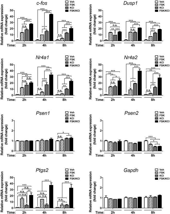
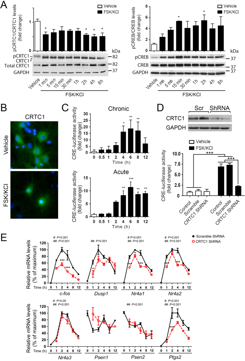
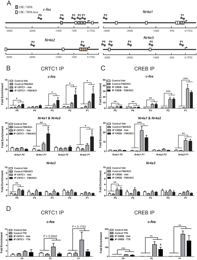
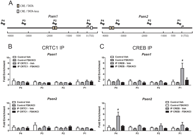
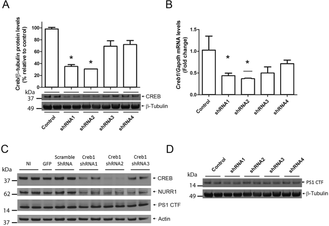
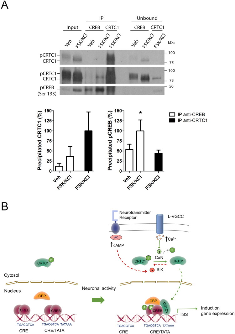
Gene expression mediated by the transcription factor cAMP-responsive element-binding protein (CREB) is essential for a wide range of brain processes. The transcriptional coactivartor CREB-regulated transcription coactivator-1 (CRTC1) is required for efficient induction of CREB target genes during neuronal activity. However, the mechanisms regulating induction of specific CREB/CRTC1-dependent genes during neuronal activity remain largely unclear. Here, we investigated the molecular mechanisms regulating activity-dependent gene transcription upon activation of the CREB/CRTC1 signaling pathway in neurons. Depolarization and cAMP signals induce preferential transcription of activity-dependent genes containing promoters with proximal CRE/TATA sequences, such as c-fos, Dusp1, Nr4a1, Nr4a2 and Ptgs2, but not genes with proximal CRE/TATA-less promoters (e.g. Nr4a3, Presenilin-1 and Presenilin-2). Notably, biochemical and chromatin immunoprecipitation analyses reveal constitutive binding of CREB to target gene promoters in the absence of neuronal activity, whereas recruitment of CRTC1 to proximal CRE/TATA promoters depends on neuronal activity. Neuronal activity induces rapid CRTC1 dephosphorylation, nuclear translocation and binding to endogenous CREB. These results indicate that neuronal activity induces a preferential binding of CRTC1 to the transcriptional complex in CRE/TATA-containing promoters to engage activity-dependent transcription in neurons.


Conflict of interest statement

The authors declare that they have no competing interests.

Figures

Figure 1
Figure 1
Neuronal activity induces CREB-dependent gene expression in primary cortical neurons. Quantitative real-time RT-PCR analysis of mRNA levels of CREB target genes in cultured cortical neurons (10 DIV). Levels of c-fos, Dusp1, Nr4a1, Nr4a2 and Ptgs2 mRNAs were differentially increased after KCl, FSK or FSK/KCl treatment in a time-dependent manner. On the contrary, Gapdh mRNA levels were not significantly different between vehicle- (Veh) and FSK/KCl-treated neurons at any time point, while Psen1 and Psen2 mRNA levels were significantly changed only after 8 hours of stimulation. Data represent fold change ± s.e.m relative to vehicle (Veh)-treated neurons from three independent experiments (n = 3). Gene expression levels were normalized to the geometric mean of Ppia, Tbp and Gapdh. N.s: non-significant. *P < 0.05, **P < 0.01, ***P < 0.001, compared to the indicated experimental group as determined by two-way ANOVA followed by Bonferroni test.
Figure 2
Figure 2
Activity-dependent CRTC1 activation in cultured neurons. (A) Activity-dependent changes on CRTC1 and CREB phosphorylation in cultured primary neurons. Western blot and quantitative analysis of pCRTC1, CRTC1, pCREB (Ser133) and CREB in 10 DIV cortical neurons treated with vehicle or FSK/KCl for 0–8 h. Values represent fold change ± s.e.m (n = 3 experiments). (B) Nuclear translocation of CRTC1 induced by neuronal activity. Representative immunofluorescence image of CRTC1 (green) and nuclear (Hoeschst 33324, blue) staining in neurons treated with vehicle or FSK/KCl for 15 min. (C) Time-dependent CRE-mediated transcription measured by a CRE-luciferase activity assay in primary neurons treated with FSK/KCl chronically (0–12 h) or acutely (1 h) and then washed and incubated for 0–12 h. Values represent fold change ± s.e.m of independent experiments (n = 3) performed in triplicate. (D) Western blot showing reduced CRTC1 levels in neurons treated with lentiviral Crtc1 ShRNA (top panel). Reduced CRE-mediated transcriptional activity in Crtc1 ShRNA-treated neurons after 4 h of treatment (bottom panel). Values represent fold change ± s.e.m (n = 3 independent transfections in duplicates). (E) CRTC1 regulates expression of CREB target genes in an activity-dependent manner. Real-time qRT-PCR analysis of mRNA levels normalized to Gapdh in neurons infected with scramble ShRNA (black dots) and Crtc1 ShRNA (red dots). Data represent mean percentage ± s.e.m of three independent experiments performed by triplicate. “#” and “##”represent statistical differences on time and treatment, respectively. *P < 0.05, ***P < 0.001, compared to scramble ShRNA-infected neurons in a specific time point. Statistical analysis was determined by two-way ANOVA followed by Bonferroni test.
Figure 3
Figure 3
Differential recruitment of CRTC1 and CREB to target gene promoters upon neuronal activity. (A) Schematic representation of gene promoters of CREB target genes and position of amplified gene regions with specific primers (P1-P5) relative to CRE/TATA-less (white boxes) and CRE/TATA (grey boxes) sites relative to the transcription start site (TSS). (B,C) Quantitative chromatin immunoprecipitation (IP) analysis showing differential binding of CRTC1 (B) and CREB (C) to c-fos, Nr4a1, 2 and 3 gene promoter regions in primary cortical neurons treated with vehicle (Veh) or FSK/KCl for 15 min. The ratio between the immunoprecipitated chromatin obtained with beads (Control), anti-CRTC1 or anti-CREB antibodies or and the input chromatin is shown as fold enrichment of the amplified target regions over an irrelevant region in chromosome 4. (D) Chomatin IP analysis showing binding of CRTC1 and  CREB to the  CRE/TATA proximal promoter region of c-fos (P1-P2). CRTC1 binding to P1 and P2 regions is reduced but not significantly after TTX treatment (12 h). Data in (BD) represent fold enrichment ± s.e.m of immunoprecipitation assays from several neuron cultures (n = 3–5). For each primer, *P < 0.05, **P < 0.01 and ***P < 0.001 represent statistical significance between groups as indicated using two-way ANOVA, whereas # P < 0.05 represent statistical significance between each group IP CRTC1- Veh and IP CRTC1-FSK/KCl in (B) and IP CREB-Veh and IP CREB-TTX in (D) using posthoc Bonferroni test.
Figure 4
Figure 4
Deficient binding of CRTC1 and CREB to Psen1 and Psen2 promoters. (A) Schematic representation of Psen1 and Psen2 promoters and position of qPCR amplified gene regions with specific primers (P1-P4) relative to CRE/TATA-less (white boxes) and CRE/TATA (gray boxes) sites. TSS: transcription start site. (B,C) Quantitative ChIP analysis showing differential binding of CRTC1 (B) and CREB (C) to distinct Psen1 and Psen2 promoter regions using anti-CRTC1 (left) and anti-CREB (right) antibodies in cortical neurons treated with vehicle or FSK/KCl for 15 min. CRTC1 does not bind to Psen1 and Psen2 promoters. Data represent fold enrichment ± s.e.m of immunoprecipitation assays from independent cultures (n = 5). # P < 0.05 represents statistical significance of IP CREB Veh vs IP CREB FSK/KCl for P1 (Psen1) or P4 (Psen2), as determined by two-way ANOVA followed by Bonferroni test.
Figure 5
Figure 5
CREB silencing does not affect basal PS1 protein levels. (A,B) Genetic inactivation of CREB in primary cortical neurons (4 DIV) infected with lentiviral particles expressing scramble (control) or Creb1 shRNAs 1-4. Creb1 shRNA-1 and shRNA-2 reduced significantly total CREB protein (A) and mRNA (B) levels in primary cortical neurons. Data represent the mean fold change ± s.e.m relative to scramble shRNA (control) from independent experiments (n = 3). Levels of mRNA were normalized to Gapdh. *P < 0.05 as determined by one-way ANOVA followed by Dunnett’s multiple comparison test. (C,D) CREB silencing does not affect levels of PS1 C-terminal fragments (CTF) in human HEK-293T cells (C) and primary mouse cortical neurons (D) but efficiently reduces NR4A2 (NURR1) protein levels.
Figure 6
Figure 6
Activity-dependent binding of CRTC1 to CREB in neurons. (A) Coimmunoprecipitation experiments showing activity-dependent binding of endogenous CRTC1 and CREB in cultured cortical neurons. Representative immunoblots of CRTC1 (top: two different exposures) and pCREB (bottom) in the lysate (input), and immunoprecipitated (IP) and unbound fractions. Endogenous CRTC1 is immunoprecipitated with an anti-CREB antibody, and pCREB is efficiently immunoprecipitated with the anti-CRTC1 antibody in neurons treated with FSK/KCl. The graphs represent the percentage of immunoprecipitated CRTC1 and pCREB normalized to the maximum immunoprecipitation with their specific antibodies in FSK/KCl conditions, which is considered  100%. Data represent percentage of immunoprecipitation ± s.e.m of four independent experiments. *P < 0.05 by one-tailed t-test. (B) Proposed model of activity-dependent gene expression mediated by CRTC1 in neurons. In basal conditions, CREB binds constitutively to both CRE/TATA- and CRE/TATA-less-containing promoters of target genes. Synaptic activity activates neurotransmitter receptors and L-type voltage gated calcium channels (L-VGCC) elevating intracellular levels of the second messengers cAMP and Ca2+, which inhibits salt-induced kinase (SIK) and activates calcineurin/protein phosphatase 2B (CaN), respectively. This results in phosphorylation (P, in green) of CREB at Ser133 and CRTC1 dephosphorylation, nuclear translocation and preferential binding to CREB/CREB binding protein (CBP) complexes into CRE/TATA-containing promoters close to the transcription start site (TSS) to engage gene transcription.

Similar articles

Cited by

References

    1. Qiu Z, Ghosh A. A brief history of neuronal gene expression: regulatory mechanisms and cellular consequences. Neuron. 2008;60:449–455. doi: 10.1016/j.neuron.2008.10.039. - DOI - PubMed
    1. Benito E, Barco A. CREB’s control of intrinsic and synaptic plasticity: implications for CREB-dependent memory models. Trends Neurosci. 2010;33:230–240. doi: 10.1016/j.tins.2010.02.001. - DOI - PubMed
    1. Sheng, M., McFadden, G. & Greenberg, M. E. Membrane depolarization and calcium induce c-fos transcription via phosphorylation of transcription factor CREB. Neuron4, 571–582, 0896-6273(90)90115-V (1990). - PubMed
    1. Chrivia JC, et al. Phosphorylated CREB binds specifically to the nuclear protein CBP. Nature. 1993;365:855–859. doi: 10.1038/365855a0. - DOI - PubMed
    1. Briand LA, Lee BG, Lelay J, Kaestner KH, Blendy JA. Serine 133 phosphorylation is not required for hippocampal CREB-mediated transcription and behavior. Learn Mem. 2015;22:109–115. doi: 10.1101/lm.037044.114. - DOI - PMC - PubMed

Publication types

MeSH terms

Substances