. 2017 Dec 6:8:1680.
doi: 10.3389/fimmu.2017.01680. eCollection 2017.

Adenosine A2A Receptor Deletion Blocks the Beneficial Effects of Lactobacillus reuteri in Regulatory T-Deficient Scurfy Mice

Affiliations

Adenosine A2A Receptor Deletion Blocks the Beneficial Effects of Lactobacillus reuteri in Regulatory T-Deficient Scurfy Mice

Baokun He et al. Front Immunol. .

Abstract

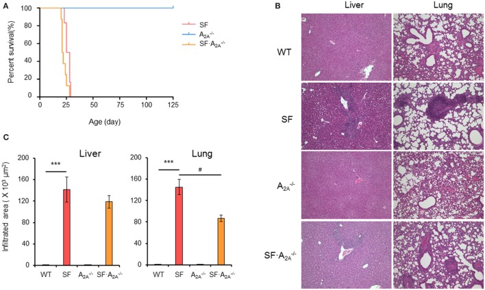
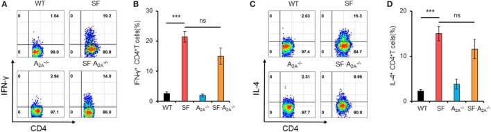
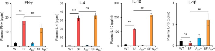
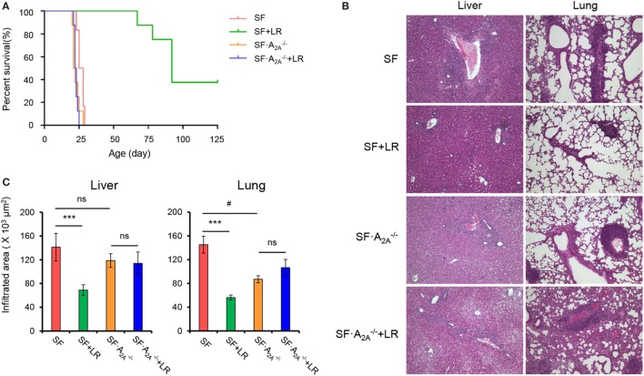
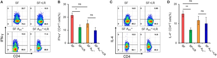
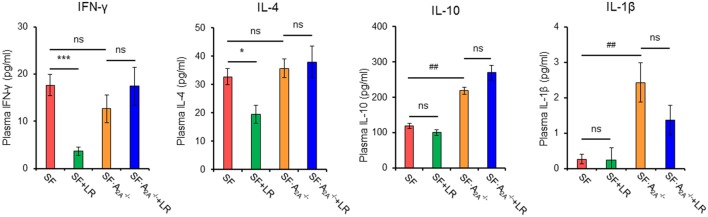
The lack of a functional Foxp3 transcription factor and regulatory T (Treg) cells causes lethal, CD4+ T cell-driven autoimmune diseases in scurfy (SF) mice and humans. Recent studies have shown that adenosine A2A receptor activation limits inflammation and tissue damage, thereby playing an anti-inflammatory role. However, the role of the adenosine A2A receptor in the development of disease in SF mice remains unclear. Using a genetic approach, we found that adenosine A2A receptor deletion in SF mice (SF[Formula: see text]) does not affect early life events, the development of a lymphoproliferative disorder, or hyper-production of pro-inflammatory cytokines seen in the Treg-deficiency state. As shown previously, Lactobacillus reuteri DSM 17938 treatment prolonged survival and reduced multiorgan inflammation in SF mice. In marked contrast, A2A receptor deletion completely blocked these beneficial effects of L. reuteri in SF mice. Altogether, these results suggest that although absence of the adenosine A2A receptor does not affect the development of disease in SF mice, it plays a critical role in the immunomodulation by L. reuteri in Treg-deficiency disease. The adenosine A2A receptor and its activation may have a role in treating other Treg dysfunction-mediated autoimmune diseases.

Keywords: IPEX; Lactobacillus reuteri; adenosine A2A receptor; autoimmunity; cytokines; probiotic; regulatory T deficiency; scurfy.


Figures

Figure 1
Figure 1
Effect of adenosine A2A receptor deletion on the development of diseases in scurfy (SF) mice. (A) Survival curves of A2A-/-, SF, and SFA2A-/- mice (n = 6–9). (B) H&E staining of representative sections of liver and lung of wild-type (WT), SF, A2A-/-, and SFA2A-/- mice (n = 6–9). (C) Quantitation of inflammatory infiltrates in liver and lung of WT, SF, A2A-/-, and SFA2A-/- mice (n = 6–9). Data are presented as mean ± SEM. ***p < 0.001. SF vs. WT. #p < 0.05. SFA2A-/- vs. SF.
Figure 2
Figure 2
Effect of adenosine A2A receptor deletion on TH1/TH2 cells in spleen of scurfy (SF) mice. (A) Representative FACS plots of IFN-γ-producing CD4+ T (TH1) cells in spleen of wild-type (WT), SF, A2A-/-, and SFA2A-/- mice. (B) Percentage of TH1 cells in spleen of WT, SF, A2A-/-, and SFA2A-/- mice (n = 6–9). (C) Representative FACS plots of IL-4-producing CD4+ T (TH2) cells in spleen of WT, SF, A2A-/-, and SFA2A-/- mice. (D) Percentage of TH2 cells in spleen of WT, SF, A2A-/-, and SFA2A-/- mice (n = 6–9). Data are presented as mean ± SEM. ***p < 0.001. SF vs. WT. ns, non-significance.
Figure 3
Figure 3
Effect of adenosine A2A receptor deletion on pro-inflammatory cytokines in scurfy (SF) mice. Plasma levels of IFN-γ, IL-1β, IL-4, and IL-10 in wild-type (WT), SF, A2A-/-, and SFA2A-/- mice were quantified by a mouse multi-spot pro-inflammatory panel kit (n = 6–9). Data are presented as mean ± SEM. **p < 0.01, ***p < 0.001. SF vs. WT. #p < 0.05, ##p < 0.01. SFA2A-/- vs. SF. ns, non-significance.
Figure 4
Figure 4
Adenosine A2A receptor deletion blocks effects of Lactobacillus reuteri on scurfy (SF) mice. (A) Survival curves of SF, SF + LR, SFA2A-/-, and SFA2A-/- + LR mice (n = 6–9). (B) H&E staining of representative sections of liver and lung of SF, SF + LR, SFA2A-/-, and SFA2A-/- + LR mice (n = 6–9). (C) Quantitation of inflammatory infiltrates in liver and lung of SF, SF + LR, SFA2A-/-, and SFA2A-/- + LR mice (n = 6–9). Data are presented as mean ± SEM. ***p < 0.001. SF + LR vs. SF. ns, non-significance. #p < 0.05.
Figure 5
Figure 5
Effect of Lactobacillus reuteri on TH1/TH2 cells in spleen of scurfy (SF) and SFA2A-/- mice. (A) Representative FACS plots of IFN-γ-producing CD4+ T (TH1) cells in spleen of SF, SF + LR, SFA2A-/-, and SFA2A-/- + LR mice. (B) Percentage of TH1 cells in spleen of SF, SF + LR, SFA2A-/-, and SFA2A-/- + LR mice (n = 6–9). (C) Representative FACS plots of IL-4-producing CD4+ T (TH2) cells in spleen of SF, SF + LR, SFA2A-/-, and SFA2A-/- + LR mice. (D) Percentage of TH2 cells in spleen of SF, SF + LR, SFA2A-/-, and SFA2A-/- + LR mice (n = 6–9). Data are presented as mean ± SEM. *p < 0.05, **p < 0.01. SF + LR vs. SF. ns, non-significance.
Figure 6
Figure 6
Effect of Lactobacillus reuteri on pro-inflammatory cytokines in scurfy (SF) and SFA2A-/- mice. Plasma levels of IFN-γ, IL-4, IL-1β, and IL-10 in SF, SF + LR, SFA2A-/-, and SFA2A-/- + LR mice were quantified by a mouse multi-spot pro-inflammatory panel kit (n = 6–9). Data are presented as mean ± SEM. *p < 0.05, ***p < 0.001. SF + LR vs. SF. ##p < 0.01. SFA2A-/- vs. SF. ns, non-significance.

Similar articles

Cited by

References

    1. Barzaghi F, Passerini L, Bacchetta R. Immune dysregulation, polyendocrinopathy, enteropathy, X-linked syndrome: a paradigm of immunodeficiency with autoimmunity. Front Immunol (2012) 3:211.10.3389/fimmu.2012.00211 - DOI - PMC - PubMed
    1. d’Hennezel E, Bin DK, Torgerson T, Piccirillo CA. The immunogenetics of immune dysregulation, polyendocrinopathy, enteropathy, X linked (IPEX) syndrome. J Med Genet (2012) 49:291–302.10.1136/jmedgenet-2012-100759 - DOI - PubMed
    1. Bennett CL, Christie J, Ramsdell F, Brunkow ME, Ferguson PJ, Whitesell L, et al. The immune dysregulation, polyendocrinopathy, enteropathy, X-linked syndrome (IPEX) is caused by mutations of FOXP3. Nat Genet (2001) 27:20–1.10.1038/83713 - DOI - PubMed
    1. Brunkow ME, Jeffery EW, Hjerrild KA, Paeper B, Clark LB, Yasayko SA, et al. Disruption of a new forkhead/winged-helix protein, scurfin, results in the fatal lymphoproliferative disorder of the scurfy mouse. Nat Genet (2001) 27:68–73.10.1038/83784 - DOI - PubMed
    1. Baris S, Schulze I, Ozen A, Aydiner EK, Altuncu E, Karasu GT, et al. Clinical heterogeneity of immunodysregulation, polyendocrinopathy, enteropathy, X-linked: pulmonary involvement as a non-classical disease manifestation. J Clin Immunol (2014) 34:601–6.10.1007/s10875-014-0059-7 - DOI - PubMed