Review
. 2018 Jun;28(2):123-175.
doi: 10.1007/s11065-017-9365-1. Epub 2017 Dec 22.

Neurodevelopmental consequences of pediatric cancer and its treatment: applying an early adversity framework to understanding cognitive, behavioral, and emotional outcomes

Affiliations
Review

Neurodevelopmental consequences of pediatric cancer and its treatment: applying an early adversity framework to understanding cognitive, behavioral, and emotional outcomes

Hilary A Marusak et al. Neuropsychol Rev. 2018 Jun.

Abstract

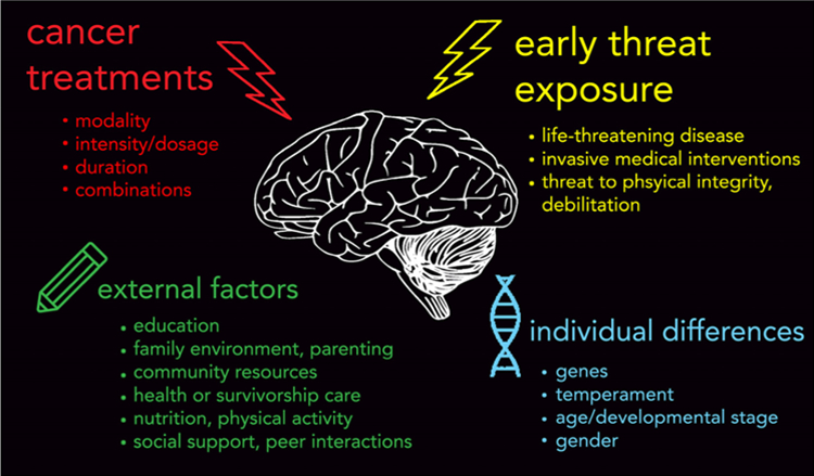
Today, children are surviving pediatric cancer at unprecedented rates, making it one of modern medicine's true success stories. However, we are increasingly becoming aware of several deleterious effects of cancer and the subsequent "cure" that extend beyond physical sequelae. Indeed, survivors of childhood cancer commonly report cognitive, emotional, and psychological difficulties, including attentional difficulties, anxiety, and posttraumatic stress symptoms (PTSS). Cognitive late- and long-term effects have been largely attributed to neurotoxic effects of cancer treatments (e.g., chemotherapy, cranial irradiation, surgery) on brain development. The role of childhood adversity in pediatric cancer - namely, the presence of a life-threatening disease and endurance of invasive medical procedures - has been largely ignored in the existing neuroscientific literature, despite compelling research by our group and others showing that exposure to more commonly studied adverse childhood experiences (i.e., domestic and community violence, physical, sexual, and emotional abuse) strongly imprints on neural development. While these adverse childhood experiences are different in many ways from the experience of childhood cancer (e.g., context, nature, source), they do share a common element of exposure to threat (i.e., threat to life or physical integrity). Therefore, we argue that the double hit of early threat and cancer treatments likely alters neural development, and ultimately, cognitive, behavioral, and emotional outcomes. In this paper, we (1) review the existing neuroimaging research on child, adolescent, and adult survivors of childhood cancer, (2) summarize gaps in our current understanding, (3) propose a novel neurobiological framework that characterizes childhood cancer as a type of childhood adversity, particularly a form of early threat, focusing on development of the hippocampus and the salience and emotion network (SEN), and (4) outline future directions for research.

Keywords: Childhood cancer; brain; brain tumor; leukemia; pediatric oncology.


Figures

Fig. 1
Fig. 1
Top: Early threat exposure and cancer treatments are both developmental insults that can alter neural development and contribute to the range of cognitive, behavioral, and emotional late and long-term effects reported in childhood cancer patients and survivors. Bottom: various potential external (e.g., environment) and individual difference factors could modify these effects. While it is largely recognized that late and long-term effects are due, in part, to injurious effects of therapeutic intervention during cancer treatment (e.g., chemotherapy, cranial irradiation) on the developing brain, we assert that the role of early threat exposure in pediatric cancer – namely, the diagnosis of a life-threatening disease and endurance of invasive medical procedures – should also be considered. Early threat exposure, a form of childhood adversity, defined as an experience that is likely to require significant psychological or neurobiological adaptation by the average child and that represent a deviation from the expectable environment (McLaughlin 2016), has been shown to strongly imprint on brain development, and is one of the strongest risk factors for virtually all commonly occurring psychological disorders. Research is needed to understand neurodevelopmental consequences of the ‘double hit’ of early threat and therapeutic intervention associated with childhood cancer, and factors that may modify outcomes. Identification of such factors will be essential for guiding early intervention to mitigate these risks
Fig. 2
Fig. 2
Altered brain structure and function in young survivors of pediatric cancer. a Reduced hippocampal volume in n = 27 ALL survivors (ages 15–22 years; in remission for 6–18 years) treated with chemotherapy-only, relative to n = 27 matched controls (Genschaft et al. 2013). b Regional increases (warm colors) and decrease (cool colors) in white matter clustered connectivity, as measured using graph theoretical analysis of DTI data in n = 31 young ALL survivors (ages 5–19 years, 6–111 months off treatment) relative to n = 39 matched controls. Of note, increased nodal clustering was noted in the ALL group in hippocampus and insula, a core SEN region, and decreased clustering in amygdala, considered a part of the SEN (Kesler et al. 2016). Abbreviations: ALL, acute lymphoblastic leukemia; SEN, salience and emotion network; DTI, diffusion tensor imaging. Image for panel B provided courtesy of Dr. Shelli Kesler. All images are adapted with permission
Fig. 3
Fig. 3
Altered brain function in young survivors of pediatric cancer. a Significant increases in response in ACC, a core SEN region, in n = 8 young ALL survivors (m = 14.54 years, SD = 2.47; 4–12 years after treatment) relative to matched controls, during a working memory (n-back) paradigm (2-back condition). No significant differences in behavioral performance (i.e., accuracy, reaction time) were observed between groups (Robinson et al. 2010). b Higher plasma methotrexate exposure during treatment for ALL is associated with lower response in ACC, a core SEN, during an attention task at more than five years (m= 7.7, SD = 1.7) post-diagnosis (n = 147; m age at scan = 13.8 years, SD = 4.8; Krull et al. 2016). Abbreviations: ALL, acute lymphoblastic leukemia; ACC, anterior cingulate cortex; SEN, salience and emotion network. All images are adapted with permission
Fig. 4
Fig. 4
Function of the salience and emotion network (SEN) and other large-scale neurocognitive networks represent key targets for psychosocial and behavioral interventions. 30 survivors of ALL or brain tumor who completed a 25-week computerized cognitive training intervention exhibited increased behavioral performance in several cognitive domains (Conklin et al. 2016). This improvement in behavioral performance was accompanied by a pre- to post-intervention reduction in SEN (anterior cingulate cortex, ACC) and CEN (lateral prefrontal cortex, LPFC) activity during a working memory task (top and bottom left panels). Additionally, lower pre-intervention activity in CEN (dorsolateral prefrontal cortex) during a working memory task predicted positive treatment response, i.e., greater positive change in working memory performance (i.e., spatial span backward; bottom right panel). Abbreviations: ALL, acute lymphoblastic leukemia; CEN, central executive network; SEN, salience and emotion network
Fig. 5
Fig. 5
Altered rsFC in young survivors of pediatric cancer. Regional increases (blue) and decreases (green-yellow) in rsFC in n = 15 young ALL (ages 8–15 years, 9–110 months off treatment) and n = 14 matched controls. In particular, ALL survivors showed reduced rsFC of amygdala (‘RAMG’) and hippocampus (‘LHIP’,’RHIP’) with attention and visual regions (e.g., occipital; Kesler et al. 2014). Abbreviations: ALL, acute lymphoblastic leukemia; rsFC, resting-state functional connectivity. All images are adapted with permission
Fig. 6
Fig. 6
Altered brain structure in adult survivors of pediatric cancer. a Relative to matched controls (n = 27), adult survivors of childhood brain tumor (n = 27, ages 18–32; average of 13.7 years [SD = 5.37] since diagnosis) demonstrate reductions in indicators of white matter integrity in frontal and temporal areas. White matter integrity in frontal areas was positively correlated with IQ (King et al. 2015b). b Altered hippocampal volume and shape in adult survivors of pediatric ALL treated with CRT (n = 39, mean age = 26.7 years [SD = 3.4]; average of 23.9 years [SD = 3.1] since diagnosis) or CT only (n = 36, mean age = 24.9 years [SD = 3.6]; average of 15 years [SD = 1.7] since diagnosis) relative to controls (n = 23, mean age = 23.1 years; Edelmann et al. 2014). Abbreviations: ALL, acute lymphoblastic leukemia; CRT, cranial radiation therapy; CT, chemotherapy. All images are adapted with permission
Fig. 7
Fig. 7
Altered brain function in adult survivors of pediatric cancer. a Abnormal elevation in hippocampal activity during encoding of later forgotten items in n = 10 adult survivors of pediatric ALL (average age = 30.8 years; 20–30 years after treatment) relative to n = 10 matched controls (“CTL”, Monje et al. 2013). Functional neural changes were accompanied by poorer recognition memory and hippocampal atrophy in the ALL relative to control group. b Adult survivors of pediatric brain tumor (n = 17, average age = 23.39 [SD = 4.46]; 15.5 years (SD = 7.6) post diagnosis) exhibited greater activation in dmPFC, a SEN region, during a working memory paradigm (2-back condition) relative to matched controls (n = 17). Higher dmPFC activation was associated with poorer performance, suggesting an ineffective compensatory neural mechanism – similar to studies in children (Fig. 3a; King et al. 2015a). (c) Increased rsFC among frontal regions in n = 16 adult survivors of childhood cerebellar tumors (ages 17–34 years; average of 14.0 years [SD = 7.3] since diagnosis) relative to n = 16 matched controls (Chen et al. 2016). Abbreviations: ALL, acute lymphoblastic leukemia; SEN, salience and emotion network; rsFC, resting-state functional connectivity; dmPFC, dorsomedial prefrontal cortex. All images are adapted with permission
Fig. 8
Fig. 8
Common alterations in brain structure and function in individuals exposed to more commonly-studied childhood experiences that involve similar elements of early threat (e.g., interpersonal violence, abuse), suggesting a role for neurotoxic effects of early threat exposure in neurodevelopmental consequences of pediatric cancer. a Age-related decreases in hippocampal volume of 27 threat-exposed youth with PTSD (mean age = 14.2 years, SD = 2.7) relative to 27 matched controls (Keding & Herringa 2015). b Increased response in ACC, a core SEN, during a behavioral inhibition task (Go/No-Go) in 16 youth with interpersonal trauma-related PTSS (ages 10–16 years) relative to 14 matched controls (Carrion et al. 2008). No group differences in task performance were observed, suggesting a compensatory neural mechanism that is also observed in pediatric cancer survivors (see Fig. 3a). c Relative to 21 unexposed matched controls, 21 youth exposed to interpersonal threat (e.g., violence, abuse) demonstrated altered rsFC between superficial (‘SF’) amygdala and various regions of SEN (e.g., ACC, insula), prefrontal cortex, and temporal and occipital lobes. Similar to a study of young survivors of pediatric ALL (see Fig. 5), threat-exposed youth demonstrate reduced (negative) rsFC between amygdala and temporo-occitiptal studies, involved in attention and visual processing (Thomason et al. 2015). d Relative to 19 matched controls, 14 youth exposed to interpersonal threat (M age = 12.61 years, SD = 2.11) demonstrate reduced SEN rsFC within the ACC, and increased response in insula, a core SEN region, during a cognitive control (conflict) task. Higher insula response was associated with poorer task performance (Marusak et al. 2015a). e Increased network centrality of the insula, a core SEN region, in 145 18–25 year-olds with histories of maltreatment exposure relative to 123 matched controls (Teicher et al. 2014). Increased prominence of the insula within a whole-brain network is similar to a report in young ALL survivors (Kesler et al. 2014; see Fig. 5). Network centrality was measured using graph theoretical analysis of structural MRI data. Abbreviations: ALL, acute lymphoblastic leukemia; ACC, anterior cingulate cortex; SEN, salience and emotion network; rsFC, resting-state functional connectivity; PTSS, posttraumatic stress symptoms. All images are adapted with permission

References

    1. Aber JL, Bennett NG, Conley DC, & Li J (1997). The effects of poverty on child health and development. Annual review of public health, 18(1), 463–483. - PubMed
    1. Ainsworth MS (1979). Infant–mother attachment. American psychologist, 34(10), 932. - PubMed
    1. Alderfer MA, & Kazak AE (2006). Family issues when a child is on treatment for cancer. In Comprehensive handbook of childhood cancer and sickle cell disease: A biopsychosocial approach (pp. 53–74). New York: Oxford University Press.
    1. Alderfer MA, Labay LE, & Kazak AE (2003). Brief report: does posttraumatic stress apply to siblings of childhood cancer survivors? Journal of pediatric psychology, 28(4), 281–286. - PubMed
    1. Anderson FS, & Kunin-Batson AS (2009). Neurocognitive late effects of chemotherapy in children: The past 10 years of research on brain structure and function. Pediatric Blood & Cancer, 52(2), 159–164. 10.1002/pbc.21700. - DOI - PubMed

LinkOut - more resources