

Matrix regeneration agents improve wound healing in non-stressed human corneal epithelial cells
- PMID: 29271418
- PMCID: PMC5898859
- DOI: 10.1038/eye.2017.277
Matrix regeneration agents improve wound healing in non-stressed human corneal epithelial cells
Abstract
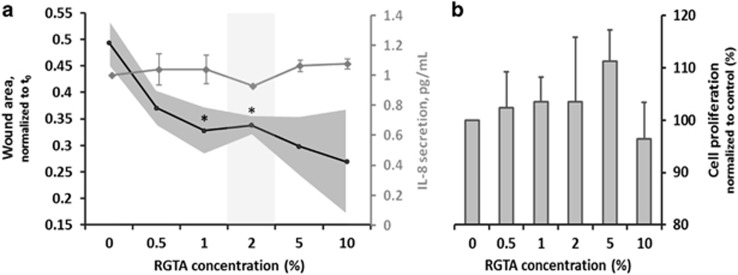
PurposeMatrix regenerating agents (RGTAs) emerged as promising in vivo wound-healing agents. These agents could prove beneficial for the treatment of dry eye disease-associated corneal micro-erosions; therefore, we aimed to evaluate the wound healing efficacy of regenerative agents (RGTAs or serum) in an in vitro model of hyperosmolarity (HO) stressed and non-stressed human corneal epithelial cells.Patients and methodsThe migration and proliferation induced by the regenerative agents was evaluated using an in vitro scratch wound assay and brome-deoxy-uridine incorporation. The inflammatory profile and effects of osmoregulators were also investigated. The two-tailed paired t-test calculated the statistical significance, with P-value<0.05 considered significant.ResultsThe most efficient inducer of re-epithelization was 2% serum, followed closely by 2% RGTA with an average improvement in cell migration of 1.8- and 1.4-fold, respectively, when compared with the non-treated control. Hyperosmolar stress significantly reduced the restorative effects of both serum and RGTAs; these effects were, however, neutralized by the osmoregulator betaine.ConclusionThese findings suggest that RGTAs could provide efficient treatment for dry-eye associated corneal micro-lesions if ocular surface HO is neutralized.
Conflict of interest statement
The authors declare no conflict of interest.
Figures


Similar articles
-
Trefoil factor family peptide 3 (TFF3) is upregulated under experimental conditions similar to dry eye disease and supports corneal wound healing effects in vitro.Invest Ophthalmol Vis Sci. 2014 May 8;55(5):3037-42. doi: 10.1167/iovs.13-13423. Invest Ophthalmol Vis Sci. 2014. PMID: 24713479
-
RGTA-based matrix therapy in severe experimental corneal lesions: safety and efficacy studies.J Fr Ophtalmol. 2013 Nov;36(9):740-7. doi: 10.1016/j.jfo.2013.01.012. Epub 2013 Aug 16. J Fr Ophtalmol. 2013. PMID: 23958066
-
Treatment with solubilized Silk-Derived Protein (SDP) enhances rabbit corneal epithelial wound healing.PLoS One. 2017 Nov 20;12(11):e0188154. doi: 10.1371/journal.pone.0188154. eCollection 2017. PLoS One. 2017. PMID: 29155856 Free PMC article.
-
Matrix regenerative therapy.Rom J Ophthalmol. 2017 Jan-Mar;61(1):2-10. doi: 10.22336/rjo.2017.2. Rom J Ophthalmol. 2017. PMID: 29450364 Free PMC article. Review.
-
[Regenerating agents (RGTAs): a new therapeutic approach].Ann Pharm Fr. 2006 Mar;64(2):135-44. doi: 10.1016/s0003-4509(06)75306-8. Ann Pharm Fr. 2006. PMID: 16568015 Review. French.
Cited by
-
Case Reports for Topical Treatment of Corneal Ulcers with a New Matrix Therapy Agent or RGTA® in Dogs.Vet Sci. 2019 Dec 13;6(4):103. doi: 10.3390/vetsci6040103. Vet Sci. 2019. PMID: 31847217 Free PMC article.
-
Development of In Vitro Dry Eye Models to Study Proliferative and Anti-Inflammatory Effects of Allogeneic Serum Eye Drops.Int J Mol Sci. 2023 Jan 13;24(2):1567. doi: 10.3390/ijms24021567. Int J Mol Sci. 2023. PMID: 36675083 Free PMC article.
-
Corneal Repair and Regeneration: Current Concepts and Future Directions.Front Bioeng Biotechnol. 2019 Jun 11;7:135. doi: 10.3389/fbioe.2019.00135. eCollection 2019. Front Bioeng Biotechnol. 2019. PMID: 31245365 Free PMC article. Review.
References
-
- Lu L, Reinach PS, Kao WW. Corneal epithelial wound healing. Exp Biol Med (Maywood) 2001; 226: 653–664. - PubMed
-
- Quinto GG, Campos M, Behrens A. Autologous serum for ocular surface diseases. Arq Bras Oftalmol 2008; 71: 47–54. - PubMed
-
- Muraine M, Gueudry J, Toubeau D, Gardea E, Verspyck E, Menguy E et al. Advantages of amniotic membrane transplantation in eye surface diseases. J Fr Ophtalmol 2006; 29: 1070–1083. - PubMed
-
- Aifa A, Gueudry J, Portmann A, Delcampe A, Muraine M. Topical treatment with a new matrix therapy agent (RGTA) for the treatment of corneal neurotrophic ulcers. Invest Ophthalmol Vis Sci 2012; 53: 8181–8185. - PubMed
MeSH terms
Substances
LinkOut - more resources
Full Text Sources
Other Literature Sources
