Comparative Study
. 2019 May;234(5):5863-5879.
doi: 10.1002/jcp.26419. Epub 2018 Dec 17.

Differential mechanisms of adenosine- and ATPγS-induced microvascular endothelial barrier strengthening

Affiliations
Comparative Study

Differential mechanisms of adenosine- and ATPγS-induced microvascular endothelial barrier strengthening

Róbert Bátori et al. J Cell Physiol. 2019 May.

Abstract

Maintenance of the endothelial cell (EC) barrier is critical to vascular homeostasis and a loss of barrier integrity results in increased vascular permeability. While the mechanisms that govern increased EC permeability have been under intense investigation over the past several decades, the processes regulating the preservation/restoration of the EC barrier remain poorly understood. Herein we show that the extracellular purines, adenosine (Ado) and adenosine 5'-[γ-thio]-triphosphate (ATPγS) can strengthen the barrier function of human lung microvascular EC (HLMVEC). This ability involves protein kinase A (PKA) activation and decreases in myosin light chain 20 (MLC20) phosphorylation secondary to the involvement of MLC phosphatase (MLCP). In contrast to Ado, ATPγS-induced PKA activation is accompanied by a modest, but significant decrease in cyclic adenosine monophosphate (cAMP) levels supporting the existence of an unconventional cAMP-independent pathway of PKA activation. Furthermore, ATPγS-induced EC barrier strengthening does not involve the Rap guanine nucleotide exchange factor 3 (EPAC1) which is directly activated by cAMP but is instead dependent upon PKA-anchor protein 2 (AKAP2) expression. We also found that AKAP2 can directly interact with the myosin phosphatase-targeting protein MYPT1 and that depletion of AKAP2 abolished ATPγS-induced increases in transendothelial electrical resistance. Ado-induced strengthening of the HLMVEC barrier required the coordinated activation of PKA and EPAC1 in a cAMP-dependent manner. In summary, ATPγS-induced enhancement of the EC barrier is EPAC1-independent and is instead mediated by activation of PKA which is then guided by AKAP2, in a cAMP-independent mechanism, to activate MLCP which dephosphorylates MLC20 resulting in reduced EC contraction and preservation.

Keywords: ATPγS; PKA; adenosine; endothelial barrier protection; myosin light chain.


Conflict of interest statement

CONFLICTS OF INTEREST

The authors declare that there are no conflicts of interest.

Figures

FIGURE 1
FIGURE 1
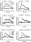
Adenosine and ATPγS enhance the EC barrier in HLMVEC. HLMVECs were treated with increasing concentrations (10–200 μM) of adenosine (a) or ATPγS (b) and TER was recorded. Results are presented as mean ± SEM of three independent experiments. Arrows indicate the time points of Ado and ATPγS administration. Ado: adenosine; ATPγS: adenosine 5′-[γ-thio]-triphosphate; EC: endothelial cell; HLMVEC: human lung microvascular endothelial cell; TER: transendothelial electrical resistance
FIGURE 2
FIGURE 2
Relative expression of adenosine (P1) and P2Y receptor mRNAs in HLMVEC. Receptor mRNA expression was determined by quantitative RT-PCR. Bar graph represents the normalized level of adenosine (P1) receptors (A1, A2A, A2B, and A3) (a) or P2Y receptors (b) by 18S rRNA and are presented as arbitrary unit (AU). mRNA: messenger RNA; rRNA: ribosomal RNA; RT-PCR: reverse-transcription polymerase chain reaction
FIGURE 3
FIGURE 3
ATPγS improves endothelial barrier function via P2Y4 and P2Y12 receptors in HLMVEC. Endothelial cells were treated either with nonspecific siRNA (nsRNA) or silencing RNA specific to different P2Y receptors for 72 hr and TER was measured upon challenge with 50 μM ATPγS. Depletion of receptors was determined by western blotting with specific antibody using β-tubulin as loading control (insets). Arrows indicate the time points of ATPγS administration. ATPγS: adenosine 5′-[γ-thio]-triphosphate; HLMVEC: human lung microvascular endothelial cell; TER: transendothelial electrical resistance; siRNA: small interfering RNA
FIGURE 4
FIGURE 4
Effect of ATPγS and adenosine on cAMP levels and PKA activity in HLMVEC. HLMVECs were treated with 100 μM Ado (a) or ATPγS (b) for 30 min and the levels of cAMP were measured by competitive enzyme linked immunosorbent assay (Cyclic AMP EIA Kit). Results are shown as mean ± SEM of three individual experiments, with three parallels each time. *p < 0.05, **p < 0.01, ***p < 0.001 versus control (0 time point, one-way ANOVA, the Newman–Keuls post hoc test). (c) HLMVECs were treated with 100 μM Ado or ATPγS for 30 min and PKA activity was measured by colorimetric protein kinase activity assay. Results are shown as mean ± SEM of three individual experiments. *p < 0.05, Veh versus Ado or Veh versus ATPγS, one-way ANOVA, the Newman–Keuls post hoc test. (d) Changes in the level of MYPT1pS695/T696 was assessed by western blotting upon vehicle, 50 μM Ado or ATPγS treatment (upper panel). Bar graphs represent the changes in the level of MYPT1pS695/Thr696 determined by densitometric analysis of immunoblots from four independent experiments (mean ± SEM, *p < 0.05, Veh versus Ado or Veh versus ATPγS, one-way ANOVA, the Newman–Keuls post hoc test). Ado: adenosine; ANOVA: analysis of variance; ATPγS: adenosine 5′-[γ-thio]-triphosphate; cAMP: cyclic adenosine monophosphate; HLMVEC: human lung microvascular endothelial cell; PKA: protein kinase A
FIGURE 5
FIGURE 5
Effect of PKA and EPAC1 depletion on adenosine and ATPγS-induced MLC20 dephosphorylation. HLMVECs were transfected with nonspecific siRNA (nsRNA), PKAcα (a), EPAC1 (b), specific silencing RNA or with both together (c). Three days after transfection the cells were treated with 50 μM ATPγS or Ado for 30 min and the level of ppMLC20T18/S19, depletion of PKAc and EPAC1 was determined by western blot analysis. The bar graphs represent the changes in the level of ppMLC20T18/S19. Densitometric analysis of blots from three to seven independent experiments (mean ± SEM). **p < 0.01, ***p < 0.001 versus nonspecific siRNA (nsRNA)-treated vehicle control, one-way ANOVA, the Newman–Keuls post-hoc test). Ado: adenosine; ANOVA: analysis of variance; ATPγS: adenosine 5′-[γ-thio]-triphosphate; EPAC1: exchange factor 3; HLMVEC: human lung microvascular endothelial cell; ns: not significant; MLC20: myosin light chain 20; PKA: protein kinase A; siRNA: small interfering RNA
FIGURE 6
FIGURE 6
Effect of PKAc and EPAC1 depletion on adenosine- and ATPγS-induced EC barrier enhancement. HLMVECs were transfected with nonspecific siRNA (nsRNA), PKAcα specific (a) or EPAC1 specific (b) silencing RNAs. Three days after transfection HLMVECs were treated with 50 μM Ado (left panel) or ATPγS (right panel) and changes in TER was recorded. Arrows indicate the time points of ATPγS and Ado administration. Data are presented as mean ± SEM. Depletion of PKA catalytic subunit or EPAC1 (insets) was confirmed by western blot analysis. Ado: adenosine; ANOVA: analysis of variance; ATPγS: adenosine 5′-[γ-thio]-triphosphate; EPAC1: exchange factor 3; HLMVEC: human lung microvascular endothelial cell; PKA: protein kinase A; TER: transendothelial electrical resistance
FIGURE 7
FIGURE 7
AKAP2 contributes to ATPγS-induced endothelial barrier function. a–c, Effect of AKAP2, AKAP9, and AKAP12 silencing on HLMVEC barrier function. HLMVECs were transfected with nonspecific siRNA (nsRNA) or AKAP2, AKAP9, and AKAP12 specific silencing RNA. Three days after transfection HLMVECs were treated with 50 μM ATPγS (right panels) or 50 μM Ado (left panels) and changes in TER was recorded. Arrows indicate the time points of ATPγS and Ado administration. Depletion of AKAPs (insets) were confirmed by western blot analysis. Data are presented as mean ± SEM of four independent experiments. Ado: adenosine; ATPγS: adenosine 5′-[γ-thio]-triphosphate; HLMVEC: human lung microvascular endothelial cell; siRNA: small interfering RNA; TER: transendothelial electrical resistance
FIGURE 8
FIGURE 8
Effect of AKAP2 depletion on adenosine and ATPγS-induced MLC20 dephosphorylation. HLMVECs were transfected with nonspecific siRNA (nsRNA) and AKAP2-specific silencing RNA. Three days after transfection the cells were treated with vehicle, 50 μM Ado or ATPγS for 30 min and the level of ppMLC20T18/S19, further depletion of AKAP2 was determined by western blot analysis. The bar graphs represent the changes in the level of ppMLC20T18/S19. Densitometric analysis of blots from four independent experiments (mean ± SEM). *p < 0.05, **p < 0.01, ***p < 0.001 versus nonspecific siRNA-treated vehicle control, one-way ANOVA, the Newman–Keuls post hoc test). ANOVA: analysis of variance; ATPγS: adenosine 5′-[γ-thio]-triphosphate; HLMVEC: human lung mic rovascular endothelial cell; MLC20: myosin light chain 20; ns: not significant; siRNA: small interfering RNA
FIGURE 9
FIGURE 9
Identification of an AKAP2-MYPT1 complex. (a) c-myc-MYPT1-containing plasmid was transfected into HEK293 cells. After 48 hr harvested cell lysate was immunoprecipitated (IP) with control IgG or anti-myc antibody. IPs were subjected to western blot analysis with specific antibodies to myc-tag (MYPT1) and AKAP 2, 9, and 12. (b) AKAP2 with HA-tag was overexpressed in HEK293 cells and after 48 hr the IP was carried out as described in Section 2. The samples were subjected to immunoblotting with antibodies against HA-tag (AKAP2), MYPT1, PKAc, and Gi2
FIGURE 10
FIGURE 10
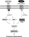
Schematic representation of PKA–, EPAC1–, and AKAP2–PKA-mediated signaling pathways induced by adenosine and ATPγS in HLMVECs. We hypothesize that adenosine stimulates increased cAMP levels in HLMVECs through A2B receptors which leads to the activation of two downstream targets, PKA and EPAC1. In contrast, ATPγS dependent signaling is mediated by P2Y4 and P2Y12 receptors leading to the cAMP-independent activation of PKA that requires AKAP2 expression. The effect of both, adenosine and ATPγS converge at the level of MLCP via increased phosphorylation of the regulatory subunit MYPT1 resulting in MLCP deinhibition, which leads to MLC20 dephosphorylation and finally to EC barrier strengthening. ATPγS: adenosine 5′-[γ-thio]-triphosphate; cAMP: cyclic adenosine monophosphate; EC: endothelial cell; EPAC1: exchange factor 3; HLMVEC: human lung mic rovascular endothelial cell; MLC20: myosin light chain 2; MLCP: myosin light chain phosphatase; PKA: protein kinase A

Similar articles

Cited by

References

    1. Aslam M, Tanislav C, Troidl C, Schulz R, Hamm C, & Gunduz D (2014). cAMP controls the restoration of endothelial barrier function after thrombin-induced hyperpermeability via Rac1 activation. Physiological Reports, 2(10), e12175 10.14814/phy2.12175 - DOI - PMC - PubMed
    1. Baron L, Gombault A, Fanny M, Villeret B, Savigny F, Guillou N, … Couillin I (2015). The NLRP3 inflammasome is activated by nanoparticles through ATP, ADP and adenosine. Cell Death & Disease, 6, e1629 10.1038/cddis.2014.576 - DOI - PMC - PubMed
    1. Batori R, Becsi B, Nagy D, Konya Z, Hegedus C, Bordan Z, … Erdodi F (2017). Interplay of myosin phosphatase and protein phosphatase-2A in the regulation of endothelial nitric-oxide synthase phosphorylation and nitric oxide production. Science Reports, 7, 44698 10.1038/srep44698 - DOI - PMC - PubMed
    1. Bilbao PS, Santillan G, & Boland R (2010). ATP stimulates the proliferation of MCF-7 cells through the PI3K/Akt signaling pathway. Archives of Biochemistry and Biophysics, 499(1–2), 40–48. 10.1016/j.abb.2010.05.001 - DOI - PubMed
    1. Birukova AA, Burdette D, Moldobaeva N, Xing J, Fu P, & Birukov KG (2010). Rac GTPase is a hub for protein kinase A and Epac signaling in endothelial barrier protection by cAMP. Microvascular Research, 79(2), 128–138. 10.1016/j.mvr.2009.11.007 - DOI - PMC - PubMed

Publication types

MeSH terms

LinkOut - more resources