Review
. 2017 Dec 22;19(1):24.
doi: 10.3390/ijms19010024.

Bio-Functional Design, Application and Trends in Metallic Biomaterials

Affiliations
Review

Bio-Functional Design, Application and Trends in Metallic Biomaterials

Ke Yang et al. Int J Mol Sci. .

Abstract

Introduction of metals as biomaterials has been known for a long time. In the early development, sufficient strength and suitable mechanical properties were the main considerations for metal implants. With the development of new generations of biomaterials, the concepts of bioactive and biodegradable materials were proposed. Biological function design is very import for metal implants in biomedical applications. Three crucial design criteria are summarized for developing metal implants: (1) mechanical properties that mimic the host tissues; (2) sufficient bioactivities to form bio-bonding between implants and surrounding tissues; and (3) a degradation rate that matches tissue regeneration and biodegradability. This article reviews the development of metal implants and their applications in biomedical engineering. Development trends and future perspectives of metallic biomaterials are also discussed.

Keywords: biodegradable metals; biological function design; biomechanical design; metal implants; porous structure.


Conflict of interest statement

The authors declare no conflict of interest.

Figures

Figure 1
Figure 1
Different clinic applications for metal implants. Metal implants are mainly used in stents and hard tissue repair, which includes maxillofacial, spine and orthopedic fixation implants. WSS: wall shear stress; B: new bones.
Figure 2
Figure 2
(a) Left: Models and grid; Right: a titanium reconstruction prosthesis. (b) The distribution of the strain of the von Mises on titanium prosthesis in different working conditions.
Figure 3
Figure 3
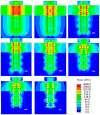
Von Mises stress distributions in models with different interference magnitudes after immediate implantation of titanium screw.
Figure 4
Figure 4
(a) Hole implantation and mesh generation; (b) Stress distribution of single hole model.
Figure 5
Figure 5
The distribution of WSS/drug concentration in different links DESs. (a) Wall shear stress (WSS), (I): Three S-type links, (II): Three U-type links, (III): Six S-type links, (IV): Six U-type links. (b) Drug concentration, (I): Three S-type links, (II): Three U-type links, (III): Six S-type links (IV): Six U-type links.
Figure 6
Figure 6
The distribution of WSS in different curvatures DESs. (a) 30°; (b) 60°; (c) 90°.
Figure 7
Figure 7
(a) Drug-coated Schematic diagram, the drug-eluting stent is nickel-titanium alloy and drug coated with rapamycin; (b) The drug distribution of four coated designs.
Figure 8
Figure 8
Some porous orthopedic implants fabricated by selective laser melting or selective laser sintering (SLM/SLS) technology. The upward arrow in SLM/SLS indicates that the platform of the printer is pushed up to provide print powders, and the downward arrows indicate the platform drop to recycle the print powders, bidirectional arrow means reciprocating pave the print powders.
Figure 9
Figure 9
The osteoinduction phenomenon of in porous Ti metals. (a) is the porous Ti specimen, (b) is the histological observation after the Ti specimen subjected to NaOH treatment (NTPT), (c) is acid-alkali treatment (AAPT) specimen, (d) is hydrogen peroxide treatment (HOPT) specimen, (e) and (f) are hydrogen peroxide solution containing tantalum chloride treatment (HTPT) and chemical and thermal treatment (CTPT) specimens. Toluidine blue dye; FT: fiber texture; B: new bones; magnification: 200×.
Figure 10
Figure 10
The explanation of degradation and apatite formation process on the surface of biodegradable metals (BMs). (a) is the metal implants just contact with body fluid, the oxidation-reduction reaction happened, the metals give away electrons formed anode, and the body fluid medium obtains electrons as cathelectrode; (b) is the corrosion happened and the metal corrosion product layer generated; (c) is the apatite layer formed and (d) is the final surface of the BMs.
Figure 11
Figure 11
Two main application products: (a) stents and (b) orthopedic implants made of BMs.

References

    1. Xu G., Fu X., Du C., Ma J., Li Z., Tian P., Zhang T., Ma X. Biomechanical comparison of mono-segment transpedicular fixation with short-segment fixation for treatment of thoracolumbar fractures: A finite element analysis. Proc. Inst. Mech. Eng. J. Part H Eng. Med. 2014;228:1005–1013. doi: 10.1177/0954411914552308. - DOI - PubMed
    1. Guo B., Lei B., Li P., Ma P.X. Functionalized scaffolds to enhance tissue regeneration. Regen. Biomater. 2015;2:47–57. doi: 10.1093/rb/rbu016. - DOI - PMC - PubMed
    1. Pownder S.L., Koff M.F., Shah P.H., Fortier L.A., Potter H.G. Magnetic resonance imaging of an equine fracture model containing stainless steel metal implants. Equine Vet. J. 2016;48:321–325. doi: 10.1111/evj.12424. - DOI - PubMed
    1. Hudetz D., Ursic Hudetz S., Harris L.G., Luginbuhl R., Friederich N.F., Landmann R. Weak effect of metal type and ica genes on staphylococcal infection of titanium and stainless steel implants. Clin. Microbiol. Infect. 2008;14:1135–1145. doi: 10.1111/j.1469-0691.2008.02096.x. - DOI - PubMed
    1. Hench L.L., Polak J.M. Third-generation biomedical materials. Science. 2002;295 doi: 10.1126/science.1067404. - DOI - PubMed

LinkOut - more resources