Review
. 2017 Dec 22;19(1):32.
doi: 10.3390/ijms19010032.

Chaperones and the Proteasome System: Regulating the Construction and Demolition of Striated Muscle

Affiliations
Review

Chaperones and the Proteasome System: Regulating the Construction and Demolition of Striated Muscle

Casey Carlisle et al. Int J Mol Sci. .

Abstract

Protein folding factors (chaperones) are required for many diverse cellular functions. In striated muscle, chaperones are required for contractile protein function, as well as the larger scale assembly of the basic unit of muscle, the sarcomere. The sarcomere is complex and composed of hundreds of proteins and the number of proteins and processes recognized to be regulated by chaperones has increased dramatically over the past decade. Research in the past ten years has begun to discover and characterize the chaperones involved in the assembly of the sarcomere at a rapid rate. Because of the dynamic nature of muscle, wear and tear damage is inevitable. Several systems, including chaperones and the ubiquitin proteasome system (UPS), have evolved to regulate protein turnover. Much of our knowledge of muscle development focuses on the formation of the sarcomere but recent work has begun to elucidate the requirement and role of chaperones and the UPS in sarcomere maintenance and disease. This review will cover the roles of chaperones in sarcomere assembly, the importance of chaperone homeostasis and the cooperation of chaperones and the UPS in sarcomere integrity and disease.

Keywords: HSP; homeostasis; misfolded protein; molecular chaperone; protein complex assembly; protein degradation; sarcomere.


Conflict of interest statement

The authors declare no conflict of interest.

Figures

Figure 1
Figure 1
Simplified schematic of the mature sarcomere in striated muscle. The major components of the mature sarcomere are shown. The Z-disc, composed mainly of α-actinin, denote the boundaries of individual sarcomeres, add stability and act as attachment sites for signaling factors, neighboring myofibrils and the myocyte cell membrane (sarcolemma). Thin filaments made of actin, nebulin and tropomodulin extend from the Z-disc to interact with the globular head domains of the myosin thick filaments that protrude out from the M-line toward the Z-disc. The binding of myosin to actin, of the thin filaments, allows the thick filaments to pull the Z-discs toward the center of the sarcomere resulting in contraction/shortening of the sarcomere length. The protein giant, titin and the elastic properties of myomesin in the M-line buffer the contraction of the sarcomere. Desmin is incorporated into the Z-discs of sarcomeres to help stabilize sarcomere structure, align sarcomeres in neighboring myofibrils and connect the contractile structures to the sarcolemma.
Figure 2
Figure 2
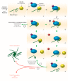
The folding of nascent client proteins by their chaperone and co-chaperones. Exemplifying the folding of the muscle myosin II globular head domain by Hsp90α1 and Unc45b as an example of molecular chaperones folding their client proteins. As proteins are synthesized by the ribosome, co-chaperones such as Unc45b can bind to the nascent polypeptide as it emerges from the ribosome (A,B). ATP-bound Hsp90α1 is recruited to nascent myosin polypeptides by Unc45b (B,C). Binding of Hsp90α1 causes a conformational change that hydrolyzes ATP to ADP that disassociates from Hsp90α1 (CE). Unc45b releases Hsp90α1 and myosin, as Hsp90α1 rebinds cytoplasmic ATP, which allows the controlled folding of segments of unbound myosin (FH). Hsp90α1 and Unc45b repeatedly rebind and fold myosin until it is completely folded (I,J; 1 & 2). If myosin is unable to be folded due to missing chaperones or incomplete binding, myosin (or any protein requiring folding) misfolds and can aggregate within the cell or be degraded via the ubiquitin proteasome system (3). Green arrows indicate the sequence of folding events that leads to a correctly folded protein to be incorporated into the sarcomere. Red arrows indicate the outcomes of misfolded proteins. Note that proteins can be discarded from any step of nascent protein folding and aggregate or be degraded.
Figure 3
Figure 3
The Premyofibril Model of Sarcomere Assembly and Necessary Chaperones. Sarcomere assembly begins with the dimerization of integrins in the sarcolemma. Integrins recruit talin and viniculin to the sarcolemma to form protocostameres (A). ZASP localizes to the protocostameres to recruit α-actinin, which is folded and incorporated by its chaperone, N-RAP (B,C). The organization of α-actinin into Z-bodies likely recruits the protein giants nebulin and titin to the developing Z-discs (D). Nebulin and titin extend out from the Z-discs to the sarcomere center as Z-discs migrate away from one another to reach mature sarcomere length (D). GimC and TRiC fold actin before incorporating into thin filaments along the nebulin scaffold (E). Titin folding and integrity is maintained, in part, by αβ-crystallin during sarcomere assembly and muscle development (E). Non-muscle myosin II is proposed to aid in the alignment and formation of the thin filaments but the factors required for non-muscle myosin folding and incorporation are unknown ((F); dotted shape). In the final stages of sarcomere assembly, non-muscle myosin is replaced by muscle myosin II to form the thick filaments, which are assembled by Hsp90α1, Unc45b and Smyd1b (G). The M-line assembles either immediately after or simultaneously to thick filament formation and incorporates the tails of myosin heavy chains and the C-terminus of titin (G).
Figure 4
Figure 4
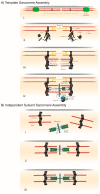
The Template and Independent Subunit Models of Sarcomere Assembly. The template model of sarcomere assembly suggests that sarcomeres require a template to form (A). The cell’s stress fibers, which have simple contractile structures, are proposed to be the templates for the formation of sarcomeres in myofibrils (A, i). Very similar to the premyofibril model, components of the Z-disc (such as α-actinin) form at stress fiber contractile sites (A, ii). Nebulin and titin extend from the Z-disc as actin is assembled into thin filaments along the nebulin scaffold (A, iii). Muscle myosin is folded and incorporated as thick filaments into the developing sarcomere by its chaperones and co-chaperones (iv). The independent subunit model for sarcomere assembly described sarcomere formation by the joining of pre-assembled subunits, or sections, of the sarcomere (B). These pre-assembled units consist of the Z-discs with attached actin thin filaments and the M-line combined with thick filaments and the protein giant, titin (B, iiii). These units come together and physically connect at the sarcolemma to create the mature contractile sarcomere (B, ii,iii).
Figure 5
Figure 5
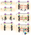
Damaged protein response in sarcomere assembly and maintenance. (A) In a healthy assembling sarcomere, chaperones (C) fold and incorporate muscle proteins; (B) when protein damage occurs during sarcomere assembly, chaperones dissociate from their complex to bind client proteins. Transcription factor (TF) translocates to the nucleus and initiates transcription of chaperones [61]. When protein damage occurs during sarcomere maintenance, either (C) a chaperone independent response occurs by which E3 ligases target damaged proteins and mark them for degradation or (D) a chaperone dependent response occurs by which chaperones and the UPS cooperate to target damaged proteins to the proteasome.
Figure 6
Figure 6
Models of chaperone and UPS cooperation in protein quality control. (a) Kinetic Model of Protein Triage. 1. Protein aggregate forms. Chaperones (C) are recruited. 2. Chaperones bind client protein and either succeeds in refolding (2′) or fail (2′′). 3. Misfolded proteins either rebinds chaperone or is targeted for degradation by the UPS; (b). cofactor mediated model of protein turnover. 1. Protein aggregate forms. Chaperones (C) are recruited. 2. Chaperone binds client protein and co-chaperones (co) are recruited and either promotes refolding of client protein (2′, 3. or degradation 2′′, 3′); (c) degradation complex model of protein triage. 1. Protein aggregate forms and Chaperones (C) are recruited. 2. Chaperone binds client protein and co-chaperones (co) are recruited, transforming the chaperone complex into the E3 ligase complex. 3. E3 complex is targeted to the proteasome for degradation.

References

    1. Bianchi T., Gelosa L. On the preventive detection of pathogenic staphylococci in the rhinopharynx of employees in the food industry in the province of Milan during the 5-year period of 1967–1971. Ann. Sclavo. 1973;15:83–98. - PubMed
    1. Van den Berg B., Wain R., Dobson C.M., Ellis R.J. Macromolecular crowding perturbs protein refolding kinetics: Implications for folding inside the cell. EMBO J. 2000;19:3870–3875. doi: 10.1093/emboj/19.15.3870. - DOI - PMC - PubMed
    1. Rose G.D., Fleming P.J., Banavar J.R., Maritan A. A backbone-based theory of protein folding. Proc. Natl. Acad. Sci. USA. 2006;103:16623–16633. doi: 10.1073/pnas.0606843103. - DOI - PMC - PubMed
    1. Kony D.B., Hunenberger P.H., van Gunsteren W.F. Molecular dynamics simulations of the native and partially folded states of ubiquitin: Influence of methanol cosolvent, pH, and temperature on the protein structure and dynamics. Protein Sci. 2007;16:1101–1118. doi: 10.1110/ps.062323407. - DOI - PMC - PubMed
    1. Schubert U., Antón L.C., Gibbs J., Norbury C.C., Yewdell J.W., Bennink J.R. Rapid degradation of a large fraction of newly synthesized proteins by proteasomes. Nature. 2000;404:770–774. doi: 10.1038/35008096. - DOI - PubMed

LinkOut - more resources