Review
. 2018 Feb 28;118(4):1460-1494.
doi: 10.1021/acs.chemrev.7b00510. Epub 2017 Dec 22.

Eight Kinetically Stable but Thermodynamically Activated Molecules that Power Cell Metabolism

Affiliations
Review

Eight Kinetically Stable but Thermodynamically Activated Molecules that Power Cell Metabolism

Christopher T Walsh et al. Chem Rev. .

Erratum in

Abstract

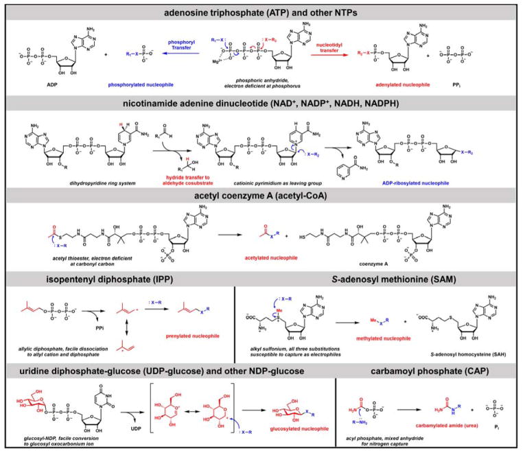
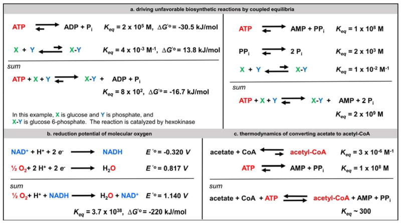
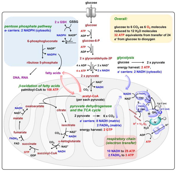
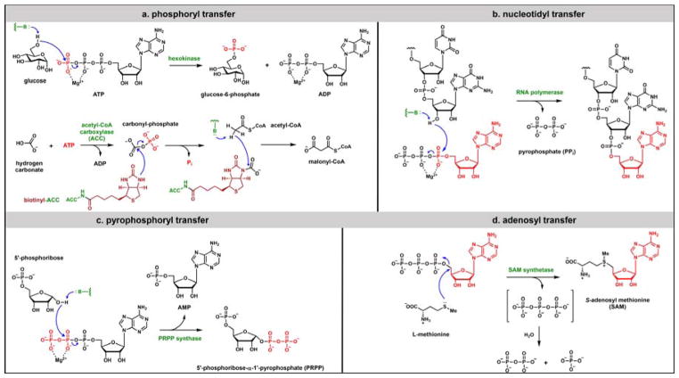
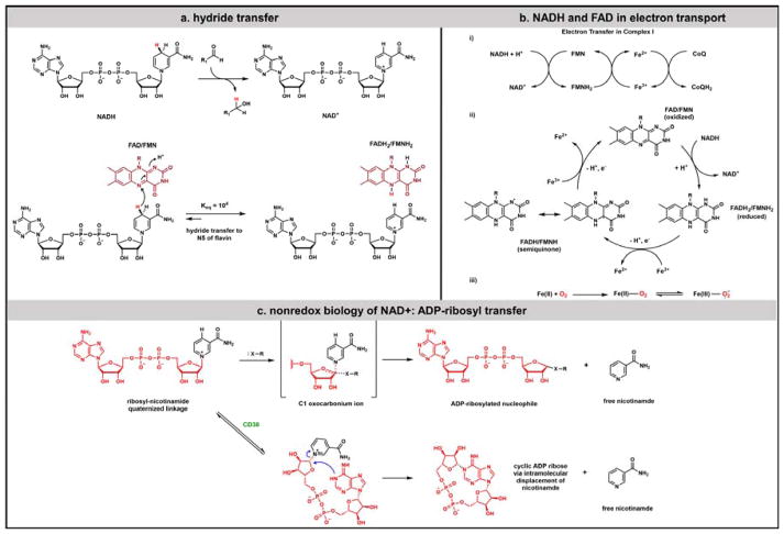
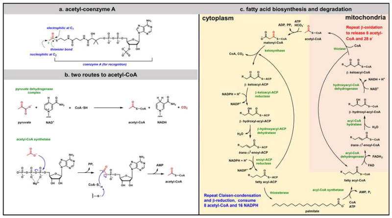
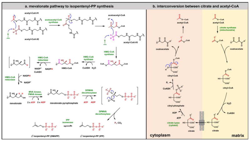
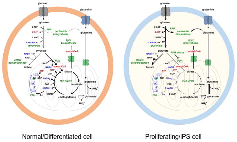
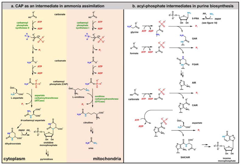
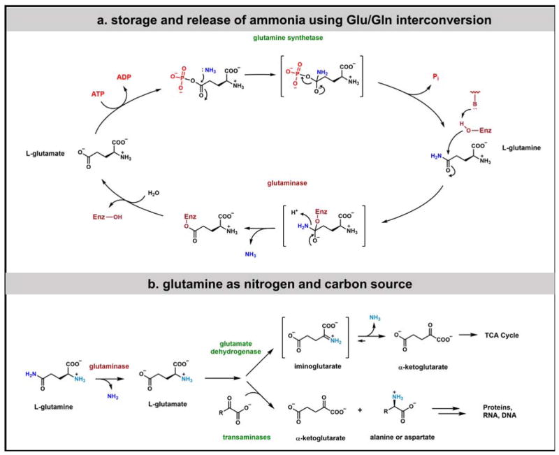
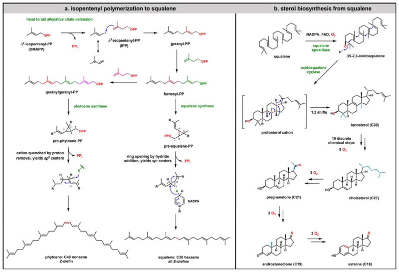
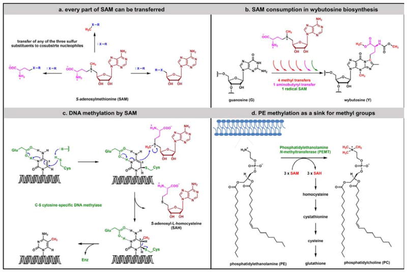
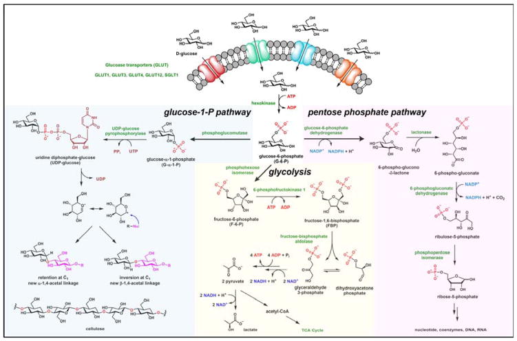
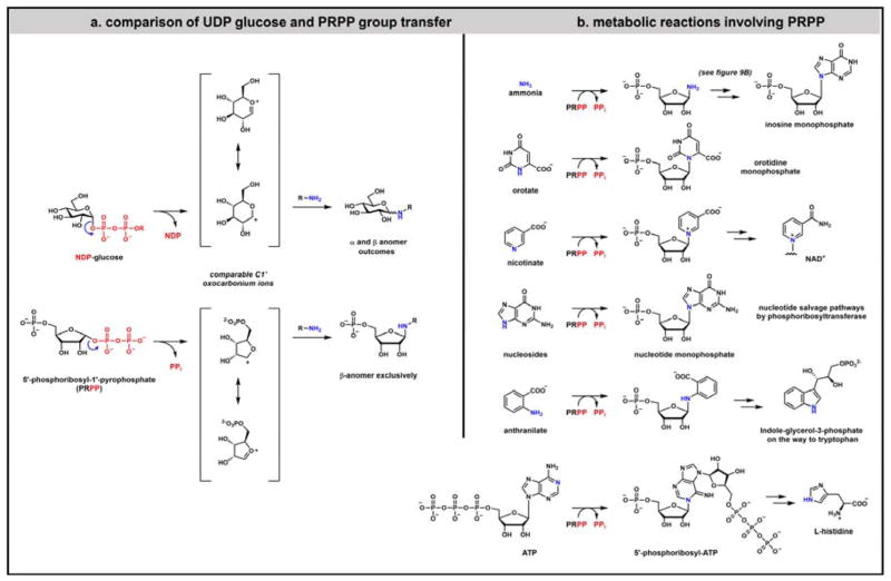
Contemporary analyses of cell metabolism have called out three metabolites: ATP, NADH, and acetyl-CoA, as sentinel molecules whose accumulation represent much of the purpose of the catabolic arms of metabolism and then drive many anabolic pathways. Such analyses largely leave out how and why ATP, NADH, and acetyl-CoA (Figure 1 ) at the molecular level play such central roles. Yet, without those insights into why cells accumulate them and how the enabling properties of these key metabolites power much of cell metabolism, the underlying molecular logic remains mysterious. Four other metabolites, S-adenosylmethionine, carbamoyl phosphate, UDP-glucose, and Δ2-isopentenyl-PP play similar roles in using group transfer chemistry to drive otherwise unfavorable biosynthetic equilibria. This review provides the underlying chemical logic to remind how these seven key molecules function as mobile packets of cellular currencies for phosphoryl transfers (ATP), acyl transfers (acetyl-CoA, carbamoyl-P), methyl transfers (SAM), prenyl transfers (IPP), glucosyl transfers (UDP-glucose), and electron and ADP-ribosyl transfers (NAD(P)H/NAD(P)+) to drive metabolic transformations in and across most primary pathways. The eighth key metabolite is molecular oxygen (O2), thermodynamically activated for reduction by one electron path, leaving it kinetically stable to the vast majority of organic cellular metabolites.


Figures

Figure 1
Figure 1
Seven organic molecules that power cell metabolism: ATP and related Nucleoside Triphosphates (NTPs); oxidized (NAD+, NADP+) and reduced (NADH, NADPH) nicotinamide adenine dinucleotides; acetyl-Coenzyme A; Δ2−isopentenyl diphosphate (also known historically as isopentenyl pyrophosphate (IPP)); S-adenosyl methionine (SAM); Uridine-diphosphoglucose (UDP-glucose) and related NDP-hexoses; carbamoyl phosphate (CAP).
Figure 2
Figure 2
Thermodynamics of coupled metabolic reactions: (a) driving unfavorable biosynthetic reactions by coupled equilibria involving phosphoryl or adenylyl (nucleotidtyl) transfers that fragment ATP; (b) the favorable thermodynamics for four electron reduction of O2 to 2 H2O from 2 NADH is revealed by the reduction potential difference. This equilibrium is silent about the kinetic barriers to reduction; (c) thermodynamics of coupled activation of acetate to acetyl-CoA as ATP is cleaved.
Figure 3
Figure 3
An overview of cellular glucose metabolism. The coupled pathways of glycolysis, TCA cycle, and electron transport down the membrane respiratory chain summarize removal of 24 electrons from each glucose molecule and ~30 ATP molecules accumulated for each four electron reduction of O2. A second major flux of glucose carbons is via the pentose phosphate pathway, yielding 2 equivalents of NADPH used for fatty acid biosynthesis and to make D-ribose-5-P for nucleic acid biosynthesis. The diagram also notes that complete oxidation of the predominant C16 fatty acyl-CoA, palmityl-CoA to 8 molecules of acetyl-CoA that are run through the TCA cycle and respiratory chain yields ~108 ATPs, emphasizing that saturated fatty acyl chains are energy storage molecules.
Figure 4
Figure 4
Mechanistic Alternatives for ATP Side Chain Cleavage to Drive Coupled Equilibria: (a) phosphoryl transfer involves attack of cosubstrate nucleophile on electrophilic γ-phosphorus atom of Mg-ATP; (b) attack of cosubstrate nucleophile on α-P of Mg-ATP constitutes nucleotidyl (here adenylyl) transfer and is the common side chain cleavage mode in RNA, DNA, and protein biosynthesis; (c) Pyrophosphoryl transfer involves cosubstrate attack on the β-P of Mg-ATP and is relatively rare, although the formation of PRPP is central to purine biosynthesis; (d) The fourth mode of ATP side chain cleavage involves attack of cosubstrate nucleophile on C5′ of the ribose moiety of ATP and, while also a metabolically rare cleavage mode, is central to the formation of S-adenosyl methionine.
Figure 5
Figure 5
Two modes of reactivity of oxidized nicotinamide coenzymes: (a) the most common role of NAD(P)+ is as a hydride acceptor during cosubstrate oxidation to form NAD(P)H. In the back direction NAD(P)H is a thermodynamically activated hydride donor for cosubstrate reductions. (b) FAD has a special place in NAD(P)H biochemistry as a favored acceptor of a hydride to give FADH2. The dihydroflavin can subsequently be reoxidized either by a reverse of the two electron pathway or by two one electron steps. FAD (and FMN) thus functions as a 2 electron/1 electron step down redox transformer and interfaces between NAD(P)H and obligate one electron acceptors such as O2 and Fe3+ proteins; (c) Nonredox biology of NAD+ involves ADP-ribosyl transfer to nucleophiles. These can be side chains on proteins or growing ADP-ribosyl chains. Also shown is the comparable intramolecular displacement on NAD+, creating cyclic ADP ribose.
Figure 6
Figure 6
Overview of Acetyl-CoA: (a) structure and chemical features of acetyl-CoA; (b) Two biosynthetic routes to acetyl-CoA involve (1) the oxidative decarboxylation of pyruvate to acetyl-CoA in the transition between glycolysis and TCA cycle catalyzed by the multi-subunit pyruvate dehydrogenase; (2) the conversion of acetate back to acetyl-CoA by nucleotidyl transfer and acetyl-AMP intermediacy. (Free acetate in mammalian cells can arise by multiple routes including histone deacetylase action); (c) in times of energy excess, the utilization of acetyl-CoA carbons for fatty acid biosynthesis occurs in cell cytoplasm while the reverse steps occur during mitochondrial beta oxidation of fatty acids to generate acetyl-CoA molecules.
Figure 7
Figure 7
Acetyl-CoA metabolism part two: (a) conversion of three molecules of acetyl-CoA to the Δ2 and Δ3 isomers of isopentenyl diphosphate (IPP) by the enzymes of the mevalonate pathway. Note the utilization of three molecules of ATP and two of NADPH as HMG-CoA is converted to Δ2-IPP; (b) acetyl-CoA and citrate can be interconverted by distinct enzymes in both cytoplasm and mitochondria and citrate can pass between the two compartments. Citryl-CoA is an intermediate common to both arms.
Figure 8
Figure 8
Comparison of metabolic fluxes from glucose and glutamine in normal/differentiating cells which are depicted with a robust TCA cycle and electron transport system vs proliferating IPS cells. Thick arrows indicate a greater flux through steps compared to thin arrows. The right hand panel indicates less energetic flux through the TCA cycle and increased aerobic glycolysis. The ability to “dump” excess electrons and regenerate NAD+ for glycolysis to continue depends substantially on high levels of lactate dehydrogenase, allowing reduction of pyruvate to lactate which can be excreted.
Figure 9
Figure 9
Carbamoyl phosphate and other acyl phosphates in purine biosynthesis: (a) carbamoyl-P is generated from carbonate, ammonia, and two molecules of ATP with carbonyl-P as an intermediate. Carbamoyl-P is central to mammalian nitrogen metabolism as a carbamoyl donor in anabolic metabolism, cytoplasmic pyrimidine biosynthesis, and in catabolic metabolism, mitochondrial urea synthesis; (b) The utility of acyl phosphates as kinetically stable, thermodynamically activated acyl transfer metabolites is emphasized in purine biosynthesis where four enzymes make glycyl-P, formyl-P, carbonyl-P, and CAIR-P as bound acyl donor species, respectively.
Figure 10
Figure 10
Glutamine as a nitrogen source: (a) glutamine and glutamate as key amino acids in balancing the storage and release of ammonia. Glutamine and glutamate are interconverted by the enzymes glutamine synthetase and glutaminase. Glutamine synthetase catalysis involves the acyl-phosphate γ-glutamyl-P; (b) exogenous glutamine can serve as both a nitrogen and carbon source; sequential action of glutaminase and glutamate dehydrogenase can liberate both amide and amino nitrogens as ammonia while transaminases that utilize glutamate as amino donor move the amino group onto many ketoacid frameworks. The deaminated α-ketoglutarate scaffold can serve as TCA cycle substrate and as cofactor for the oxygenases noted in Figures 15 and 16.
Figure 11
Figure 11
Isoprenoid metabolism: (a) isopentenyl-PP regioisomers are the key building blocks for the C30 hexaene squalene and the C40 nonaene phytoene. Chain elongations go by C5 head to tail elongations via iterative ally cation chemistry up to the C15 and C20 stages before C-C bond formation switches to head to head C1 to C1 connections in squalene and phytoene, with cyclopropyl cation formations and rearrangements involved in presqualene and prephytoene formation and breakdown; (b) sterol biosynthesis from squalene involves prior epoxidation of the 2,3-double bond, a cyclization cascade to create the familiar tetracyclic framework and a series of 1,2 H and CH3 migrations before release of the C30 lanosterol. A series of oxygenases then act in different tissues, on unactivated carbon sites, to convert the C30 lanosterol to C27 cholesterol, to C21 pregnenelone to C19 androstenedione. The cytochrome P450 enzyme aromatase carves out the angular A/B ring methyl group of androstenedione to aromatize the A ring as male sex hormone is converted to the C18 female sex hormone estrone.
Figure 12
Figure 12
S-adenosyl methionine (SAM) as thermodynamically activated alkyl transfer metabolite: (a) the sulfonium group in SAM activates all three substituents for transfer as electrophilic alkyl fragments, although methyl group transfer is by far the most common; (b) the versatility of SAM is evident in formation of the wybutosine modified base in tRNA. Six equivalents of SAM are consumed, four are moved as [CH3+] equivalents, the fifth as a [CH3•] equivalent. The sixth molecule of SAM instead donates an electrophilic aminobutyryl group; (c) DNA methylation at C5 of Cytidine residues occurs by the indicated addition/elimination mechanism with SAM as the one carbon donor; (d) SAM is also involved in membrane phospholipid maturation, converting hundreds of thousands to millions of phosphatidylethanolamine to phosphatidylcholine molecules via three consecutive N-methyl transfers in mammalian cells.
Figure 13
Figure 13
Three main pathways for cellular glucose utilization: in addition to the glycolysis and pentose phosphate pathways for flux of glucose-6-P (refer also back to Figure 3), the third pathway branches off from glucose-1-P, via action of phosphoglucomutase. The resultant UDP-glucose is the proximal donor of glucosyl moieties as glucosyl C1 oxocarbonium ion equivalents in oligosaccharide, polysaccharide, and glycoprotein biosynthesis, resulting in C1 connectivity of all such transferred glycosyl moieties. Gluconeogenesis is not included in this figure.
Figure 14
Figure 14
5-Phosphoribose-1-pyrophosphate (PRPP) as activated phosphoribosyl donor (a) UDP-glucose and PRPP are metabolites activated by the C1-pyrophosphate groups for C1-O bond cleavage via oxocarbonium ion transition states; (b) PRPP serves as an electrophilic phosphoribosyl donor to a set of cellular amine cosubstrates, including de novo purine and pyrimidine nucleotide biosynthesis, purine and pyrimidine salvage, NAD+ biosynthesis, and also to provide some of the carbon atoms in both tryptophan and histidine biogenesis.
Figure 15
Figure 15
Two main types of iron-based monooxygenases: (a) heme-based Cytochromes P450 use high valent ferryl oxygen intermediates as strong oxidants for one electron chemistry to effect C-H bond homolysis at unactivated carbon sites and OH• rebound for net hydroxylation; (b) the maturation of arachidonate into PGG2 and PGH2 involves an initial allyl radical conversion to a pair of peroxy radicals one of whch closes to the cyclopentane endoperoxide characteristic of PGG2 and PGH2; (c) the typical two His, one Glu/Asp coordination set for nonheme mononuclear iron oxygenases also includes cosubstrate α-ketoglutarate as the 4th and 5th ligands to Fe. An analogous FeIV=O high valent oxoiron species performs comparable homolytic C-H bond cleavage and OH• equivalent transfer; (d) oxidative demethylation of 5-MeC residues in DNA is mediated by the TET subfamily of nonheme mononuclear iron oxygenases, in three sequential steps as shown.
Figure 16
Figure 16
Product profile from wild type and mutant form of isocitrate dehydrogenase (IDH-1): isocitrate is oxidized to the oxalosuccinate β-keto acid that is then subjected to actve site-mediated regiospecific decarboxylation of one of the three carboxylates to yield α-ketoglutarate. In addition to its role in the TCA cycle, α-KG is the oxidizable cosubstrate for nonheme mononuclear iron monoxygenases, some of which are shown in the top panel (also see Figure 14). In the mutIDH-1 catalytic cycle, product release is slowed long enough for the nascent α-KG to be reduced by the incipient NADPH product to yield R-2-hydroxyglutarate. This alternate product is an inhibitor of the mononuclear iron oxygenases by competition at the α-KG binding site and has been termed an oncometabolite.
Figure 17
Figure 17
Schematic of six inner mitochondrial membrane transporters for ATP/ADP, aspartate/glutamate, α-ketoglutarate/malate. Citrate, acyl-carnitine/carnitine, SAM/SAH.
Figure 18
Figure 18
The seven central metabolites that power small molecule metabolism also dominate posttranslational modification of proteins: (a) the two most common histone lysine H3 and H4 tail modifications involve acetyl-CoA for acetylations and SAM for methylations; (b) The small GTPase family member Ras is processed at the C-terminus for alkylation by a C15 Δ2-prenyl-PP (farnesyl-PP). This sets up proteolytic processing by Ras converting enzyme 1. The third posttranslational modification is a SAM-dependent carboxy-O-methyltransferase; (c) Glucose-6-P dehydrogenase, the gatekeeper enzyme to the pentose phosphate pathway, undergoes interconversion between acetylated inactive forms and deacetylated active forms, where the deacetylase is the NAD+-dependent sirtuin SIRT2; Reprinted with permission from Wang et al, “Regulation of G6PD acetylation by SIRT2 and KAT9 modulates NADPH homeostasis and cell survival during oxidative stress”, EMBO J, 33, Copyright (2014) John Wiley and Sons. (d) Another small GTPase family member Rho undergoes a series of PTMs that use three of the key seven metabolites: UDP-glucose to glucosylate the side chain –OH of Thr37, ADP ribosylation from NADP+ on the side chain amide of Asn41, and carboxy-O-methylation of Cys190 from SAM after prenylation and proteolysis to reveal Cys190 as the new C-terminus.

References

    1. Frolkis A, Knox C, Lim E, Jewison T, Law V, Hau DD, Liu P, Gautam B, Ly S, Guo AC, et al. SMPDB: the small molecule pathway database. Nucleic Acids Res. 2010;38:D480–487. - PMC - PubMed
    1. Patti GJ, Yanes O, Siuzdak G. Innovation: Metabolomics: the apogee of the omics trilogy. Nat Rev Mol Cell Biol. 2012;13:263–269. - PMC - PubMed
    1. Hackett SR, Zanotelli VR, Xu W, Goya J, Park JO, Perlman DH, Gibney PA, Botstein D, Storey JD, Rabinowitz JD. Systems-level analysis of mechanisms regulating yeast metabolic flux. Science. 2016;354:aaf2786. - PMC - PubMed
    1. Jencks WP. Catalysis in chemistry and enzymology. Dover Publications; 1987.
    1. Canto C, Menzies KJ, Auwerx J. NAD(+) metabolism and the control of energy homeostasis: a balancing act between mitochondria and the nucleus. Cell Metab. 2015;22:31–53. - PMC - PubMed

Publication types

LinkOut - more resources