. 2017 Dec 22;12(12):e0190131.
doi: 10.1371/journal.pone.0190131. eCollection 2017.

Vitamin D receptor expression is essential during retinal vascular development and attenuation of neovascularization by 1, 25(OH)2D3

Affiliations

Vitamin D receptor expression is essential during retinal vascular development and attenuation of neovascularization by 1, 25(OH)2D3

Nasim Jamali et al. PLoS One. .

Abstract

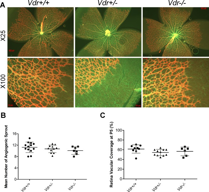
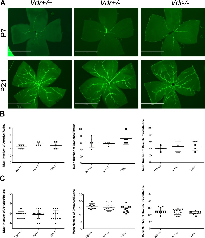
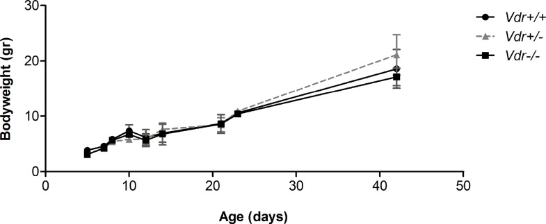
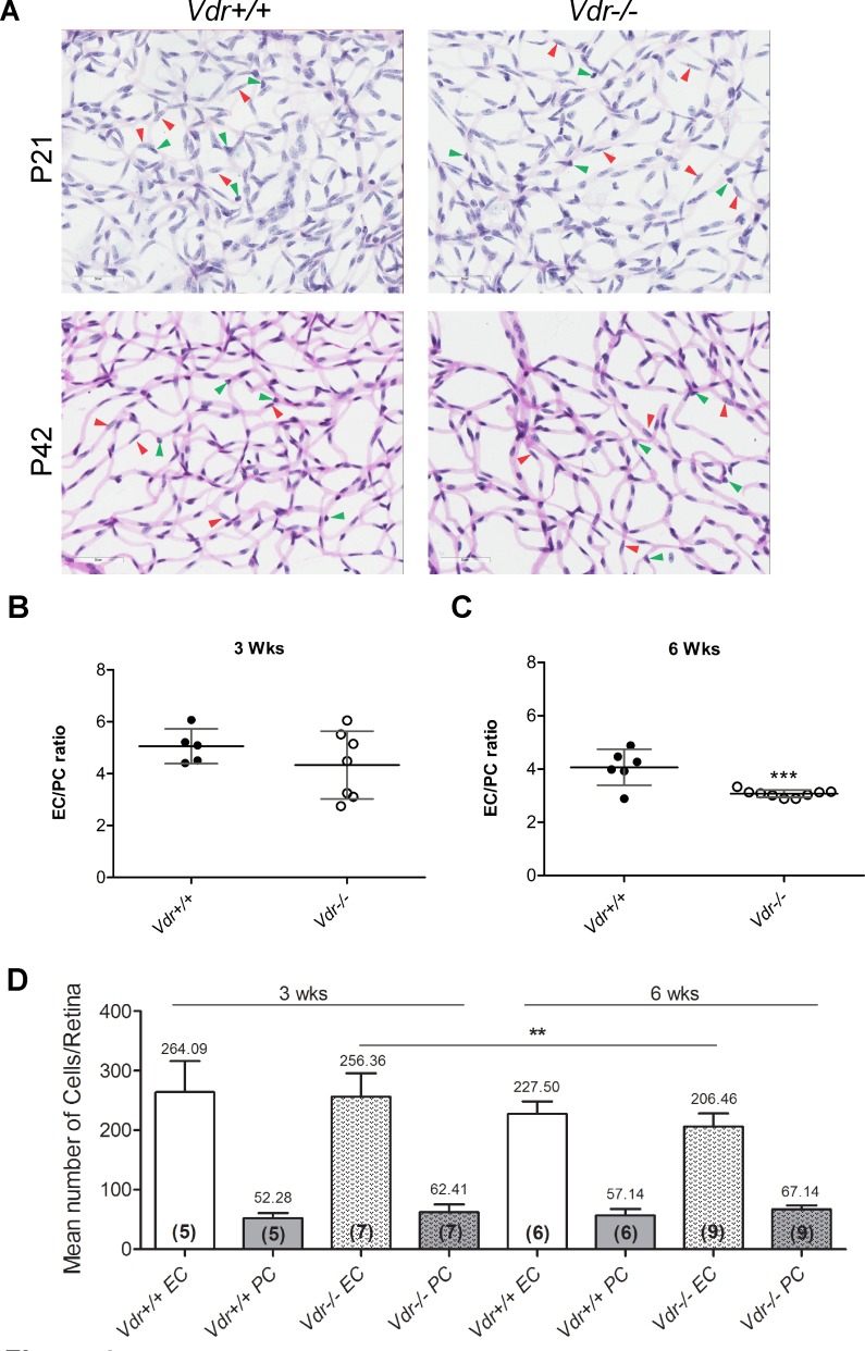
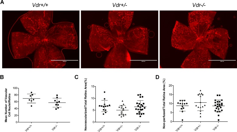
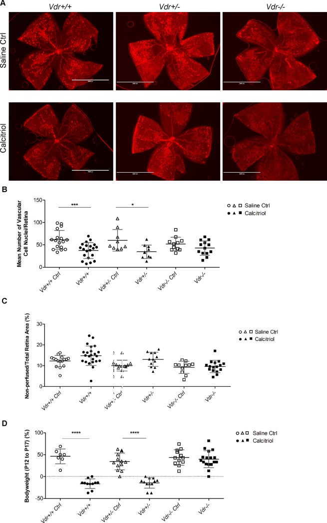
Vitamin D provides a significant benefit to human health, and its deficiency has been linked to a variety of diseases including cancer. Vitamin D exhibits anticancer effects perhaps through inhibition of angiogenesis. We previously showed that the active form of vitamin D (1, 25(OH)2D3; calcitriol) is a potent inhibitor of angiogenesis in mouse model of oxygen-induced ischemic retinopathy (OIR). Many of vitamin D's actions are mediated through vitamin D receptor (VDR). However, the role VDR expression plays in vascular development and inhibition of neovascularization by 1, 25(OH)2D3 remains unknown. Here using wild type (Vdr +/+) and Vdr-deficient (Vdr -/-) mice, we determined the impact of Vdr expression on postnatal development of retinal vasculature and retinal neovascularization during OIR. We observed no significant effect on postnatal retinal vascular development in Vdr -/- mice up to postnatal day 21 (P21) compared with Vdr +/+ mice. However, we observed an increase in density of pericytes (PC) and a decrease in density of endothelial cells (EC) in P42 Vdr -/- mice compared with Vdr +/+ mice, resulting in a significant decrease in the EC/PC ratio. Although we observed no significant impact on vessel obliteration and retinal neovascularization in Vdr -/- mice compared with Vdr +/+ mice during OIR, the VDR expression was essential for inhibition of retinal neovascularization by 1, 25(OH)2D3. In addition, the adverse impact of 1, 25(OH)2D3 treatment on the mouse bodyweight was also dependent on VDR expression. Thus, VDR expression plays a significant role during retinal vascular development, especially during maturation of retinal vasculature by promoting PC quiescence and EC survival, and inhibition of ischemia-mediated retinal neovascularization by 1, 25(OH)2D3.


Conflict of interest statement

Competing Interests: The authors have declared that no competing interests exist.

Figures

Fig 1
Fig 1. The development of superficial layer of retinal vasculature is independent of Vdr expression.
(A) Demonstrates GFAP and Col IV stained retinal vessels prepared from postnatal day 5 (P5) Vdr +/+ and Vdr -/- mice. Please note similar expansion of astrocytes (green, GFAP) and progression of expanding vessels (red, Col IV). Scale bar = 200 μm for x25 and Scale bar = 50 μm for x100 images. (B) The mean number of angiogenic sprouts at the angiogenic fronts were quantified per field (x100) in each retina. (C) Coverage of retinal vasculature relative to total retina area were measured for each retina and is shown as a percentage. (n≥ 5; each point represents one mouse).
Fig 2
Fig 2. The organization of major blood vessels and development of primary retinal vascular plexus is not affected by Vdr-deficiency.
(A) Retinas from P7 and P21 mice were wholemount stained with anti-α-smooth muscle actin and imaged at x12.5. Mean number of major arteries, branches, and branch points were quantified per retina and shown, respectively, in (B) for P7 and (C) for P21. (n≥ 5; each point represents one mice) Scale bar = 2,000 μm.
Fig 3
Fig 3. Vdr-deficiency minimally affected the mice bodyweight.
Mice bodyweight were determined as detailed in Methods up to 6-weeks of age. We observed no significant differences in postnatal mice bodyweight (gram) at each time point (n≥ 4).
Fig 4
Fig 4. Altered vascular cell density and EC/PC ratios in Vdr -/- mice.
Retinas from P21 and P42 mice were prepared by trypsin digest and H&E/PAS staining. Slides were then scanned and images captured at x400. (A) Representative images are shown; Scale bar = 50 μm. Number of EC (red arrow head) and PC (green arrow head) were counted for at least 6 images per mice, and EC/PC ratio calculated for P21 (B) and for P42 (C) mice; (***P = 0.0008). (D) The quantitative assessment of this data, and the number of EC and PC along with the number of retinas counted in each group in parentheses, are shown. (n≥ 5; **P = 0.0043).
Fig 5
Fig 5. Similar degree of ischemia-driven retinal neovascularization in Vdr+/+ and Vdr-/- during OIR.
(A) Representative images (x20) of wholemount retinal neovascularization isolated from P17 mice exposed to a cycle of hyperoxia and room air (OIR) and stained with collagen IV. Retinas from Vdr +/+, Vdr +/-, and Vdr -/- littermates were wholemount stained with anti-collagen IV to visualize the vasculature. Scale bar = 2,000 μm. Quantitative assessment of the neovascularization (histological evaluation and quantitative analysis of images) and area of vessel obliteration are shown in (B), (C), and (D) respectively. (n≥ 7; each point represents one mice).
Fig 6
Fig 6. Vdr expression is required for significant inhibition of retinal neovascularization by1, 25(OH)2D3.
(A) Representative images (x20) of collagen IV stained wholemount retinal neovascularization from P17 OIR with and without 1, 25(OH)2D3 treatment from Vdr +/+, Vdr +/-, and Vdr -/- mice. Scale bar = 2,000 μm. Quantitative assessment of the neovascularization (histological evaluation) and area of vessel obliteration from these groups are shown in (B) and (C), respectively; (***P = 0.0006, *P = 0.0121). (D) The evaluated mice bodyweight (gr) comparison from the above groups is shown; (****P<0.0001). (n≥ 7; each point represents one mouse).

Similar articles

Cited by

References

    1. Zittermann A. Vitamin D-Cholecalciferol In: Herrmann W, Obeid R, editors. Vitamins in the Prevention of Human Diseases. Germany: De Gruyter; 2011. p. 363–95.
    1. Crew KD. Vitamin D: Are We Ready to Supplement for Breast Cancer Prevention and Treatment? ISRN Oncology. 2013;2013:483687 10.1155/2013/483687. PMC3600307. - DOI - PMC - PubMed
    1. Reins RY, McDermott AM. Vitamin D: Implications for ocular disease and therapeutic potential. Exp Eye Res. 2015;134:101–10. 10.1016/j.exer.2015.02.019. 10.1016/j.exer.2015.02.019 - DOI - DOI - PMC - PubMed
    1. Johnson JA, Grande JP, Roche PC, Campbell RJ, Kumar R. Immuno-localization of the calcitriol receptor, calbinclin-D28k and the plasma membrane calcium pump in the human eye. Curr Eye Res. 1995;14(2):101–8. 10.3109/02713689508999921 - DOI - PubMed
    1. Jamali N, Sorenson C, Sheibani N. Vitamin D attenuates proangiogenic properties of pericytes through increased production of VEGF (679.1). The FASEB Journal. 2014;28(1 Supplement).

Publication types

MeSH terms

LinkOut - more resources