. 2018 Feb 1;7(2):gix131.
doi: 10.1093/gigascience/gix131.

eHistology image and annotation data from the Kaufman Atlas of Mouse Development

Affiliations

eHistology image and annotation data from the Kaufman Atlas of Mouse Development

Richard A Baldock et al. Gigascience. .

Abstract

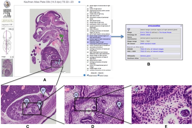
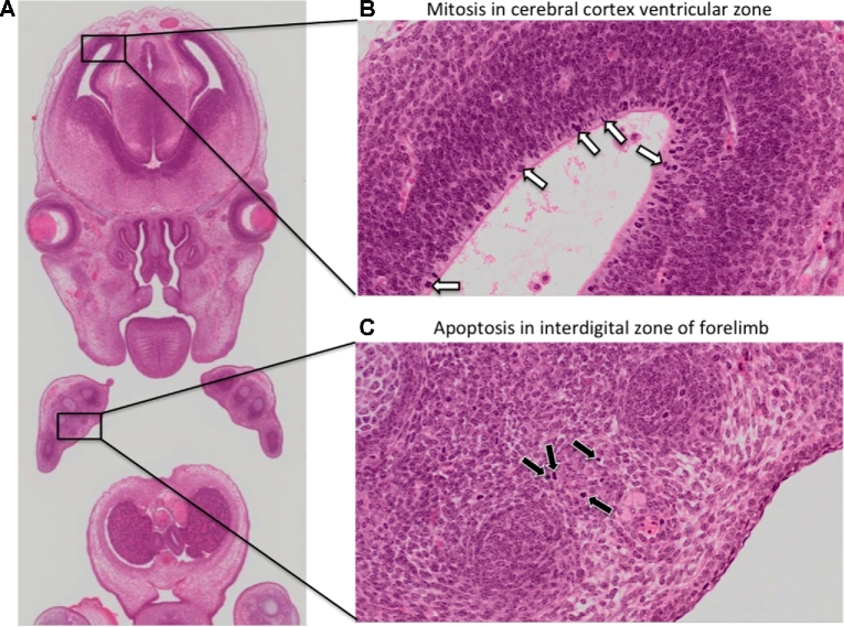
"The Atlas of Mouse Development" by Kaufman is a classic paper atlas that is the de facto standard for the definition of mouse embryo anatomy in the context of standard histological images. We have redigitized the original haematoxylin and eosin-stained tissue sections used for the book at high resolution and transferred the hand-drawn annotations to digital form. We have augmented the annotations with standard ontological assignments (EMAPA anatomy) and made the data freely available via an online viewer (eHistology) and from the University of Edinburgh DataShare archive. The dataset captures and preserves the definitive anatomical knowledge of the original atlas, provides a core image set for deeper community annotation and teaching, and delivers a unique high-quality set of high-resolution histological images through mammalian development for manual and automated analysis.


Figures

Figure 1:
Figure 1:
Kaufman Atlas eHistology Viewer. A, Screen shot showing the user interface with annotation list. The 3 selected terms showing as a blue “flags” numbers 37, 38, 39 are over the developing kidney. B, The pop-up dialog with extra information on selecting the “37.” C–E, Progressively higher-resolution images corresponding to zooming-in on the image. At full resolution, the pixel spacing is 0.34 × 0.34 microns and reveals cellular architecture and arrangements.
Figure 2:
Figure 2:
Observing mitosis and apoptosis in cellular resolution eHistology atlas images. A key advantage of capturing histology images at high resolution is the ability to morphologically identify mitotically dividing cells and apoptotic cells in embryo atlas images. A, Zoomed-out view of a coronal image of an E14.5 embryo. B, On the zoomed-in view, neuroblasts in the ventricular zone of the cerebral cortex show intense haematoxylin staining (white arrows), a morphological feature associated with chromosome condensation in mitotically dividing cells. C, On the zoomed-in view, scattered cells in the interdigital zone of the forelimb are pyknotic (black arrows), a morphological feature associated with nuclear condensation. The pyknotic cells additionally show signs of cell shrinkage. These are morphological hallmarks of apoptosis.

References

    1. Kaufman MH. The Atlas of Mouse Development. Amsterdam, the Netherlands: Elsevier Academic Press; 1994.
    1. Richardson L, Venkataraman S, Stevenson P et al. . EMAGE mouse embryo spatial gene expression database: 2014 update. Nucl Acids Res 2014;42(D1):D835–44. - PMC - PubMed
    1. Hayamizu TF, Wicks MN, Davidson DR et al. . EMAP/EMAPA ontology of mouse developmental anatomy: 2013 update. J Biomed Sem 2013;4(1):15. - PMC - PubMed
    1. Hayamizu TF, Baldock RA, Ringwald M. Mouse anatomy ontologies: enhancements and tools for exploring and integrating biomedical data. Mamm Genome 2015;26(9–10):422–30. - PMC - PubMed
    1. Brown SD, Moore MW. Towards an encyclopaedia of mammalian gene function: the International Mouse Phenotyping Consortium. Dis Model Mech 2012;5(3):289–92. - PMC - PubMed

Publication types