Comparative Study
. 2018 May;175(9):1419-1438.
doi: 10.1111/bph.14132. Epub 2018 Mar 23.

Different contributions of chemokine N-terminal features attest to a different ligand binding mode and a bias towards activation of ACKR3/CXCR7 compared with CXCR4 and CXCR3

Affiliations
Comparative Study

Different contributions of chemokine N-terminal features attest to a different ligand binding mode and a bias towards activation of ACKR3/CXCR7 compared with CXCR4 and CXCR3

Martyna Szpakowska et al. Br J Pharmacol. 2018 May.

Abstract

Background and purpose: Chemokines and their receptors form an intricate interaction and signalling network that plays critical roles in various physiological and pathological cellular processes. The high promiscuity and apparent redundancy of this network makes probing individual chemokine/receptor interactions and functional effects, as well as targeting individual receptor axes for therapeutic applications, challenging. Despite poor sequence identity, the N-terminal regions of chemokines, which play a key role in their activity and selectivity, contain several conserved features. Thus far little is known regarding the molecular basis of their interactions with typical and atypical chemokine receptors or the conservation of their contributions across chemokine-receptor pairs.

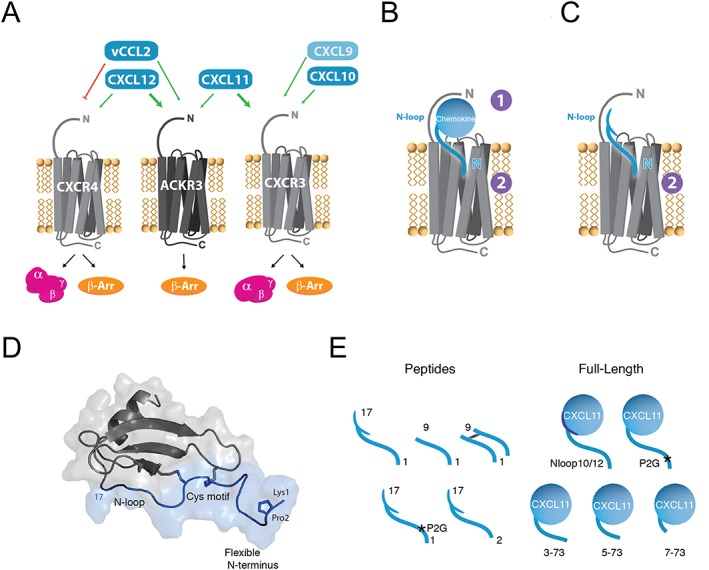
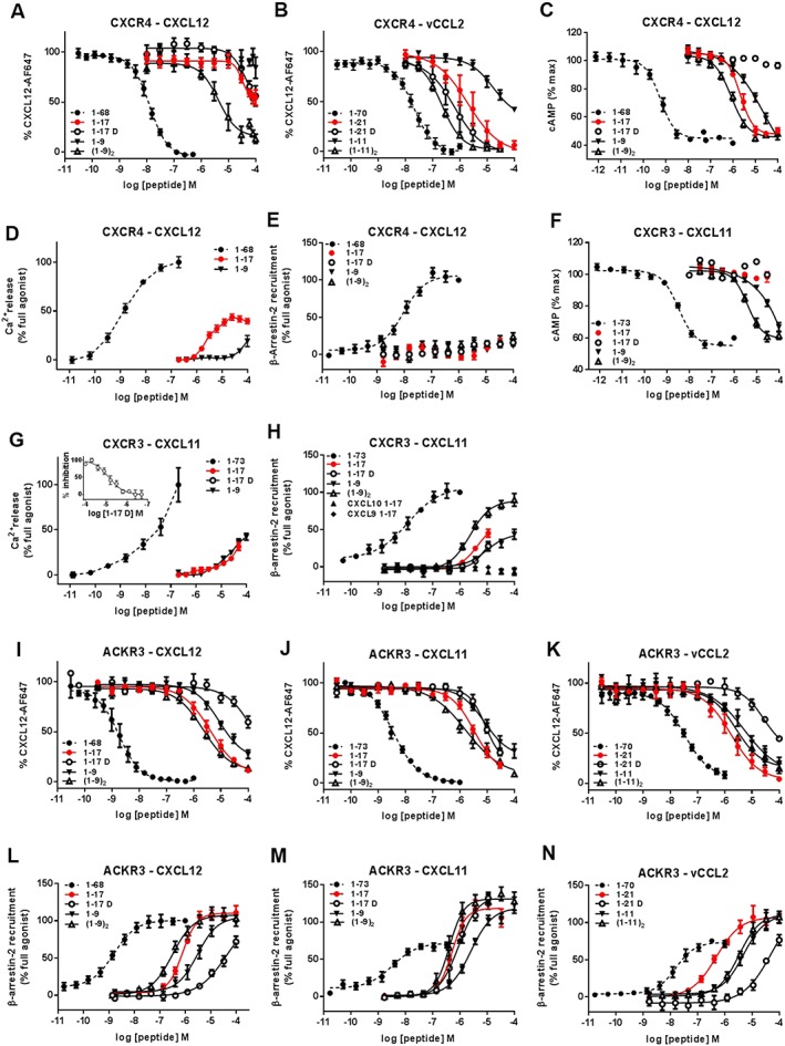
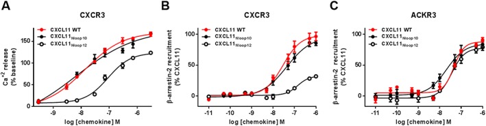
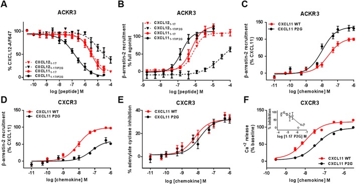
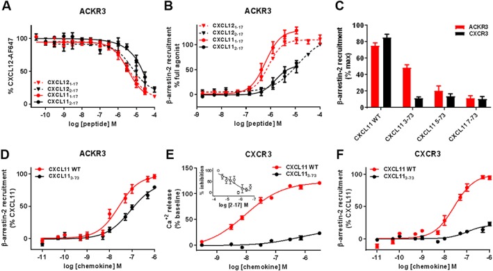
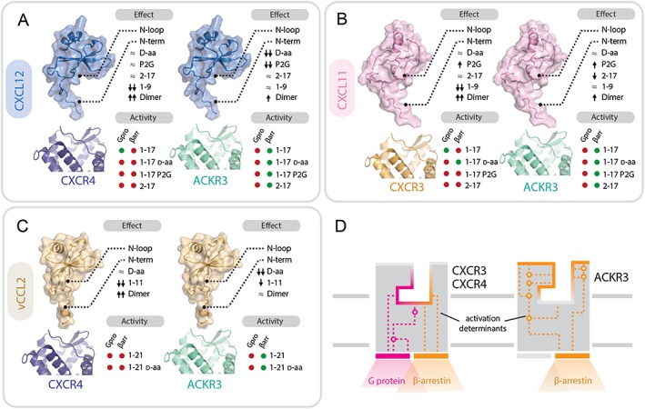
Experimental approach: We used a broad panel of chemokine variants and modified peptides derived from the N-terminal region of chemokines CXCL12, CXCL11 and vCCL2, to compare the contributions of various features to binding and activation of their shared receptors, the two typical, canonical G protein-signalling receptors, CXCR4 and CXCR3, as well as the atypical scavenger receptor CXCR7/ACKR3, which shows exclusively arrestin-dependent activity.

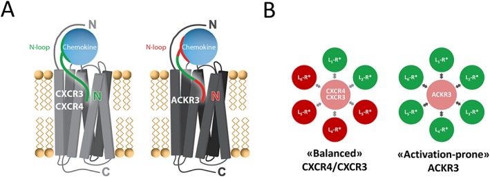
Key results: We provide molecular insights into the plasticity of the ligand-binding pockets of these receptors, their chemokine binding modes and their activation mechanisms. Although the chemokine N-terminal region is a critical determinant, neither the most proximal residues nor the N-loop are essential for binding and activation of ACKR3, as distinct from binding and activation of CXCR4 and CXCR3.

Conclusion and implications: These results suggest a different interaction mechanism between this atypical receptor and its ligands and illustrate its strong propensity to activation.


Figures

Figure 1
Figure 1
CXCR4‐ACKR3‐CXCR3 receptor‐ligand interaction network and chemokine N‐terminal features. (A) Selectivity and crosstalk between the two canonical G protein‐signalling chemokine receptors, CXCR4 and CXCR3, the atypical β‐arrestin‐biased receptor ACKR3 and their shared ligands. CXCL12 is the only endogenous chemokine ligand for CXCR4. It is also the highest affinity chemokine for ACKR3 but does not bind CXCR3. The viral chemokine vCCL2 is a CXCR4 antagonist but acts as an agonist of ACKR3. CXCL11 is the dominant ligand for CXCR3 and binds also to ACKR3. CXCL10 and CXCL9 bind and activate CXCR3, but not ACKR3. (B) Two‐site/two‐step model for the interactions of full‐length chemokines with their cognate receptors. In the first step, the body of the chemokine and the N‐loop are specifically recognized by the N terminus of the receptor (CRS1). During the second step, the insertion of the chemokine N terminus into the receptor‐binding pocket (CRS2) stabilizes its active form triggering downstream signalling. (C) Peptides derived from the N‐terminal region of chemokines, which represent useful probes to investigate the interaction between chemokines and receptors. (D) Schematic representation of CXCL12 and location of the N‐terminal features investigated in this study (blue). The N‐terminal region encompasses the flexible N terminus (1–8), the CXC cysteine motif (9–11) and the N‐loop (13–17). (E) Chemokine‐derived peptides and CXCL11 variants investigated in this study.
Figure 2
Figure 2
Binding, G protein signalling and β‐arrestin‐2 recruitment to CXCR4, CXCR3 and ACKR3 induced by full‐length chemokines and chemokine N‐terminal peptides. Binding and modulation of CXCR4 (A–E), CXCR3 (F–H) and ACKR3 (I–N) by full‐length CXCL12, CXCL11, vCCL2 and peptides derived from their N‐terminal regions. Binding to CXCR4 (A and B) and ACKR3 (I–K) was assessed by binding competition studies with Alexa Fluor 647‐coupled CXCL12 in U87 cells stably expressing the receptors and analysed by flow cytometry. G protein signalling induced by full‐length chemokines and peptides derived from their N‐terminal regions towards CXCR4 (C and D) and CXCR3 (F and G) was evaluated by measuring the modulation of the basal intracellular cAMP concentration using Glo‐cAMP sensor (C and F) or the release of intracellular calcium using Fluo‐2 dye and FLIPR platform (D and G). (G‐inset) Antagonist properties of peptide CXCL111–17D monitored in calcium assay. β‐arrestin‐2 recruitment to CXCR4 (E), CXCR3 (H) and ACKR3 (L–N) induced by full‐length chemokines and N‐terminal peptides was monitored using a Nanoluciferase‐based complementation assay (NanoBIT). Each experiment was performed five times, and the data shown are means ± SEM.
Figure 3
Figure 3
ACKR3 and CXCR3 activation by CXCL11 N‐loop variants. (A) CXCR3 activation by CXCL11 WT and N‐loop variants monitored in HEK293 cells through intracellular calcium release using FLIPR 4 Calcium Flux kit dye. (B and C) Comparison of β‐arrestin‐2 recruitment to CXCR3 (B) and ACKR3 (C) induced by CXCL11WT and N‐loop variants monitored in HEK293 cells by BRET using receptor‐YFP fusion constructs and β‐arrestin‐2‐Rluc. Each experiment was performed five times, and the data shown are means ± SEM.
Figure 4
Figure 4
Binding and activation of ACKR3 and CXCR3 by N‐terminal peptides and full‐length CXCL11 bearing the P2G mutation. (A and B) Comparison of the effects of the P2G mutation in CXCL12‐ and CXCL11‐derived peptides on (A) binding to ACKR3 assessed by competition studies with Alexa Fluor 647‐coupled CXCL12 and (B) β‐arrestin‐2 recruitment to ACKR3 monitored by NanoLuc complementation in U87 cells. (C and D) Comparison of β‐arrestin‐2 recruitment to ACKR3 (C) and CXCR3 (D) induced by CXCL11WT and P2G mutant in HEK293 cells monitored by BRET using receptor‐YFP fusion constructs and β‐arrestin‐2‐Rluc. (E and F) CXCR3 activation by CXCL11WT and P2G mutant monitored in HEK293 cells through (E) adenylate cyclase inhibition using BRET reporter GFP10‐Epac‐ Rluc3 and (F) intracellular calcium release using FLIPR 4 Calcium Flux kit dye. (F‐inset) Antagonist properties of peptide CXCL111–17/P2G monitored in calcium assay. Each experiment was performed five times, and the data shown are means ± SEM.
Figure 5
Figure 5
β‐arrestin‐2 recruitment and G protein signalling induced by N‐terminally truncated CXCL11 variants towards ACKR3 and CXCR3. (A and B) Comparison of the effects of N‐terminal residue truncation in CXCL12‐ and CXCL11‐derived peptides on (A) binding to ACKR3 assessed by competition studies with Alexa Fluor 647‐coupled CXCL12 and (B) β‐arrestin‐2 recruitment to ACKR3 monitored by NanoLuc complementation in U87 cells. (C) Impact of progressive N‐terminal truncation on the ability of CXCL11 (100 nM) to recruit β‐arrestin‐2 to ACKR3 and CXCR3. Values are expressed as percentage of the maximum β‐arrestin‐2 recruitment monitored with saturating concentrations of CXCL11WT. (D and F) Comparison of β‐arrestin‐2 recruitment to ACKR3 (D) and CXCR3 (F) induced by CXCL11WT and variants lacking the first two residues (CXCL113–73) monitored by BRET. (E) Comparison of maximum CXCR3 mediated intracellular calcium release induced by CXCL11WT and CXCL113–73 variant in HEK cells using FLIPR 4 Calcium Flux kit dye. (E‐inset) Antagonist properties of peptide CXCL112–17 monitored in calcium assay. Each experiment was performed five times, and the data shown are means ± SEM.
Figure 6
Figure 6
Differential contribution of chemokine N‐terminal features to interactions with CXCR3/CXCR4 and ACKR3 and distribution between receptor active/inactive states. (A) The entire chemokine N‐terminal region (green) is critical for the binding and activation of the canonical receptors CXCR3 and CXCR4, whereas the most N‐terminal residues (red) as well as the N‐loop (red) do not appear important for activation of the atypical receptor ACKR3. (B) Comparison of the distribution of active (R* green) and inactive (R* red) conformations of CXCR4/CXCR3 and ACKR3 stabilized by ligands (L) targeting the receptor transmembrane ligand‐binding pocket (CRS2). CXCR4 and CXCR3 have low basal activity (light red) and are ‘balanced’ receptors as ligand binding to CRS2 can stabilize either the active state (green) or inactive state (red). ACKR3 is an ‘activation‐prone’ receptor as ligand binding preferentially stabilizes the active state (green) of the receptor leading to β‐arrestin‐2 recruitment. Indeed, so far, no ligand targeting the transmembrane pocket of ACKR3 without displaying agonist activity has been reported.
Figure 7
Figure 7
Contributions of chemokine and receptor regions to binding and activation of ACKR3, CXCR3 and CXCR4. (A–C) Importance of structural determinants of chemokines CXCL12 (A), CXCL11 (B) and vCCL2 (C) for binding and activity towards the receptors. Upper diagram: effects of the different modifications of chemokine N‐terminal domains on receptor binding and/or activity: ≈ no effect or <5‐fold change; ↑ or ↓ increase or decrease by >5 fold; ↑↑ or ↓↓ increase or decrease by >50 fold. Lower diagram: activity (shown as green circles) and lack of activity (shown as red circles) in G protein signalling (Gpro) and β‐arrestin recruitment (βarr) of peptides comprising the flexible N terminus and the N‐loop (1–17/21), their D stereoisomers and proximally modified variants (P2G, 2–17). (D) Comparison of ACKR3 and CXCR3/4 binding pockets. The activation determinants of ACKR3 are localized closer to the surface compared with CXCR3 and CXCR4. Ligand binding to ACKR3 pocket triggers a limited signalling repertoire (i.e. β‐arrestin‐2 recruitment). In contrast, CXCR4 and CXCR3 activation determinants are localized deeper in the ligand binding pocket with chemokine triggering G protein signalling and, subsequently, arrestin recruitment.

References

    1. Alexander SPH, Christopoulos A, Davenport AP, Kelly E, Marrion NV, Peters JA et al (2017a). The Concise Guide to PHARMACOLOGY 2017/18: G protein‐coupled receptors. Br J Pharmacol 174: S17–S129. - PMC - PubMed
    1. Alexander SPH, Fabbro D, Kelly E, Marrion NV, Peters JA, Faccenda E et al (2017b). The Concise Guide to PHARMACOLOGY 2017/18: Enzymes. Br J Pharmacol 174: S272–S359. - PMC - PubMed
    1. Allen SJ, Crown SE, Handel TM (2007). Chemokine: receptor structure, interactions, and antagonism. Annu Rev Immunol 25: 787–820. - PubMed
    1. Bachelerie F, Ben‐Baruch A, Burkhardt AM, Combadiere C, Farber JM, Graham GJ et al (2014a). International Union of Basic and Clinical Pharmacology. [corrected]. LXXXIX. Update on the extended family of chemokine receptors and introducing a new nomenclature for atypical chemokine receptors. Pharmacol Rev 66: 1–79. - PMC - PubMed
    1. Bachelerie F, Graham GJ, Locati M, Mantovani A, Murphy PM, Nibbs R et al (2014b). New nomenclature for atypical chemokine receptors. Nat Immunol 15: 207–208. - PubMed

Publication types