. 2017 Dec 22;7(1):18099.
doi: 10.1038/s41598-017-18291-0.

The crucial role of the TRPM7 kinase domain in the early stage of amelogenesis

Affiliations

The crucial role of the TRPM7 kinase domain in the early stage of amelogenesis

Kayoko Ogata et al. Sci Rep. .

Abstract

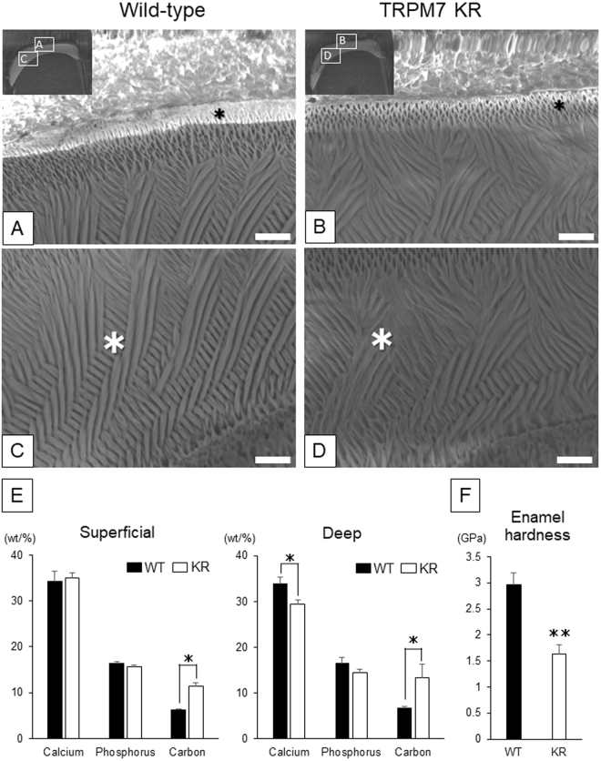
Transient receptor potential melastatin-7 (TRPM7) is a bi-functional protein containing a kinase domain fused to an ion channel. TRPM7 is highly expressed in ameloblasts during tooth development. Here we show that TRPM7 kinase-inactive knock-in mutant mice (TRPM7 KR mice) exhibited small enamel volume with opaque white-colored incisors. The TRPM7 channel function of ameloblast-lineage cells from TRPM7 KR mice was normal. Interestingly, phosphorylation of intracellular molecules including Smad1/5/9, p38 and cAMP response element binding protein (CREB) was inhibited in ameloblasts from TRPM7 KR mice at the pre-secretory stage. An immunoprecipitation assay showed that CREB was bound to TRPM7, suggesting that direct phosphorylation of CREB by TRPM7 was inhibited in ameloblast-lineage cells from TRPM7 KR mice. These results indicate that the function of the TRPM7 kinase domain plays an important role in ameloblast differentiation, independent of TRPM7 channel activity, via phosphorylation of CREB.


Conflict of interest statement

The authors declare that they have no competing interests.

Figures

Figure 1
Figure 1
(AF) Anterior (A and C) and lateral (B and D) views of the maxillary incisors and occlusal views of the mandibular molars (E and F) of 20-week-old wild-type (WT) (A,B and E) and TRPM7 KR (C,D and F) mice. (G,H) Sagittal views of the maxillary incisors of 16-week-old WT (G) and TRPM7 KR (H) mice, obtained using high-definition radiography. (IL) 3D Reconstruction of upper incisor enamel (yellow) with/without dentin (white) from WT (I,J) and TRPM7 KR (K,L) mice using micro-CT analysis of (G) and (H), respectively. (M) The volume of the enamel of the maxillary and mandibular incisors of WT and TRPM7 KR mice aged 12–16 weeks, calculated from analysis of the micro-CT images. Data are presented as the mean ± standard deviation (n = 3). *P < 0.05. KR: TRPM7 KR mutant; WT: wild-type.
Figure 2
Figure 2
(AD) Scanning electron microscopy (SEM) images showing the superficial (A,B) and deep (C,D) layers of the maxillary incisor enamel from 7-week-old wild-type (A,C) and TRPM7 KR (B,D) mice. The inset in each of (A) and (B) indicates the regions that were magnified to obtain (A,C) and (B,D), respectively. Scale bar: 10 μm (AD). *Regions investigated using element mapping at the microstructural level. (E) The contents of calcium, phosphorus and carbon in the superficial and deep layers of incisor enamel from wild-type (WT) and TRPM7 KR mice. The content of each element is shown as the normalized concentration in weight percent (wt/%). (F) Measurements of enamel micro-hardness in WT and TRPM7 KR mice. The mean micro-hardness was 2.95 ± 0.24 GPa in WT mice and 1.64 ± 0.18 GPa in TRPM7 KR mice. Data in (E,F) are presented as the mean ± standard deviation (n = 3). KR: TRPM7 KR mutant; WT: wild-type. *P < 0.05, **P < 0.01.
Figure 3
Figure 3
(A) The pattern of TRPM7 mRNA expression in a whole-body sagittal section at E18.5. (BG) High-magnification images showing TRPM7 mRNA expression in the lung (B), intestine (C), liver (D), kidney (E) and tooth germ (F,G). (H) Quantitative RT-PCR analysis of TRPM7 and TRPM6 mRNA expression levels in various organs from 8-week-old wild-type mice. The mRNA expression levels of TRPM7 (white bars) and TRPM6 (black bars) are normalized to that of actin and presented as the mean ± standard deviation (n = 3). Am: ameloblast; DP: dental papilla; Od: odontoblast. Scale bars: 5 mm (A), 100 μm (BF), 50 μm (G).
Figure 4
Figure 4
(AD) TRPM7 protein expression in E14.5 (A,B) and E16.5 (C,D) molars, detected by immunohistochemistry. (B) and (D) are higher magnifications of the tooth germ shown in (A) and (C), respectively. (EL) Adjacent sections of molars at E18.5 stained using different techniques. (E,I) Hematoxylin and eosin staining. (F,J) TRPM7 protein expression detected by immunohistochemistry. (G,K) Expression of nestin protein (a marker of differentiated odontoblasts) detected by immunohistochemistry. (H,L) Expression of amelogenin protein (a marker of differentiated ameloblasts) detected by immunohistochemistry. (M) Hematoxylin and eosin staining of an incisor from a wild-type mouse at P10. (N) TRPM7 protein expression, detected by immunohistochemistry, in an incisor from a wild-type mouse at P10. (O) Higher magnification of the box in (M). (P) Higher magnification of the box in (N). (Q) TRPM7 protein expression, detected by immunohistochemistry, in an incisor from a control (heterozygous) mouse. (R) Higher magnification of the box in (Q), showing pre-ameloblasts and odontoblasts. (S) TRPM7 protein expression, detected by immunohistochemistry, in an incisor from a TRPM7 KR mouse. (T) Higher magnification of the box in (S), illustrating the pre-ameloblasts and odontoblasts. White arrowheads indicate the cervical region. Scale bars: 100 μm (AQ, S), 50 μm (R, T). Am: ameloblast, d: dentin matrix, DM: dental mesenchyme, DP: dental papilla; IEE: inner enamel epithelium; Od: odontoblast; OEE: outer enamel epithelium; pAm: pre-ameloblast.
Figure 5
Figure 5
(AD) Mg2+-inhibited cation (MIC) currents recorded from inner enamel epithelium-lineage cells from wild-type mice. (A) Time course of the change in membrane current at +90 mV and −100 mV during intracellular and extracellular Mg2+ depletion and following the application of 3 mM MgCl2 to the extracellular solution. (B) Current-voltage (I-V) relationships obtained at the time points indicated by the vertical bars in (A), i.e. before (black line) and after (gray line) application of 3 mM MgCl2. (C) I-V relationships obtained before (black line) and after (gray line) application of 2-aminoethoxyphenylborate (2-APB; 500 μM), a non-specific modulator of several TRP channels including TRPM7. (D) I-V relationships obtained before (black line) and after (gray line) application of FTY720 (10 μM), also an inhibitor of TRPM7 channel activity. (E,F) I-V relationships recorded from inner enamel epithelium-lineage cells from wild-type and TRPM7 KR mutant mice. The I-V relationships were obtained before (Mg2+-free; black line) and after (gray line) application of 3 mM MgCl2. (G) The graph on the right compares the amplitudes of the MIC currents (net current inhibited by 3 mM MgCl2) at +90 mV and −100 mV. Data are expressed as the mean ± standard error of the mean (n = 4). KR: TRPM7 KR mutant; m.p.: membrane potential; N.S.: not significant (P ≥ 0.05); WT: wild-type.
Figure 6
Figure 6
(A,B) Hematoxylin and eosin staining of incisors from control (heterozygous) (A) and TRPM7 KR (B) mice at P10. (C,D) Immunohistochemical detection of the expression of P-Smad1/5/9 in incisors from control (C) and TRPM7 KR (D) mice at P10. (E,F) Immunohistochemical detection of the expression of P-p38 in incisors from control (E) and TRPM7 KR (F) mice at P10. (GJ) Higher magnification views of regions in (A) and (B) to show the morphology of the incisors from control (G,I) and TRPM7 KR (H,J) mice at the pre-secretory (G,H) and secretory (I,J) stages. (KN) Immunohistochemical detection of P-Smad1/5/9 expression in incisors from control (K,M) and TRPM7 KR (L,N) mice at the pre-secretory (K,L) and secretory (M,N) stages. (OR) Immunohistochemical detection of P-p38 expression in incisors from control (O,Q) and TRPM7 KR (P,R) mice at the pre-secretory (O,P) and secretory (Q,R) stages. (S) Immunoblotting analysis of extracts from mHAT9d cells, using anti-Smad1, anti-Smad5 or anti-p38 antibodies after immunoprecipitation by anti-TRPM7 antibody. Input: total cell lysate; IP: immunoprecipitation. Full-length blots are presented in Supplementary Figure 1. Am: ameloblast; d: dentin matrix; DP: dental papilla; e: enamel matrix; HE: hematoxylin and eosin; pAm: pre-ameloblast. Scale bars: 100 μm (AF), 50 μm (GR).
Figure 7
Figure 7
(AD) Immunohistochemical detection of the expressions of CREB (A,B) and P-CREB (C,D) in incisors from control (heterozygous) (A,C) and TRPM7 KR (B,D) mice at P10. (EL) Higher magnification views of regions in (AD) to show the expression of CREB (EH) and P-CREB (IL) in incisors from control (E,G,I,K) and TRPM7 KR (F,H,J,L) mice at the pre-secretory stage (E,F,I,J) and secretory stage (G,H,K,L). (M) Western blot analysis of CREB and P-CREB expression in first molar tooth germ at P2. Full-length blots are presented in Supplementary Figure 2. (N) Immunoblotting analysis of extracts from mHAT9d cells, using anti-CREB antibody after immunoprecipitation by anti-TRPM7 antibody (upper lane) or anti-TRPM7 antibody after immunoprecipitation by anti-CREB antibody (lower lane). Input: total cell lysate; IP: immunoprecipitation. Full-length blots are presented in Supplementary Figure 3. Am: ameloblast; d: dentin matrix; DP: dental papilla; e: enamel matrix; KR: TRPM7 KR mutant; pAm: pre-ameloblast; WT: wild-type. Scale bars: 100 μm (AD), 50 μm (EL).

References

    1. Simmer JP, et al. Regulation of dental enamel shape and hardness. J. Dent. Res. 2010;89:1024–1038. doi: 10.1177/0022034510375829. - DOI - PMC - PubMed
    1. Hu JC, Chun YH, Al Hazzazzi T, Simmer JP. Enamel formation and amelogenesis imperfecta. Cells Tissues Organs. 2007;186:78–85. doi: 10.1159/000102683. - DOI - PubMed
    1. Josephsen K, Fejerskov O. Ameloblast modulation in the maturation zone of the rat incisor enamel organ. A light and electron microscopic study. J. Anat. 1977;124:45–70. - PMC - PubMed
    1. Gritli-Linde A, et al. Shh signaling within the dental epithelium is necessary for cell proliferation, growth and polarization. Development. 2002;129:5323–5337. doi: 10.1242/dev.00100. - DOI - PubMed
    1. Josephsen K, et al. Ion transporters in secretory and cyclically modulating ameloblasts: a new hypothesis for cellular control of preeruptive enamel maturation. Am. J. Physiol. Cell Physiol. 2010;299:C1299–1307. doi: 10.1152/ajpcell.00218.2010. - DOI - PubMed

Publication types

Substances

LinkOut - more resources