. 2018 Feb:115:82-93.
doi: 10.1016/j.yjmcc.2017.12.007. Epub 2017 Dec 20.

Calcium phosphate particles stimulate interleukin-1β release from human vascular smooth muscle cells: A role for spleen tyrosine kinase and exosome release

Affiliations

Calcium phosphate particles stimulate interleukin-1β release from human vascular smooth muscle cells: A role for spleen tyrosine kinase and exosome release

Yana Dautova et al. J Mol Cell Cardiol. 2018 Feb.

Abstract

Aims: Calcium phosphate (CaP) particle deposits are found in several inflammatory diseases including atherosclerosis and osteoarthritis. CaP, and other forms of crystals and particles, can promote inflammasome formation in macrophages leading to caspase-1 activation and secretion of mature interleukin-1β (IL-1β). Given the close association of small CaP particles with vascular smooth muscle cells (VSMCs) in atherosclerotic fibrous caps, we aimed to determine if CaP particles affected pro-inflammatory signalling in human VSMCs.

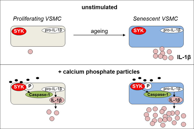
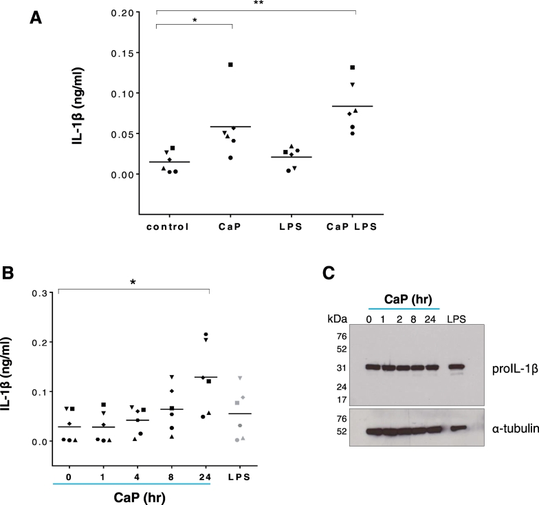
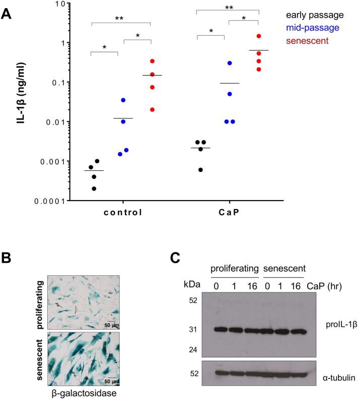
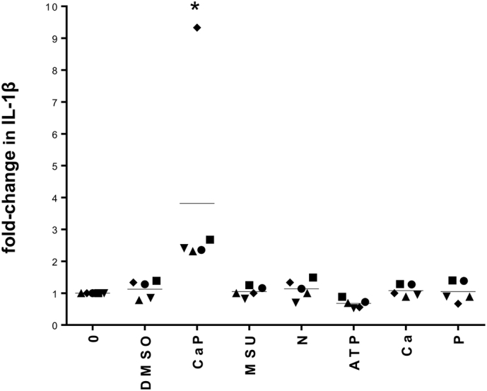
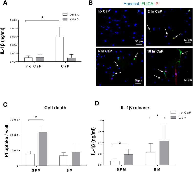
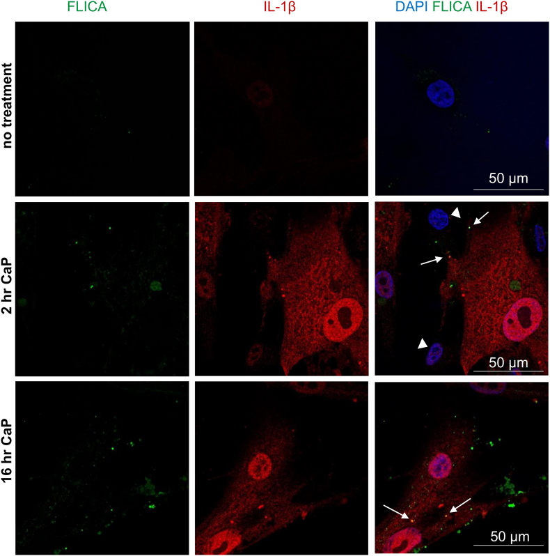
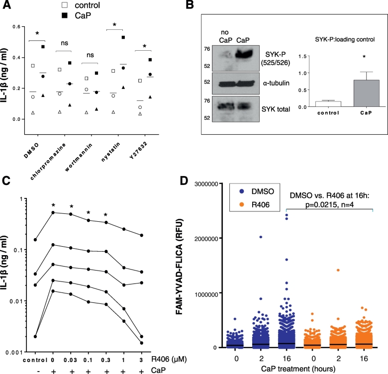
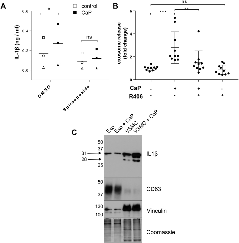
Methods and results: Using ELISA to measure IL-1β release from VSMCs, we demonstrated that CaP particles stimulated IL-1β release from proliferating and senescent human VSMCs, but with substantially greater IL-1β release from senescent cells; this required caspase-1 activity but not LPS-priming of cells. Potential inflammasome agonists including ATP, nigericin and monosodium urate crystals did not stimulate IL-1β release from VSMCs. Western blot analysis demonstrated that CaP particles induced rapid activation of spleen tyrosine kinase (SYK) (increased phospho-Y525/526). The SYK inhibitor R406 reduced IL-1β release and caspase-1 activation in CaP particle-treated VSMCs, indicating that SYK activation occurs upstream of and is required for caspase-1 activation. In addition, IL-1β and caspase-1 colocalised in intracellular endosome-like vesicles and we detected IL-1β in exosomes isolated from VSMC media. Furthermore, CaP particle treatment stimulated exosome secretion by VSMCs in a SYK-dependent manner, while the exosome-release inhibitor spiroepoxide reduced IL-1β release.

Conclusions: CaP particles stimulate SYK and caspase-1 activation in VSMCs, leading to the release of IL-1β, at least in part via exosomes. These novel findings in human VSMCs highlight the pro-inflammatory and pro-calcific potential of microcalcification.

Keywords: Calcium phosphate particles; Caspase-1; Cytokines; Exosomes; SYK; Vascular smooth muscle.


Figures

Image 2
Graphical abstract
Fig. 1
Fig. 1
CaP particles stimulate IL-1β release. A. ELISA measurement of IL-1β release from 6 different human VSMC isolates (indicated by 6 different symbols) over 16 h with or without CaP particles (12.5 μg/mL) and with or without a 24-h pre-treatment with LPS (0.1 μg/mL). The control contained equivalent PBS. Values with means are displayed and one-way ANOVA followed by Holm-Sidak's multiple comparisons test was used to compare groups with the control. Significant differences are indicated as * (P < 0.05) or ** (P < 0.001). B. IL-1β release from 6 different human VSMC isolates exposed to CaP particles (12.5 μg/mL) at time points up to 24 h or LPS (0.1 μg/mL) for 24 h. Values with means are displayed and were analysed by one-way ANOVA followed Holm-Sidak's multiple comparisons test; significant differences from the no additions ‘0’ control are indicated as * (P < 0.01). C. Western analysis of pro-IL-1β levels in VSMC lysates treated as in B. In C, blots are representative of 4 independent experiments in cell isolates from different individuals. Results from 3 further VSMC isolates are displayed in the Supplement Fig. S2B.
Fig. 2
Fig. 2
IL-1β release at different levels at different stages of VSMC culture. A. ELISA measurement of IL-1β release from early passage (3–5), mid-passage (6–13) or senescent VSMCs (14–18) with or without CaP particles (12.5 μg/mL) incubated over 16 h. Data are displayed on a log-scale and mean levels for each treatment are indicated. Data were log transformed prior to statistical analysis. A two-way ANOVA followed by a Holm-Sidak's multiple comparisons test was used to compare groups. Significant differences are indicated by * (P < 0.01) or ** (P < 0.001). 4 different VSMC isolates were used for each cell culture stage. B. β-galactosidase staining of proliferating (mid-passage) or senescent VSMCs, showing higher expression in senescent cells. C. Western analysis of pro-IL-1β levels in proliferating or senescent VSMC lysates treated with CaP particles (12.5 μg/mL) for indicated times. Blots showing results from 3 different cell isolates are shown in the Supplement Fig. S2C and S2D.
Fig. 3
Fig. 3
Inflammasome activators MSU, nigericin (N) and ATP do not induce IL-1β release from human VSMCs. ELISA measurement of IL-1β release from 5 different VSMCs isolates (indicated by 5 different symbols), displayed as fold-change related to ‘0’ control values for each cell isolate. Cells were treated with either CaP particles (12.5 μg/mL), MSU crystals (12.5 μg/mL), nigericin (25 μM), ATP (10 mM), CaCl2 (5.4 mM, ‘Ca’), Na2HPO4 (2 mM, ‘P’), vehicle control (DMSO) or no additions (0) for 16 h. Mean levels for each treatment are indicated. Raw data are displayed in Supplement Fig. S4. A one-way ANOVA on log-transformed raw data followed by a Holm-Sidak's multiple comparisons test determined that CaP treatment significantly differed from each of the other treatments (*P < 0.05).
Fig. 4
Fig. 4
CaP particles stimulate caspase-1 activity. A. ELISA measurement of IL-1β release from 3 different VSMC isolates over 16 h with or without CaP particles (12.5 μg/mL) and with or without YVAD (20 μM). A two-way ANOVA followed by Holm-Sidak's multiple comparisons test was used to determine differences between means. Results are presented as mean ± SEM, *P < 0.05. B. VSMCs were treated with or without CaP particles at indicated times then labelled with FAM-YVAD-FLICA (active caspase-1 reagent), PI and Hoechst and imaged live. Arrows indicate foci/cells containing active caspase-1. Images are representative of 5 experiments in different cell isolates, quantitation of active caspase-1 and PI in live cells is displayed in Fig. 6D and Supplement Figs. S8 and S9. C and D. PI uptake (C) or IL-1β release (D) from 3 different VSMCs isolates in serum-free conditions (SFM) or in basal medium containing 5% FBS (BM) with or without CaP particles (12.5 μg/mL) over 16 h. Mean values ± SEM are displayed. In C, a two-way ANOVA followed by Holm-Sidak's multiple comparisons test was performed and significant differences between corresponding no CaP and CaP-treated samples are indicated by * (P < 0.05). In D, a two-way ANOVA was performed on log-transformed data followed by Holm-Sidak's multiple comparison's test and significant differences between corresponding no CaP and CaP-treated samples are indicated by * (P < 0.05).
Fig. 5
Fig. 5
Caspase-1 activity colocalises with IL-1β. VSMCs were treated with or without CaP particles (12.5 μg/mL) for indicated times, labelled with FAM-YVAD-FLICA (active caspase-1 reagent), then fixed and incubated with antibodies recognising IL-1β. Arrows indicate areas in merged images where active caspase-1 and IL-1β overlap. Arrowheads indicate some cells treated with CaP particles that did not contain IL-1β. Confocal images are representative of experiments in 3 different cell isolates.
Fig. 6
Fig. 6
CaP uptake and SYK phosphorylation. A. ELISA measurement of IL-1β release from VSMCs that were exposed to chlorpromazine (1 μg/mL), wortmannin (100 ng/mL), nystatin (10 μg/mL), Y27632 (10 μM) or vehicle control (DMSO) for 1 h, then exposed to CaP particles (12.5 μg/mL) for 16 h. A two-way ANOVA followed by Sidak's multiple comparisons test was used to compare groups. Significant differences are indicated by * (P < 0.01), n = 3, i.e. VSMC isolates from 3 different individuals (indicated by 3 different symbols). B. VSMCs were treated with or without CaP particles (12.5 μg/mL) for 10 min and analysed for SYK or phosphorylated SYK (525/526) by Western analysis. Western blots shown are representative of experiments from 3 different cell isolates (see Supplement Fig. S7) Ratios of SYK-P to loading control were higher in CaP-treated cells, compared with non-treated controls (paired t-test of log-transformed data, P < 0.01, n = 3 different isolates). C. ELISA measurement of IL-1β release from 5 different VSMC isolates exposed to CaP particles (12.5 μg/mL) and R406 at concentrations indicated or vehicle control (DMSO). One-way ANOVA of log-transformed data was performed followed by Holm-Sidak's multiple comparisons tests; indicated treatments differed significantly from control values, *P < 0.05. D. Quantitation of active caspase-1 in live VSMCs after treatment with CaP particles (12.5 μg/mL) for 0, 2 or 16 h with either a 1 h pre-treatment with R406 (1 μM) or equivalent DMSO. Results shown are representative of 4 independent experiments in different cell isolates. Scatter plots display fluorescence levels of the caspase-1 substrate (FAM-YVAD-FLICA) for approximately 10,000 live VSMCs for each treatment group. Each dot represents the active caspase-1 level for 1 cell. As indicated, when comparing VSMCs from 4 different individuals, CaP particles increased active caspase-1 activity and this effect was reduced in the presence of R406, when comparing the top 5th percentile of VSMCs at the 16 h time point (P = 0.0215).
Fig. 7
Fig. 7
Release of IL-1β via exosomes. A. VSMCs were treated with or without CaP particles (12.5 μg/mL) for 16 h with or without a pre-treatment with spiroepoxide (10 μM). A two-way ANOVA followed by Sidak's multiple comparisons test was used to compare groups. Significant differences are indicated by * (P < 0.05), n = 3, i.e. VSMC isolates from 3 different individuals (indicated by 3 different symbols). B. Exosome secretion by VSMCs. VSMCs were incubated in 2.5%FBS/M199 media with or without CaP particles (12.5 μg/mL) and with or without R406 (1 μM) for 16 h. Conditioned media were harvested and exosomes were quantified using an anti-CD63 bead-capturing assay which detects CD63/CD81 exosomes. Statistical significance was tested by one-way ANOVA followed by Sidak's multiple comparisons test. Significant differences are indicated by **(P < 0.01), ***(P < 0.001), n = 10. C. Western analysis of exosomes isolated by differential ultracentrifugation from conditioned media of VSMCs treated with or without CaP particles for 16 h. Exo, exosomes; VSMC, whole cell lysates. The membrane was probed with IL-1β antibodies (R and D), CD63 (BD Pharminigen), with vinculin and Coomassie brilliant blue staining to demonstrate protein loading.

Similar articles

Cited by

References

    1. Virmani R., Burke A.P., Kolodgie F.D., Farb A. Pathology of the thin-cap fibroatheroma: a type of vulnerable plaque. J. Interv. Cardiol. 2003;16(3):267–272. - PubMed
    1. Ehara S., Kobayashi Y., Yoshiyama M., Shimada K., Shimada Y., Fukuda D. Spotty calcification typifies the culprit plaque in patients with acute myocardial infarction: an intravascular ultrasound study. Circulation. 2004;110(22):3424–3429. - PubMed
    1. Vengrenyuk Y., Carlier S., Xanthos S., Cardoso L., Ganatos P., Virmani R. A hypothesis for vulnerable plaque rupture due to stress-induced debonding around cellular microcalcifications in thin fibrous caps. Proc. Natl. Acad. Sci. U. S. A. 2006;103(40):14678–14683. - PMC - PubMed
    1. Hutcheson J.D., Maldonado N., Aikawa E. Small entities with large impact: microcalcifications and atherosclerotic plaque vulnerability. Curr. Opin. Lipidol. 2014;25(5):327–332. - PMC - PubMed
    1. Maldonado N., Kelly-Arnold A., Vengrenyuk Y., Laudier D., Fallon J.T., Virmani R. A mechanistic analysis of the role of microcalcifications in atherosclerotic plaque stability: potential implications for plaque rupture. Am. J. Physiol. Heart Circ. Physiol. 2012;303(5):H619–28. - PMC - PubMed

Publication types