. 2019 Jan:44:20-26.
doi: 10.1016/j.mito.2017.12.008. Epub 2017 Dec 20.

Conditional MitoTimer reporter mice for assessment of mitochondrial structure, oxidative stress, and mitophagy

Affiliations

Conditional MitoTimer reporter mice for assessment of mitochondrial structure, oxidative stress, and mitophagy

Rebecca J Wilson et al. Mitochondrion. 2019 Jan.

Abstract

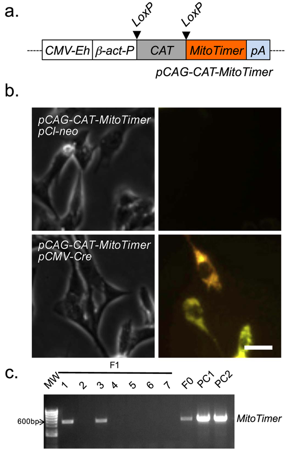
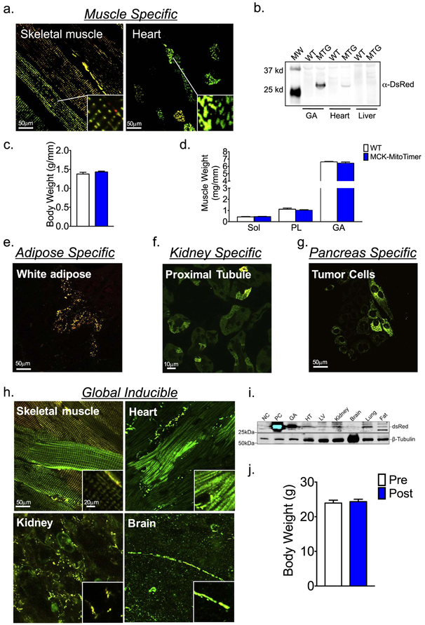
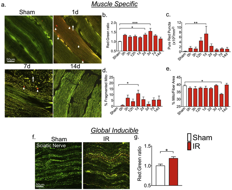
Assessment of structural and functional changes of mitochondria is vital for biomedical research as mitochondria are the power plants essential for biological processes and tissue/organ functions. Others and we have developed a novel reporter gene, pMitoTimer, which codes for a redox sensitive mitochondrial targeted protein that switches from green fluorescence protein (GFP) to red fluorescent protein (DsRed) when oxidized. It has been shown in transfected cells, transgenic C. elegans and Drosophila m., as well as somatically transfected adult skeletal muscle that this reporter gene allows quantifiable assessment of mitochondrial structure, oxidative stress, and lysosomal targeting of mitochondria-containing autophagosomes. Here, we generated CAG-CAT-MitoTimer transgenic mice using a transgene containing MitoTimer downstream of LoxP-flanked bacterial chloramphenicol acetyltransferase (CAT) gene with stop codon under the control of the cytomegalovirus (CMV) enhancer fused to the chicken β-actin promoter (CAG). When CAG-CAT-MitoTimer mice were crossbred with various tissue-specific (muscle, adipose tissue, kidney, and pancreatic tumor) or global Cre transgenic mice, the double transgenic offspring showed MitoTimer expression in tissue-specific or global manner. Lastly, we show that hindlimb ischemia-reperfusion caused early, transient increases of mitochondrial oxidative stress, mitochondrial fragmentation and lysosomal targeting of autophagosomes containing mitochondria as well as a later reduction of mitochondrial content in skeletal muscle along with mitochondrial oxidative stress in sciatic nerve. Thus, we have generated conditional MitoTimer mice and provided proof of principle evidence of their utility to simultaneously assess mitochondrial structure, oxidative stress, and mitophagy in vivo in a tissue-specific, controllable fashion.


Figures

Fig. 1.
Fig. 1.
Development of pCAG-CAT-MitoTimer construct. A plasmid DNA containing conditional expression unit of MitoTimer was constructed by genetic engineering and used for conditional expression In cultured cells to confirm its inducibility. (a) A schematic presentation of the recombinant pCAG-CAT-Mitotimer construct generated by subcloning MitoTimer coding region Into a plasmid DNA containing the CMV enhanced β-actin promoter; (b) Representative confocal Images of C2C12 cells co-transfected of pCAG-CAT-MitoTimer with either empty vector pCI-neo or pCMV-Cre. MitoTimer expression was only detected In the presence of Cre; and (c) Genotyping of the F1 progeny following the crossbredding of CAG-CAT-MitoTimer mice with wild type (WT) mice. pCAG-CAT-MitoTimer and isolated DNA fragment of CAG-CAT-MitoTimer expression unit were Included to serve as positive controls 1 and 2 (PC1 and PC2), respectively.
Fig. 2.
Fig. 2.
Generation of MitoTimer transgenic mice. A transgenic mouse line was generated by pronclear injection of isolated DNA fragment containing the conditional MitoTimer expression unit as illustrated in Fig. 1a. (a) Representative confocal images of MitoTimer expression in the skeletal muscle and heart from muscle specific MitoTimer mice (MTG; MCK-Cre;CAG-CAT-MitoTimer); (b) Immunoblot analysis of MitoTimer protein expression gastrocnemius muscle (GA), heart and liver tissues in MTG and the wild type littermates (WT); (c) Body weight was not different between MCK-MitoTimer and their WT littermates; (d) Muscle weights for Soleus (Sol, Plantaris (PL)), and Gastrocnemius (GA), normalized to tibia length, was not different between MCK-MitoTimer and their WT littermates; (e) Representative confocal images of MitoTimer expression in white adipose tissue of adipose tissue-specific inducible MitoTimer mice (adiponectin-rtTA-TRE-Cre;CAG-CAT-MitoTimer); (f) Representative confocal images of MitoTimer expression in kidney proximal tubule-specific mice (PepCK-Cre;CAG-CAT-MitoTimer); (g) Representative confocal images of MitoTimer expression in primary cells isolated from pancreatic tumor cells from pancreatic ductal adenocarcinoma MitoTimer mice (LSL-KrasG12D/+;Trp53flox/flox;Pdx-1-CreERTg/+;CAG-CAT-MitoTimer); (h) Representative confocal images of MitoTimer expression in skeletal muscle, heart, kidney, and brain in global inducible MitoTimer mice (CAG-CreERT2;CAG-CAT-MitoTimer); and (i) Immunoblot analysis of different tissues. Negative (NC) and positive (PC) controls are homogenates of the GA muscle from wild type mice and MTG mice, respectively. (j) Body weights in MTG mice was not affected by 7 days of tamoxifen injection.
Fig. 3.
Fig. 3.
MitoTimer reporter mice are sensitive for detection of mitochondrial changes in response to pathological stress in multiple tissues. MitoTimer transgenic mice were subjected to 1-h unilateral ischemia-reperfusion injury using a rubber band tourniquet followed confocal microscopy analysis. (a) Representative images of planatris muscles in muscle-specific MitoTimer mice at different time points over 14 days following IR injury. White arrows indicate fibers with a fragmented mitochondrial network, and arrow heads indicate pure red puncta. (b–e) Quantification of MitoTimer Red:Green ratio, pure red puncta, percent of fibers with mitochondrial network fragmentation, and mitochondrial content, respectively. *, **, *** denote p < 0.05, p < 0.01. p < 0.001, respectively; n = 5–7; (f) Representative images of MitoTimer signal in sciatic nerve in global inducible MitoTimer mice 3 h after ischemia-reperfusion (IR) comparing with the sham control (Sham); and (g) quantification of MitoTimer Red:Green ratio. * denotes p < 0.05. (For interpretation of the references to color in this figure legend, the reader is referred to the web version of this article.)

References

    1. Bruning JC, Michael MD, Winnay JN, Hayashi T, Accili D, Goodyear LJ, Kahn CR, 1998. A muscle-specific insulin receptor knockout exhibits features of the metabolic syndrome of NIDDM without altering glucose tolerance. Mol. Cell 2, 559–569. - PubMed
    1. Call JA, Wilson RJ, Laker RC, Zhang M, Kundu M, Yan Z, 2017. Ulk1-mediated autophagy plays an essential role in mitochondrial remodeling and functional regeneration of skeletal muscle. Am. J. Phys. Cell Physiol 312, C724–C732. - PMC - PubMed
    1. Dorweiler B, Pruefer D, Andrasi TB, Maksan SM, Schmiedt W, Neufang A, Vahl CF, 2007. Ischemia-reperfusion injury pathophysiology and clinical implications. Eur. J. Trauma Emerg. Surg 600–612. 10.1007/s00068-007-7152-z. - DOI - PubMed
    1. Drake JC, Wilson RJ, Yan Z, 2015. Molecular mechanisms for mitochondrial adaptation to exercise training in skeletal muscle. FASEB J. 1–10. 10.1096/fj.15-276337. - DOI - PMC - PubMed
    1. Friedlander SYG, Chu GC, Snyder EL, Girnius N, Dibelius G, Crowley D, Vasile E, Depinho RA, Jacks T, 2009. Article context-dependent transformation of adult pancreatic cells by oncogenic K-Ras. Cancer Cell 16, 379–389. 10.1016/j.ccr.2009.09.027. - DOI - PMC - PubMed

Publication types

Substances

LinkOut - more resources