. 2018 Oct;142(4):1066-1079.e6.
doi: 10.1016/j.jaci.2017.10.044. Epub 2017 Dec 21.

Low-density lipoprotein receptor-related protein 1 attenuates house dust mite-induced eosinophilic airway inflammation by suppressing dendritic cell-mediated adaptive immune responses

Affiliations

Low-density lipoprotein receptor-related protein 1 attenuates house dust mite-induced eosinophilic airway inflammation by suppressing dendritic cell-mediated adaptive immune responses

Amarjit Mishra et al. J Allergy Clin Immunol. 2018 Oct.

Abstract

Background: Low-density lipoprotein receptor-related protein 1 (LRP-1) is a scavenger receptor that regulates adaptive immunity and inflammation. LRP-1 is not known to modulate the pathogenesis of allergic asthma.

Objective: We sought to assess whether LRP-1 expression by dendritic cells (DCs) modulates adaptive immune responses in patients with house dust mite (HDM)-induced airways disease.

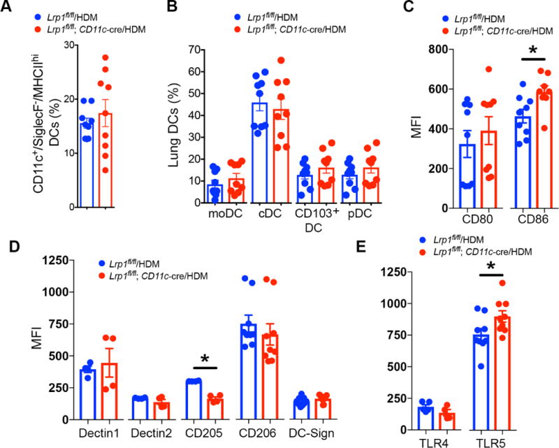
Methods: LRP-1 expression on peripheral blood DCs was quantified by using flow cytometry. The role of LRP-1 in modulating HDM-induced airways disease was assessed in mice with deletion of LRP-1 in CD11c+ cells (Lrp1fl/fl; CD11c-Cre) and by adoptive transfer of HDM-pulsed CD11b+ DCs from Lrp1fl/fl; CD11c-Cre mice to wild-type (WT) mice.

Results: Human peripheral blood myeloid DC subsets from patients with eosinophilic asthma have lower LRP-1 expression than cells from healthy nonasthmatic subjects. Similarly, LRP-1 expression by CD11b+ lung DCs was significantly reduced in HDM-challenged WT mice. HDM-challenged Lrp1fl/fl; CD11c-Cre mice have a phenotype of increased eosinophilic airway inflammation, allergic sensitization, TH2 cytokine production, and mucous cell metaplasia. The adoptive transfer of HDM-pulsed LRP-1-deficient CD11b+ DCs into WT mice generated a similar phenotype of enhanced eosinophilic inflammation and allergic sensitization. Furthermore, CD11b+ DCs in the lungs of Lrp1fl/fl; CD11c-Cre mice have an increased ability to take up HDM antigen, whereas bone marrow-derived DCs display enhanced antigen presentation capabilities.

Conclusion: This identifies a novel role for LRP-1 as a negative regulator of DC-mediated adaptive immune responses in the setting of HDM-induced eosinophilic airway inflammation. Furthermore, the reduced LRP-1 expression by circulating myeloid DCs in patients with eosinophilic asthma suggests a possible role for LRP-1 in modulating type 2-high asthma.

Keywords: Low-density lipoprotein receptor–related protein 1; dendritic cells; eosinophilic airway inflammation; house dust mite; type 2–high asthma.


Figures

Figure 1
Figure 1. Cell surface LRP-1 expression is reduced on myeloid dendritic cell subsets from eosinophilic asthmatics
A) Asthmatics (n = 14) and healthy, non-asthmatics (n = 12) with low peripheral blood eosinophil counts (< 260 cells/l), or asthmatics with high (> 260 cells/μl) peripheral blood eosinophil counts (n = 12) (* P< 0.0001, one-way ANOVA with Sidak’s multiple comparison test). B) Mean fluorescence intensity (MFI) of LRP-1 expression was assessed by flow cytometry on viable/Lin/HLA-DR+ peripheral blood dendritic cell subsets that express CD11c+/CD1c+/CD141 (MDC1), CD11c+/CD1c/CD141+ (MDC2), CD11c+/CD16+ (CD16+ DCs), and CD11c/CD123+ (pDCs). Data shown as means ± SEM (*P<0.05, one-way ANOVA with Sidak’s multiple comparison test). C) The percentage of LRP-1+ CD11c+/CD11b+/Siglec F/MHC IIhi/Mar-1/CD64 conventional myeloid DCs (cDCs) and CD11c+/CD11b+/Siglec F/MHC IIhi/Mar-1+/CD64+ monocyte-derived DCs (moDCs) from saline- and house dust mite (HDM)-challenged wild-type C57BL6 mice were quantified (n = 9 mice, * P< 0.0001, saline- versus HDM-challenged, Mann-Whitney test).
Figure 2
Figure 2. Eosinophilic airway inflammation and mucous cell metaplasia are increased in HDM-challenged Lrp1fl/fl; CD11c-Cre mice
A) Lrp1fl/fl and Lrp1fl/fl; CD11c-Cre mice were sensitized with two intraperitoneal injections of 100 μg HDM (with alum 40 mg ml−1) on day 0 and day 4 and challenged on days 8, 10, 12 and 15 by intranasal administration of HDM (50 μg) before harvest and endpoint analysis on day 17. B) The number of total BALF inflammatory cells and inflammatory cell types (eosinophils (Eos), alveolar macrophages (AM), neutrophils (PMN) and lymphocytes (Lymph)) from saline- and HDM-challenged Lrp1fl/fl; CD11c-Cre and Lrp1fl/fl mice (n = 7 – 16 mice per group, * P< 0.01, HDM-challenged Lrp1fl/fl; CD11c-Cre versus Lrp1fl/fl, one-way ANOVA with Sidak’s multiple comparison test). C) Representative lung histology sections stained with hematoxylin and eosin (H&E) or Periodic acid-schiff (PAS). Scale bars 100 μm for the 200× images and 20 μm for the 1,000× images. D) Quantification of mucous cell metaplasia as assessed by the percentage of airways that contained PAS+ cells. Data are mean ± SEM (n = 5 – 7 mice per group, * P < 0.05, HDM- challenged Lrp1fl/fl; CD11c-Cre versus Lrp1fl/fl mice, Mann-Whitney test). Results shown are pooled data from two independent experiments.
Figure 3
Figure 3. Th2 cytokine production and allergic sensitization are increased in HDM- challenged Lrp1fl/fl; CD11c-Cre mice
A) Airway resistance (cm H2O per ml s−1) to inhaled methacholine from HDM-challenged Lrp1fl/fl; CD11c-Cre and Lrp1fl/fl mice (n = 4 – 6 mice per group). (P = NS, two-way ANOVA). B) Cytokine secretion by ex vivo cultures of MLN cells that had been re-stimulated with saline or HDM (100 μg ml−1, n = 6 mice per group, * P < 0.05, HDM-challenged Lrp1fl/fl; CD11c-Cre versus Lrp1fl/fl mice, one-way ANOVA with Sidak’s multiple comparison test). C) The percentage of CD4+/CD25+/FoxP3+ Tregs in the lungs of HDM-challenged Lrp1fl/fl; CD11c-Cre mice and Lrp1fl/fl mice (n = 5 mice per group, *P < 0.008, Mann-Whitney test). D) Serum levels of HDM-specific IgE and IgG1 (n = 9 – 10 mice per group), * P< 0.05, HDM-challenged Lrp1fl/fl; CD11c-Cre versus Lrp1fl/fl mice, one-way ANOVA with Sidak’s multiple comparison test).
Figure 4
Figure 4. The adoptive transfer of CD11b+ DCs from Lrp1fl/fl; CD11c-Cre mice increases HDM-mediated eosinophilic airway inflammation
A) BMDCs from Lrp1fl/fl; CD11c-Cre and Lrp1fl/fl donor mice were pulsed ex vivo with saline or HDM (100 μg ml−1) for 16 hours. 2.5 × 10⁴ CD11c+/CD11b+/MHC IIhi DCs were adoptively transferred to wild-type (WT) C57BL6 recipient mice by intranasal administration on day 0 and intranasal HDM challenges (50 μg) were administered on alternate days, from day 9 through day 15, to all recipient mice, prior to endpoint analysis on day 16. B) The number of total BALF inflammatory cells and inflammatory cell subtypes in recipient WT mice (n = 6 – 10 mice per group, *P< 0.05, HDM-pulsed Lrp1fl/fl; CD11c-Cre DCs versus HDM-pulsed Lrp1fl/fl DCs, one-way ANOVA with Sidak’s multiple comparison test). C) Representative histologic lung sections stained with H&E and PAS. Scale bars indicate 100 μm for the 200× images and 20 μm for the 1,000× images. D) Quantification of mucous cell metaplasia as assessed by the percentage of airways that contained PAS+ cells. (n = 5 mice per group, * P < 0.008, HDM-pulsed Lrp1fl/fl; CD11c-Cre DCs versus HDM- pulsed Lrp1fl/fl DCs, Mann-Whitney test). E) Serum levels HDM-specific IgE and IgG1 (n = 9 – 10 mice per group, *P < 0.01, Mann-Whitney test, HDM-pulsed Lrp1fl/fl; CD11c-Cre DCs versus HDM-pulsed Lrp1fl/fl DCs). F) BALF levels of CCL24 from WT recipient mice that received HDM-pulsed Lrp1fl/fl; CD11c-Cre DCs or HDM-pulsed Lrp1fl/fl DCs (n = 6 – 10 mice per group, *P < 0.05, unpaired t test). G) Cytokine secretion by ex vivo cultures of MLN cells that had been re-stimulated with saline or HDM (100 μg ml−1, n = 6 – 10 mice per group, * P < 0.01, HDM-pulsed Lrp1fl/fl; CD11c-Cre DCs versus HDM-pulsed Lrp1fl/fl DCs, one-way ANOVA with Sidak’s multiple comparison test). Results shown are pooled data from two independent experiments.
Figure 5
Figure 5. Increased expression of CD86 and TLR5 by CD11c+/CD11b+ DCs from HDM- challenged Lrp1fl/fl; CD11c-Cre mice
A) The percentage of CD11c+/Siglec F/MHC IIhi total lung DCs and B) the percentage of lung DC monocyte-derived dendritic cells (moDCs), conventional myeloid dendritic cells (cDCs), CD103+ DCs, and plasmacytoid dendritic cells (pDCs) subsets as quantified by flow cytometry (P = NS, HDM-challenged Lrp1fl/fl; CD11c-Cre versus Lrp1fl/fl mice, n= 9 mice per group, unpaired t test). C) Cell surface expression of CD80 and CD86 by lung CD11c+/CD11b+/SSClo/SiglecF/MHC IIhi DCs from HDM-challenged Lrp1fl/fl; CD11c-Cre versus Lrp1fl/fl mice (n = 8 – 9 mice per group, * P< 0.01, Mann-Whitney test). D) Cell surface expression of Dectin1, Dectin 2, CD205, CD206, and DC-SIGN by lung CD11c+/CD11b+/SSClo/SiglecF/MHC IIhi DCs from HDM-challenged Lrp1fl/fl; CD11c-Cre versus Lrp1fl/fl mice (n = 4 – 9 mice per group, * P = 0.029, Mann-Whitney test). E) Cell surface expression of TLR4 and TLR5 by lung CD11c+/CD11b+/SSClo/SiglecF/MHC IIhi DCs from HDM-challenged Lrp1fl/fl; CD11c-Cre versus Lrp1fl/fl mice (n= 4 – 9 mice per group, *P < 0.05, unpaired t test).
Figure 6
Figure 6. Lrp1-deficient DCs have augmented antigen uptake and presentation capabilities
A and B) Uptake of Alexa Fluor 647-labeled HDM (50 μg) by CD11c+/SSClo/SiglecF/MHCIIhi CD11b+ or CD11b DCs in the lungs (A) and MLNs (B) of Lrp1fl/fl; CD11c-Cre and Lrp1fl/fl mice 72 hours after intranasal administration (n= 10 mice per group, * P < 0.05, unpaired t test). C) CD11c+ BMDCs from Lrp1fl/fl and Lrp1fl/fl; CD11c-Cre mice were pulsed with the OVA 323-339 peptide and incubated at a 1:5 ratio with CFSE- labelled splenic CD4+ DO11.10 T cells for 4 days. OVA-specific proliferation is presented as % divided OT-II cells (n = 12 – 15 per group, * P < 0.0001, one-way ANOVA with Sidak’s multiple comparison test). D) Th2 cytokines released by co-cultures of OVA 323-339 peptide-pulsed BMDCs and CFSE-labelled CD4+ DO11.10 T cells (n= 10 per group, * P < 0.05, Lrp1fl/fl; CD11c-Cre versus Lrp1fl/fl, unpaired t test). Results shown are pooled data from two independent experiments.

Similar articles

Cited by

References

    1. Herz J, Strickland DK. LRP: a multifunctional scavenger and signaling receptor. Journal of Clinical Investigation. 2001;108:779–84. - PMC - PubMed
    1. Lillis AP, Van Duyn LB, Murphy-Ullrich JE, Strickland DK. LDL receptor-related protein 1: Unique tissue-specific functions revealed by selective gene knockout studies. Physiological Reviews. 2008;88:887–918. - PMC - PubMed
    1. Gaultier A, Hollister M, Reynolds I, Hsieh EH, Gonias SL. LRP1 regulates remodeling of the extracellular matrix by fibroblasts. Matrix Biology. 2010;29:22–30. - PMC - PubMed
    1. Krieger M, Herz J. Structures and functions of multiligand lipoprotein receptors: macrophage scavenger receptors and LDL receptor-related protein (Lrp) Annual Review of Biochemistry. 1994;63:601–37. - PubMed
    1. Kanekiyo T, Bu G. The low-density lipoprotein receptor-related protein 1 and amyloid- beta clearance in Alzheimer's disease. Frontiers in Aging Neuroscience. 2014;6:93. - PMC - PubMed

Publication types

MeSH terms