. 2018 Feb 2;293(5):1702-1712.
doi: 10.1074/jbc.RA117.000764. Epub 2017 Dec 22.

Structural basis for substrate specificity of methylsuccinyl-CoA dehydrogenase, an unusual member of the acyl-CoA dehydrogenase family

Affiliations

Structural basis for substrate specificity of methylsuccinyl-CoA dehydrogenase, an unusual member of the acyl-CoA dehydrogenase family

Thomas Schwander et al. J Biol Chem. .

Abstract

(2S)-methylsuccinyl-CoA dehydrogenase (MCD) belongs to the family of FAD-dependent acyl-CoA dehydrogenase (ACD) and is a key enzyme of the ethylmalonyl-CoA pathway for acetate assimilation. It catalyzes the oxidation of (2S)-methylsuccinyl-CoA to α,β-unsaturated mesaconyl-CoA and shows only about 0.5% activity with succinyl-CoA. Here we report the crystal structure of MCD at a resolution of 1.37 Å. The enzyme forms a homodimer of two 60-kDa subunits. Compared with other ACDs, MCD contains an ∼170-residue-long N-terminal extension that structurally mimics a dimer-dimer interface of these enzymes that are canonically organized as tetramers. MCD catalyzes the unprecedented oxidation of an α-methyl branched dicarboxylic acid CoA thioester. Substrate specificity is achieved by a cluster of three arginines that accommodates the terminal carboxyl group and a dedicated cavity that facilitates binding of the C2 methyl branch. MCD apparently evolved toward preventing the nonspecific oxidation of succinyl-CoA, which is a close structural homolog of (2S)-methylsuccinyl-CoA and an essential intermediate in central carbon metabolism. For different metabolic engineering and biotechnological applications, however, an enzyme that can oxidize succinyl-CoA to fumaryl-CoA is sought after. Based on the MCD structure, we were able to shift substrate specificity of MCD toward succinyl-CoA through active-site mutagenesis.

Keywords: X-ray crystallography; acyl-CoA dehydrogenase; bioengineering; enzyme catalysis; enzyme kinetics; enzyme mutation; enzyme purification; enzyme structure; ethylmalonyl-CoA pathway; flavin adenine dinucleotide (FAD); glutaryl-CoA dehydrogenase; isobutyryl-CoA dehydrogenase; mesaconyl-CoA.


Conflict of interest statement

The authors declare that they have no conflicts of interest with the contents of this article

Figures

Figure 1.
Figure 1.
Reaction catalyzed by MCD. The natural substrate of MCD is the branched (2S)-methylsuccinyl-CoA, which is converted to mesaconyl-CoA. MCD also accepts the unbranched succinyl-CoA, but with a ∼200-fold lower catalytic rate.
Figure 2.
Figure 2.
Overall structure of PdMCD. A, one monomeric subunit of PdMCD consists of five domains. The central fold found in other ACDs consists of an initial α-helical domain (green), an intermediary β-barrel domain (pink), and a C-terminal α-helical domain (blue). The N-terminal extension of PdMCD is shown in orange with the unique β-hairpin motif in red. The labeling of the secondary structure elements is in accordance with other ACDs. B, PdMCD forms a homodimer, and each subunit contains one FAD (yellow). The subunits are shown in orange and cyan with the N-terminal extension in darker shades, respectively. Adenosyl phosphate moieties were found to be bound on the surface of each subunit between Arg-81 and Trp-476. This interaction might stabilize helix H, which is involved in a crystal contact. The dimeric form of PdMCD is strengthened by the β-hairpin motif in the N-terminal extension (highlighted in the box), which forms hydrogen bonds with the opposite subunit.
Figure 3.
Figure 3.
Sequence alignment of PdMCD, RsMCD, and ACDs with known structures. The sequences were aligned, and the secondary structure elements are indicated (α-helices with barrels, β-sheets with arrows, and turns in red). The labeling of the secondary structure element follows the convention for canonical ACDs. The N-terminal domain of MCD is shown in orange. The central fold of all known ACDs is divided into two α-helical domains (green and blue) and an intermediary β-barrel domain (cyan). The secondary structure elements of the C-terminal extension of VLCAD is indicated in gray. Identical residues are shaded in gray, and the active-site glutamate is highlighted in black. The α-helix E shows a single amino acid deletion like in IVD and MCAD (indicated by a blue arrow). The Phe-287 in PdMCD and Phe-284 in RsMCD are shifted by two residues toward the C terminus, and the otherwise conserved phenylalanine/tyrosine at this position is replaced with an alanine (indicated in red and blue letters). BSCAD, short/branched chain acyl-CoA dehydrogenase.
Figure 4.
Figure 4.
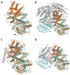
Superposition of PdMCD with other ACDs. A, structural alignment of a PdMCD monomer in orange (N-terminal domain in dark orange) with the monomer of MCAD in cyan (PDB code 3MDE) (1.55 Å over 350 Cα atoms per MCD monomer). The central fold of ACDs is conserved in the C-terminal domain of PdMCD. An extended loop region between β-strands 5 and 6 is indicated with an arrow. B, superposition of the PdMCD monomer with the tetramer (dimer of dimers in cyan and gray, respectively) of MCAD. The N-terminal extension of PdMCD aligns with the opposite monomer of MCAD and complements the dimeric interface. The unique β-hairpin motif (red) is located in a cavity at the interface, which can be found in all other ACDs known so far. C, superposition of a PdMCD monomer in orange (N-terminal domain in dark orange) with a monomer of VLCAD in cyan (PDB code 3B96) (1.85 Å over 313 Cα atoms). VLCADs have a C-terminal extension (gray), which aligns with the N-terminal extension from PdMCD and also complements the dimeric interface. D, overlay of a PdMCD monomer with a VLCAD dimer (N-terminal domain in cyan; C-terminal domain in gray). The subunits of VLCAD dimers also interact via α-helix O (pink). This feature is lacking in PdMCD, but the neighboring subunits additionally interact via the β-hairpin motif (red).
Figure 5.
Figure 5.
Superposition of FoFc electron density simulated annealing omit maps on a refined FAD and Thr-320. A, omit map at 3.0 σ for the FAD cofactor and Thr-320 of PdMCD. Thr-320 forms a hydrogen bond to the N5 of the FAD. The electron density around this residue (cyan) indicates that the threonine can assume a different rotamer conformation (purple) where the hydrogen bond is broken. The FAD cofactor forms additional hydrogen bonds with the polypeptide backbone. B, the isolalloxazine ring of FAD assumes a distinct “butterfly-like” conformation (angle of 165°), which can be observed in other ACDs as well. This conformation is assumed to indicate a shift in the electron potential of the cofactor.
Figure 6.
Figure 6.
Active-site architecture of PdMCD. A, wall-eyed stereo view of the active-site residues. The acyl moiety of (2S)-methylsuccinyl-CoA was fitted into the active site according to the conserved positioning of substrates found in other ACDs. The carbonyl group of the thioester bond forms a hydrogen bond to the 2′-hydroxyl of the FAD ribityl chain and the backbone amine of the catalytic Glu-535. The C2 of the substrate is positioned below Glu-535 for proton abstraction, and the C3 is positioned above the N5 of FAD for the hydride transfer. The base of the active-site cavity is lined by Arg-252, Arg-376, and Arg-421 for binding of the carboxylic acid group of the substrate. B, the interior surface of the active site shows a distinct pocket for the accommodation of the C2-methyl group, and the base of the cavity is positively charged for the binding of the carboxylic acid of the substrate. C, overlay of PdMCD (orange) with VLCAD (PDB code 3B96) (cyan). The α-helix E of PdMCD is shifted away from the active site and a single residue deletion (as can be found in IVD and MCAD) prevents a perturbation of this helix. A conserved phenylalanine/tyrosine in other ACDs is shifted in PdMCD. This shift allows the formation of the cavity to accommodate the C2-methyl branch of the substrate by Ala-285. Moreover, Phe-287 partially assumes an analogous position to the conserved phenylalanine/tyrosine in other ACDs. D, overlay of PdMCD (orange) with MCAD (PDB code 3MDE) (cyan), indicating a repositioned loop between β-strands 6 and 7. This leads to the formation of a salt bridge between Glu-380 and Arg-376, which is thereby positioned within the active site. E, overlay of PdMCD (orange) with GDH (PDB code 3MPI) (cyan). The conserved catalytic glutamate is found in all ACDs (except IVD). In GDH, a salt bridge to the substrate glutaryl-CoA is formed with an arginine from α-helix E. In the case of PdMCD, Arg-421 is in an analogous position but originates from α-helix G. Arg-421 is held in place by Ser-248 and Thr-418. The Arg-252 from α-helix E is within interaction distance of the fitted methylsuccinyl-CoA and may assume a different conformation upon substrate binding.
Figure 7.
Figure 7.
Kinetic properties of RsMCD WT, A282V variant, and the A282F/F284A double variant (corresponding to Ala-285 and Phe-287 in PdMCD). A, Michaelis–Menten kinetics of RsMCD WT and A282V variant with (2S)-methylsuccinyl-CoA and succinyl-CoA as substrate. The variant shows a decreased efficiency with (2S)-methylsuccinyl-CoA but has a slightly increased efficiency with succinyl-CoA. B, Michaelis–Menten kinetics of RsMCD A282F/F284A swapping mutant with (2S)-methylsuccinyl-CoA. This mutant only achieved 0.2% of relative catalytic efficiency with (2S)-methylsuccinyl-CoA and did not show measurable activity with succinyl-CoA. Error bars, S.D.

Similar articles

Cited by

References

    1. Erb T. J., Fuchs G., and Alber B. E. (2009) (2S)-Methylsuccinyl-CoA dehydrogenase closes the ethylmalonyl-CoA pathway for acetyl-CoA assimilation. Mol. Microbiol. 73, 992–1008 10.1111/j.1365-2958.2009.06837.x - DOI - PubMed
    1. Ikeda Y., Dabrowski C., and Tanaka K. (1983) Separation and properties of five distinct acyl-CoA dehydrogenases from rat liver mitochondria: identification of a new 2-methyl branched chain acyl-CoA dehydrogenase. J. Biol. Chem. 258, 1066–1076 - PubMed
    1. Izai K., Uchida Y., Orii T., Yamamoto S., and Hashimoto T. (1992) Novel fatty acid β-oxidation enzymes in rat liver mitochondria. I. Purification and properties of very-long-chain acyl-coenzyme A dehydrogenase. J. Biol. Chem. 267, 1027–1033 - PubMed
    1. Nguyen T. V., Andresen B. S., Corydon T. J., Ghisla S., Abd-El Razik N., Mohsen A. W., Cederbaum S. D., Roe D. S., Roe C. R., Lench N. J., and Vockley J. (2002) Identification of isobutyryl-CoA dehydrogenase and its deficiency in humans. Mol. Genet. Metab. 77, 68–79 10.1016/S1096-7192(02)00152-X - DOI - PubMed
    1. Besrat A., Polan C. E., and Henderson L. M. (1969) Mammalian metabolism of glutaric acid. J. Biol. Chem. 244, 1461–1467 - PubMed

Publication types

MeSH terms

LinkOut - more resources