. 2017 Dec;22(12):1-9.
doi: 10.1117/1.JBO.22.12.121719.

Application of optical coherence tomography for in vivo monitoring of the meningeal lymphatic vessels during opening of blood-brain barrier: mechanisms of brain clearing

Affiliations

Application of optical coherence tomography for in vivo monitoring of the meningeal lymphatic vessels during opening of blood-brain barrier: mechanisms of brain clearing

Oxana Semyachkina-Glushkovskaya et al. J Biomed Opt. 2017 Dec.

Abstract

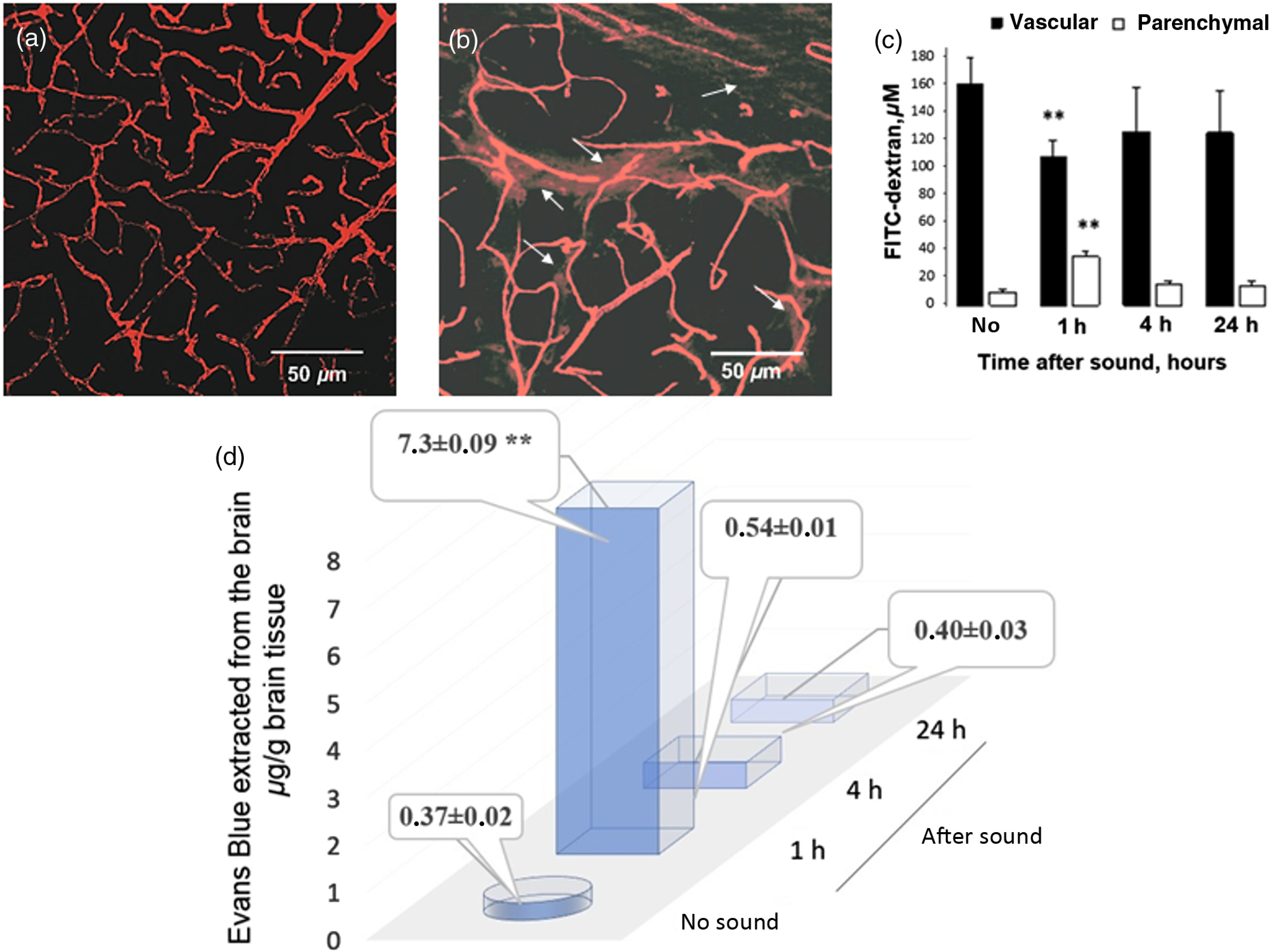
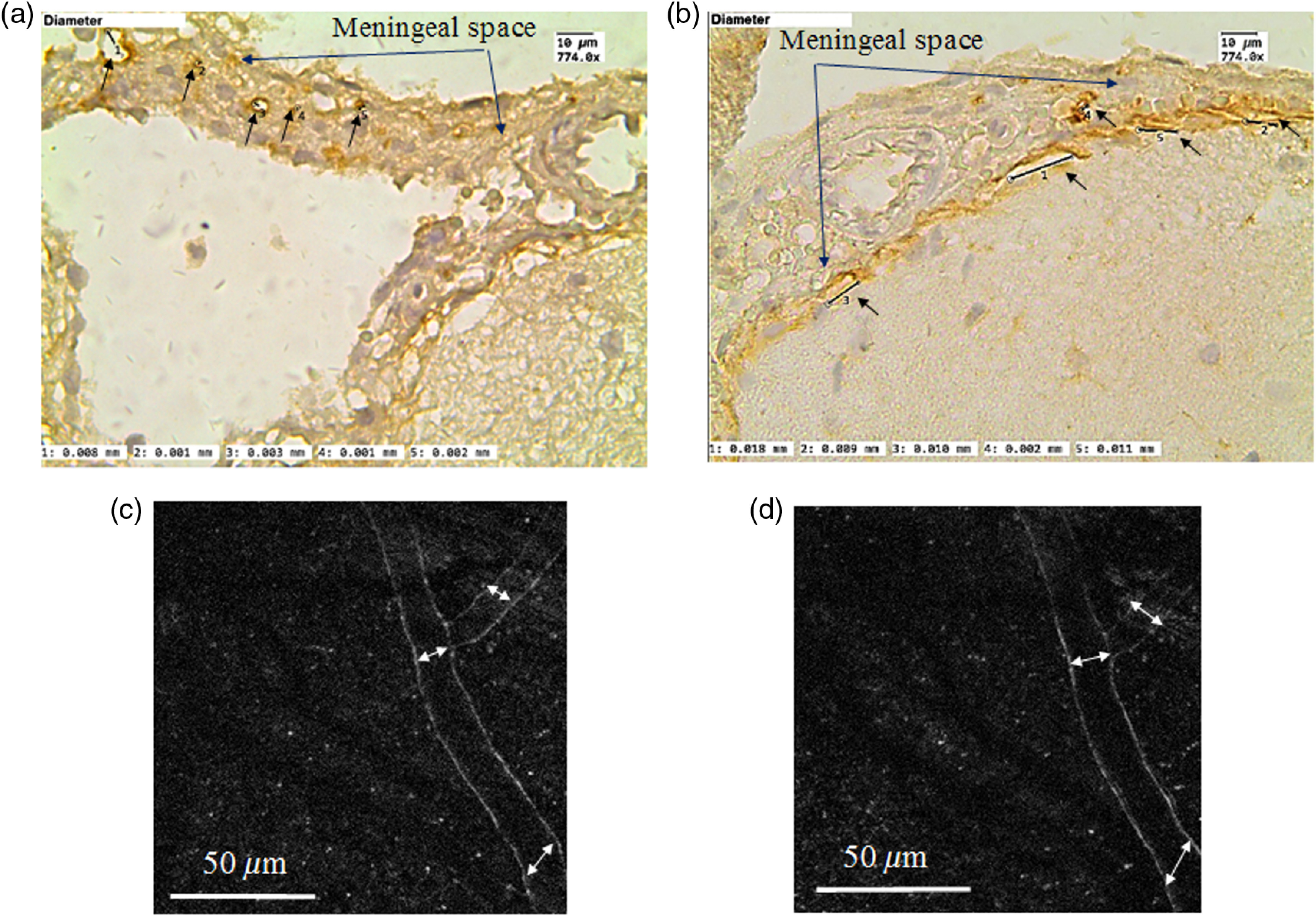
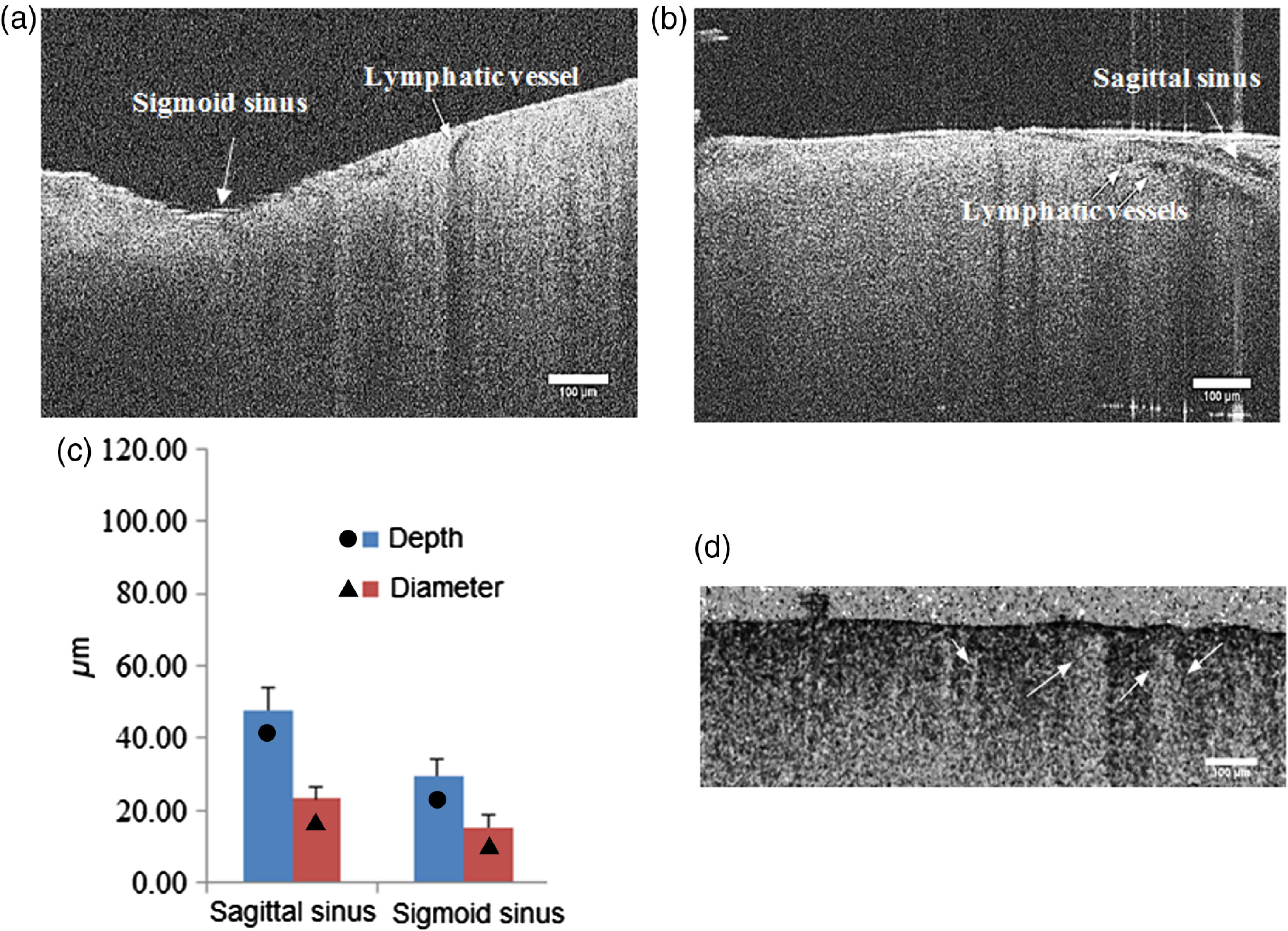
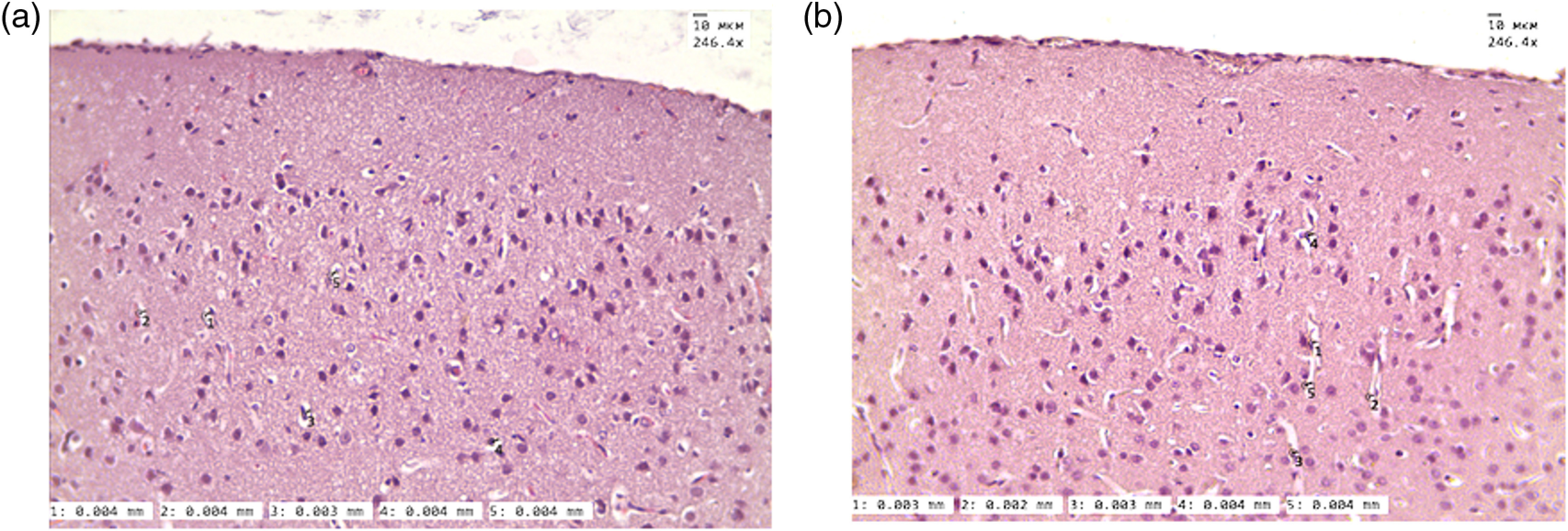
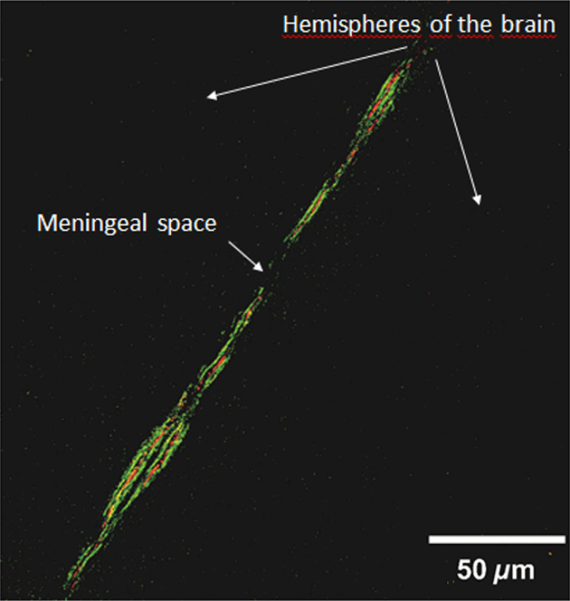
The meningeal lymphatic vessels were discovered 2 years ago as the drainage system involved in the mechanisms underlying the clearance of waste products from the brain. The blood-brain barrier (BBB) is a gatekeeper that strongly controls the movement of different molecules from the blood into the brain. We know the scenarios during the opening of the BBB, but there is extremely limited information on how the brain clears the substances that cross the BBB. Here, using the model of sound-induced opening of the BBB, we clearly show how the brain clears dextran after it crosses the BBB via the meningeal lymphatic vessels. We first demonstrate successful application of optical coherence tomography (OCT) for imaging of the lymphatic vessels in the meninges after opening of the BBB, which might be a new useful strategy for noninvasive analysis of lymphatic drainage in daily clinical practice. Also, we give information about the depth and size of the meningeal lymphatic vessels in mice. These new fundamental data with the applied focus on the OCT shed light on the mechanisms of brain clearance and the role of lymphatic drainage in these processes that could serve as an informative platform for a development of therapy and diagnostics of diseases associated with injuries of the BBB such as stroke, brain trauma, glioma, depression, or Alzheimer disease.

Keywords: blood–brain barrier; cerebral lymphatics; optical coherence tomography.


Figures

Fig. 1
Fig. 1
BBB permeability to FITC-dextran 70 kDa and EBd with albumin complex 68 kDa: (a) before sound exposure (no extravasation of FITC-dextran); (b) 1 h after sound exposure [strong extravasation of FITC-dextran (arrowed), which was defined as bright red clouds associated with a group of capillaries]; (c) the estimation of FITC-dextran concentration in brain parenchyma and microvessels 20 min after the injection. 2PLSM data are presented as mean±SEM, n=7, **p<0.01; (d) EBd in μg/g of mouse brain tissue before and after sound. Spectrofluorometric assay of EBd extravasation into the brain parenchyma shows an increase in the BBB permeability to EBd 1 h after the sound with rapid restoring of the BBB afterward.
Fig. 2
Fig. 2
ICH (a and b) and in vivo two-photon laser scanning (c and d) visualization of the meningeal lymphatic vessels: (a) and (b) the meningeal lymphatic vessels stained with LYVE1 (specific marker of endothelial cells of lymphatic vessels, the brown color is positive staining); (a) before sound-induced opening of the BBB, the average diameter of lymphatic vessels is 3.00±0.01  μm; (b) after opening of the BBB (1 h after sound), the average diameter of some lymphatic vessels is 10.00±0.07  μm; (c) and (d) the meningeal lymphatic vessels before and after opening of the BBB, respectively. 2PLSM data show the increase of diameters of the lymphatic vessels after opening of the BBB: (12.00±0.03  μm versus 17.00±0.08  μm, p<0.05).
Fig. 3
Fig. 3
The OCT images of the meningeal lymphatic vessels (black empty spaces) surrounding the sigmoid (a) and sagittal (b) sinuses. (c) The OCT analysis of diameter and depth of the meningeal lymphatic vessels. Scale bar is 100  μm (Video 1, MP4, 7.07 MB [URL: https://doi.org/10.1117/1.JBO.22.12.121719.1]).
Fig. 4
Fig. 4
The histological analysis of size of PVS (a) before and (b) 1 h after sound-induced opening of the BBB 3.8  μm—in the normal state and 3.2  μm 1 h after the opening of the BBB.
Fig. 5
Fig. 5
The confocal image of meningeal lymphatic vessels after the sound-induced opening of the BBB: the dextran (red color) is obseved in the meningeal lymphatic vessels (green color) 30 min after the opening of the BBB.

References

    1. Louveau A., et al. , “Structural and functional features of central nervous system lymphatic vessels,” Nature 523(7560), 337–341 (2015). 10.1038/nature14432 - DOI - PMC - PubMed
    1. Aspelund A., et al. , “A dural lymphatic vascular system that drains brain interstitial fluid and macromolecules,” J. Exp. Med. 212(7), 991–999 (2015). 10.1084/jem.20142290 - DOI - PMC - PubMed
    1. Mascagni P., Bellini G. B., Istoria completa dei vasi linfatici, Vol. 2, Presso Eusebio Pacini e Figlio, Florence: (1816).
    1. Belayev L., et al. , “Quantitative evaluation of blood–brain barrier permeability following middle cerebral artery occlusion in rats,” Brain Res. 739(1–2), 88–96 (1996). 10.1016/S0006-8993(96)00815-3 - DOI - PubMed
    1. Fernandez-Lopez D., et al. , “Blood–brain barrier permeability is increased after acute adult stroke but not neonatal stroke in the rat,” J. Neurosci. 32(28), 9588–9600 (2012). 10.1523/JNEUROSCI.5977-11.2012 - DOI - PMC - PubMed

Publication types