

Physiological and molecular triggers for SARS-CoV membrane fusion and entry into host cells
- PMID: 29275820
- PMCID: PMC7112017
- DOI: 10.1016/j.virol.2017.12.015
Physiological and molecular triggers for SARS-CoV membrane fusion and entry into host cells
Abstract
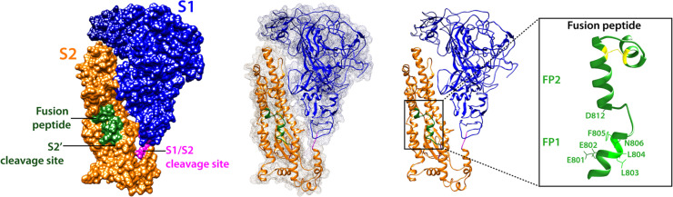
During viral entry, enveloped viruses require the fusion of their lipid envelope with host cell membranes. For coronaviruses, this critical step is governed by the virally-encoded spike (S) protein, a class I viral fusion protein that has several unique features. Coronavirus entry is unusual in that it is often biphasic in nature, and can occur at or near the cell surface or in late endosomes. Recent advances in structural, biochemical and molecular biology of the coronavirus S protein has shed light on the intricacies of coronavirus entry, in particular the molecular triggers of coronavirus S-mediated membrane fusion. Furthermore, characterization of the coronavirus fusion peptide (FP), the segment of the fusion protein that inserts to a target lipid bilayer during membrane fusion, has revealed its particular attributes which imparts some of the unusual properties of the S protein, such as Ca2+-dependency. These unusual characteristics can explain at least in part the biphasic nature of coronavirus entry. In this review, using severe acute respiratory syndrome coronavirus (SARS-CoV) as model virus, we give an overview of advances in research on the coronavirus fusion peptide with an emphasis on its role and properties within the biological context of host cell entry.
Keywords: Calcium; Coronavirus; Endosomes; Fusion peptide; SARS; Spike protein; Virus entry.
Copyright © 2017 Elsevier Inc. All rights reserved.
Figures

References
-
- Bertram S., Dijkman R., Habjan M., Heurich A., Gierer S., Glowacka I., Welsch K., Winkler M., Schneider H., Hofmann-Winkler H., Thiel V., Pöhlmann S. TMPRSS2 activates the human coronavirus 229E for cathepsin-independent host cell entry and is expressed in viral target cells in the respiratory epithelium. J. Virol. 2013;87:6150–6160. - PMC - PubMed
Publication types
MeSH terms
Substances
Grants and funding
LinkOut - more resources
Full Text Sources
Other Literature Sources
Miscellaneous
