. 2018 Jan 2;26(1):130-136.e2.
doi: 10.1016/j.str.2017.11.021. Epub 2017 Dec 21.

Structure of the G119S Mutant Acetylcholinesterase of the Malaria Vector Anopheles gambiae Reveals Basis of Insecticide Resistance

Affiliations

Structure of the G119S Mutant Acetylcholinesterase of the Malaria Vector Anopheles gambiae Reveals Basis of Insecticide Resistance

Jonah Cheung et al. Structure. .

Abstract

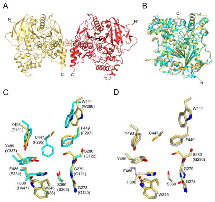
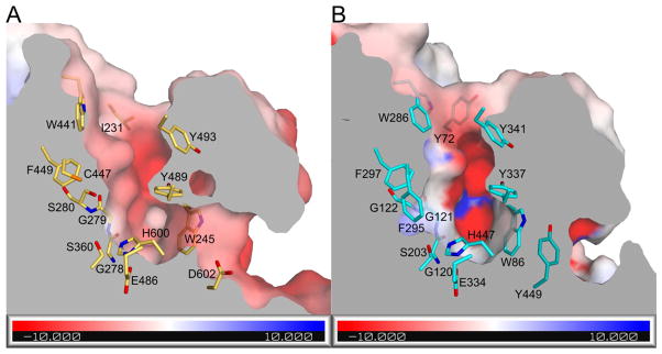
Malaria is a devastating disease in sub-Saharan Africa and is transmitted by the mosquito Anopheles gambiae. While indoor residual spraying of anticholinesterase insecticides has been useful in controlling the spread of malaria, widespread application of these compounds has led to the rise of an insecticide-resistant mosquito strain that harbors a G119S mutation in the nervous system target enzyme acetylcholinesterase. We demonstrate the atomic basis of insecticide resistance through structure determination of the G119S mutant acetylcholinesterase of An. gambiae in the ligand-free state and bound to a potent difluoromethyl ketone inhibitor. These structures reveal specific features within the active-site gorge distinct from human acetylcholinesterase, including an open channel at the base of the gorge, and provide a means for improving species selectivity in the rational design of improved insecticides for malaria vector control.

Keywords: acetylcholinesterase; channel; difluoromethyl ketone; insecticide; malaria; resistance; selectivity.


Conflict of interest statement

DECLARATION OF INTERESTS

The authors declare no competing interests.

Figures

Figure 1
Figure 1. Structural analysis of apoAgAChEG280S
(A) The structure of the apoAgAChEG280S biological dimer is drawn as yellow and red ribbons. (B) As ribbons, the catalytic domain of apoAgAChEG280S (yellow) is shown superimposed with hAChE (turquoise). Ordered N-linked carbohydrates are shown as sticks. (C) Active site gorge residues in superimposed apoAgAChEG280S (yellow carbons) and hAChE (turquoise carbons) structures are drawn as sticks with hAChE residue numbering in parentheses. (D) Active site gorge residues of superimposed apoAgAChEG280 (yellow carbons) and WT AgAChE (gray carbons) structures are shown as sticks (WT AgAChE residue type in parenthesis). Panels B, C, and D show the same protein orientation and O, N, and S atoms are colored red, blue, and orange respectively. See also Figure S1.
Figure 2
Figure 2. Surface analysis of apoAgAChEG280S
(A), (B) In a cut-away view through the active site gorge, the molecular surface is drawn with gray interiors. Solvent-exposed surfaces of apoAgAChEG280 (left) and hAChE (right) are colored semi-transparently by electrostatic potential from red to blue (−10 to +10 kBT/ec). Select residues are drawn as sticks and colored in the same scheme as Figure 1. See also Figure S2.
Figure 3
Figure 3. Structural analysis of PRC1214:AgAChEG280S
(A) The chemical structures of PRC1214 and An. gambiae-selective carbamates 1a–c are shown with non-hydrogen atoms of PRC1214 numbered in red. (B) The active site of PRC1214:AgAChEG280S is shown with residues drawn as sticks and colored by atom type (C, O, N, S, and F colored green, red, blue, orange, and light purple, respectively). Water is drawn as a turquoise sphere and hydrogen bonds are drawn as magenta dashes with distances (Å units) labeled. (C) In the color scheme of B, a cut-away view through the active site gorge shows the molecular surface with gray interiors colored semi-transparent green in solvent exposed areas. (D) Similar to panel C, the structure of DmAChE (PDB ID: 1DX4) is shown in a superimposed orientation with carbons and solvent exposed areas of the molecular surface colored purple. See also Figure S3.

Similar articles

Cited by

References

    1. Adams PD, Afonine PV, Bunkóczi G, Chen VB, Davis IW, Echols N, Headd JJ, Hung LW, Kapral GJ, Grosse-Kunstleve RW, et al. PHENIX: a comprehensive Python-based system for macromolecular structure solution. Acta Crystallogr D Biol Crystallogr. 2010;66:213–221. - PMC - PubMed
    1. Alout H, Weill M. Amino-acid substitutions in acetylcholinesterase 1 involved in insecticide resistance in mosquitoes. Chem Biol Interact. 2008;175:138–141. - PubMed
    1. Aslanidis C, de Jong PJ. Ligation-independent cloning of PCR products (LIC-PCR) Nucleic Acids Res. 1990;18:6069–6074. - PMC - PubMed
    1. Baker NA, Sept D, Joseph S, Holst MJ, McCammon JA. Electrostatics of nanosystems: Application to microtubules and the ribosome. Proc Natl Acad Sci. 2001;98:10037–10041. - PMC - PubMed
    1. Bar-On P, Millard CB, Harel M, Dvir H, Enz A, Sussman JL, Silman I. Kinetic and structural studies on the interaction of cholinesterases with the anti-Alzheimer drug rivastigmine. Biochemistry (Mosc) 2002;41:3555–3564. - PubMed

Publication types

MeSH terms

LinkOut - more resources