. 2018 Jun:90:71-118.
doi: 10.1016/j.simyco.2017.11.002. Epub 2017 Nov 21.

A preliminary account of the Cucurbitariaceae

Affiliations

A preliminary account of the Cucurbitariaceae

W M Jaklitsch et al. Stud Mycol. 2018 Jun.

Abstract

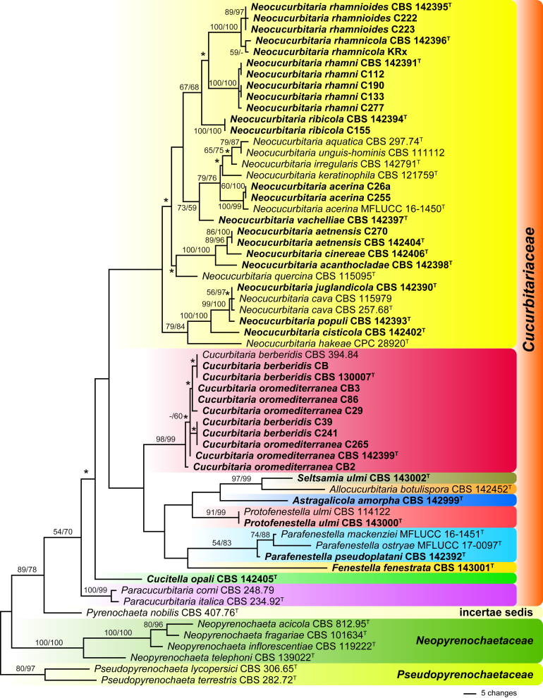
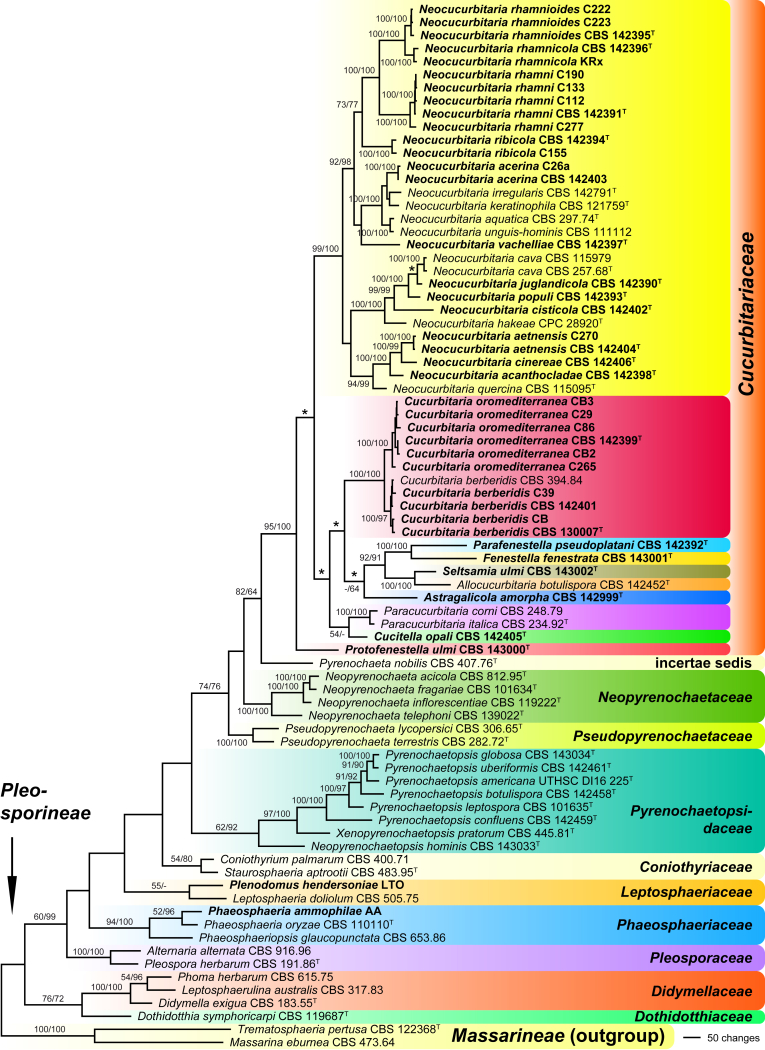
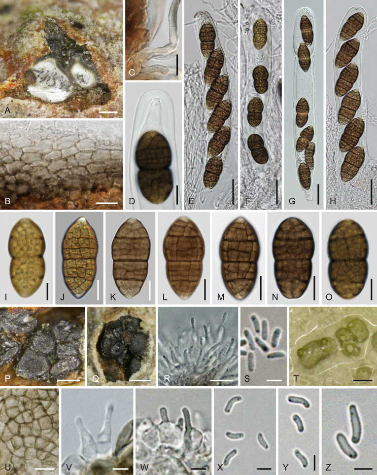
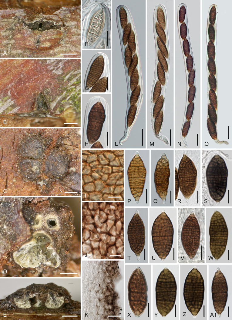
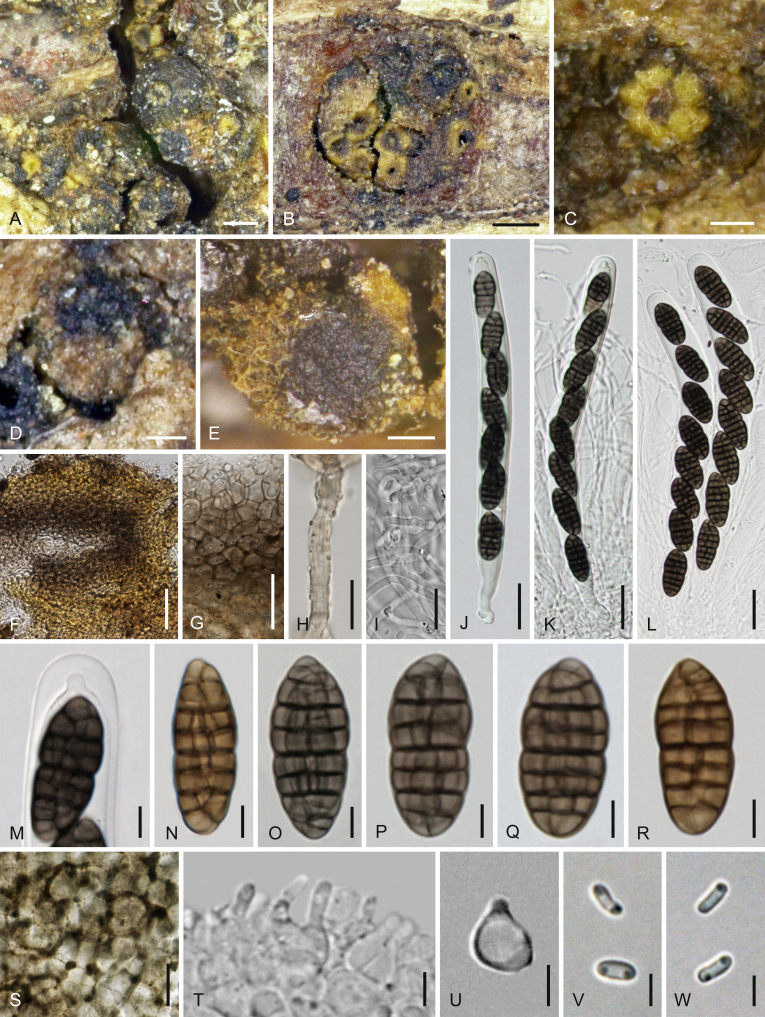
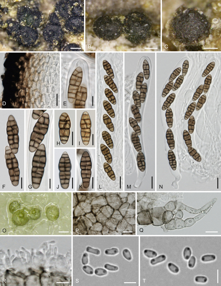
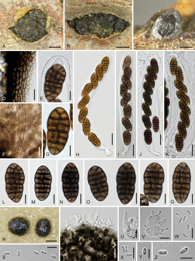
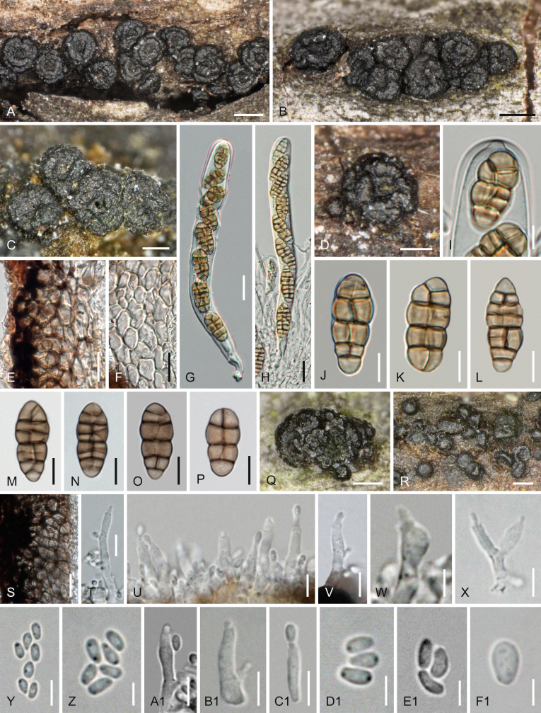
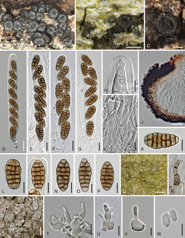
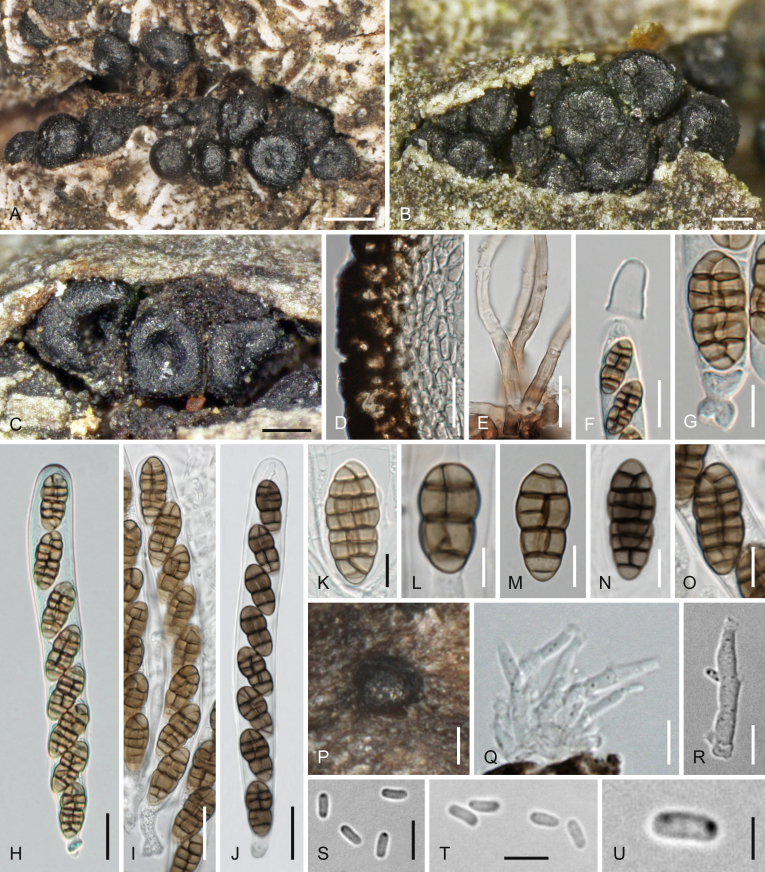
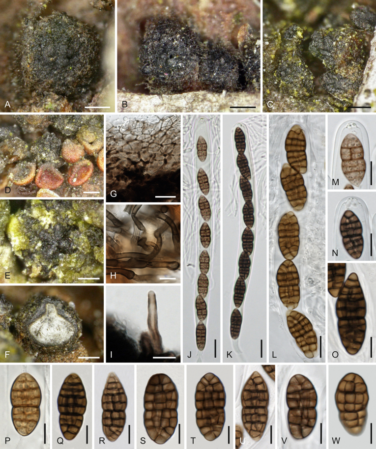
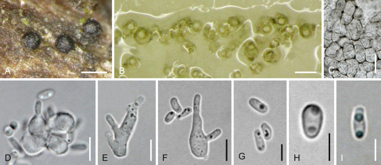
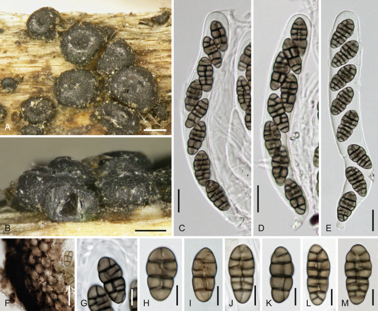
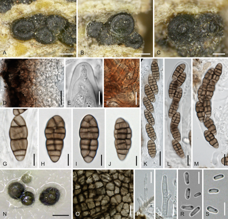
Fresh collections, type studies and molecular phylogenetic analyses of a multigene matrix of partial nuSSU-ITS-LSU rDNA, rpb2, tef1 and tub2 sequences were used to evaluate the boundaries of Cucurbitaria in a strict sense and of several related genera of the Cucurbitariaceae. Two species are recognised in Cucurbitaria and 19 in Neocucurbitaria. The monotypic genera Astragalicola, Cucitella, Parafenestella, Protofenestella, and Seltsamia are described as new. Fenestella is here included as its generic type F. fenestrata (= F. princeps), which is lecto- and epitypified. Fenestella mackenzei and F. ostryae are combined in Parafenestella. Asexual morphs of Cucurbitariaceae, where known, are all pyrenochaeta- or phoma-like. Comparison of the phylogenetic analyses of the ITS-LSU and combined matrices demonstrate that at least rpb2 sequences should be added whenever possible to improve phylogenetic resolution of the tree backbone; in addition, the tef1 introns should be added as well to improve delimitation of closely related species.

Keywords: Ascomycota; Astragalicola Jaklitsch & Voglmayr; Astragalicola amorpha Jaklitsch & Voglmayr; Cucitella Jaklitsch & Voglmayr; Cucitella opali Jaklitsch & Voglmayr; Cucurbitaria oromediterranea Jaklitsch & Voglmayr; Dothideomycetes; Fenestella princeps Tul. & C. Tul.; N. aetnensis Jaklitsch & Voglmayr; N. cinereae Jaklitsch & Voglmayr; N. cisticola Jaklitsch & Voglmayr; N. juglandicola Jaklitsch & Voglmayr; N. populi Jaklitsch & Voglmayr; N. rhamnicola Jaklitsch & Voglmayr; N. rhamnioides Jaklitsch & Voglmayr; N. ribicola Jaklitsch & Voglmayr; N. vachelliae Jaklitsch & Voglmayr; Neocucurbitaria acanthocladae Jaklitsch & Voglmayr; Neocucurbitaria rhamni (Nees : Fr.) Jaklitsch & Voglmayr; Parafenestella Jaklitsch & Voglmayr; Parafenestella mackenziei (Wanas. et al.) Jaklitsch & Voglmayr; Parafenestella ostryae (Wanas. et al.) Jaklitsch & Voglmayr; Parafenestella pseudoplatani Jaklitsch & Voglmayr; Phoma; Pleosporales; Protofenestella Jaklitsch & Voglmayr; Protofenestella ulmi Jaklitsch & Voglmayr; Pyrenochaeta; Seltsamia Jaklitsch & Voglmayr; Seltsamia ulmi Jaklitsch & Voglmayr; Sphaeria rhamni Nees; Valsa fenestrata Berk. & Broome; new taxa; phylogenetic analysis; pyrenomycetes.


Figures

Fig. 1
Fig. 1
Phylogram of one of 2720 MP trees 843 steps long (CI = 0.518, RI = 0.750), obtained by PAUP from an analysis of the ITS-LSU matrix of Cucurbitariaceae, Neopyrenochaetaceae and Pseudopyrenochaetaceae, with the latter selected as outgroup according to Fig. 2. MP and ML bootstrap support above 50 % are given at the first and second position, respectively, above or below the branches. Strains formatted in bold were isolated and sequenced in the current study; ex-type strains are indicated by a superscript T. Nodes that collapsed in the strict consensus of all 2 720 MP trees are marked by an asterisk (*). Note the lack of internal support for most backbone nodes, and the lack of resolution for closely related taxa.
Fig. 2
Fig. 2
Phylogram of one of 64 MP trees 9 817 steps long (CI = 0.345, RI = 0.625), obtained by PAUP from an analysis of the combined matrix (SSU-ITS-LSU, rpb2, tef1, tub2) of Cucurbitariaceae and selected Pleosporales. MP and ML bootstrap support above 50 % are given at the first and second position, respectively, above or below the branches. Strains formatted in bold were isolated and sequenced in the current study; ex-type strains are indicated by a superscript T. Nodes that collapsed in the strict consensus of the 64 MP trees are marked by an asterisk (*).
Fig. 3
Fig. 3
A–I, KW, YF1.Cucurbitaria oromediterranea. J, X.C. berberidis (WU 35987). A, B. Ascomata in face view. C, D. Ascoma in vertical section showing thickened basal peridium. E, F. Peridium in vertical section, showing opaque warts in E. G. Subicular hyphae. HJ. Asci. K, L. Ascus tips showing ocular chamber. M. Hamathecium. NX. Ascospores. Y. Pycnidium on natural substrate. ZF1. Asexual morph on CMD at 22 °C after 2–3 wk. Z. Pycnidium. A1. Setae. B1–D1. Conidiophores and phialides. E1, F1. Conidia. A, CG, L, M, QT, W. WU 35988; B, NP, U, V, YF1. WU 35991/C86; H, I. WU 35989; K. WU 35990. Scale bars: A, B = 0.5 mm; C, D = 150 μm; E, Y, Z = 50 μm; F, M–X, C1 = 10 μm; G, K, L, B1 = 7 μm; H–J, A1 = 20 μm; D1–F1 = 5 μm.
Fig. 4
Fig. 4
Astragalicola amorpha (WU 35994). A, B. Pycnidia in face view on the natural substrate. C. Sectioned pycnidia showing olivaceous interior. D. Peridium in vertical section. EK. Conidiophores and phialides. L, M. Conidia. D, J, L, M. in 3 % KOH. Scale bars: A, B = 300 μm; C = 150 μm; D, F, H = 7 μm; E = 10 μm; G, I, L = 5 μm; J, K, M = 3 μm.
Fig. 5
Fig. 5
Cucitella opali (WU 35995/CBS 142405). AO. Sexual morph (WU 35995). A. Pustule of ascomata in bark with two ascomata in vertical section. B. Peridium in vertical section. C. Subicular hyphae. D. Ascus apex. EH. Asci (exotunica broken in G). IO. Ascospores (I. young). PS. Asexual morph on natural substrate. P, Q. Pycnidia. R. Cluster of short conidiophores with phialides. S. Conidia. TZ. Asexual morph in culture (CBS 142405 on CMD after 5–6 d at 22 °C). T. Pycnidia. U. Pycnidial wall. V, W. Conidiogenous cells. XZ. Conidia. D, F, I, O. in 3 % KOH. Scale bars: A, P = 150 μm; B–D, J–O, U = 10 μm; E–H = 25 μm; I, R = 7 μm; Q = 250 μm; S, V–Z = 3 μm; T = 70 μm.
Fig. 6
Fig. 6
Fenestella fenestrata. A, B. Pseudostroma pustules in face view (B. vertically cut half pustule). C. Valsoid group of exposed ascomata in remnants of subiculum. D, E. Sectioned pseudostromata in (oblique) side view showing ascomata and eroded ostioles (shiny patches in E are due to a glue previously used for attachment to a sheet). FH. Ascus apices (F. immature). IK. Peridium (I, J. in face view; K. in vertical section). LO. Asci. PA1. Ascospores (in R, S, V with hamathecial threads in the background and free end in S; in X without appendage cells). K, S, W, Y, Z. in 3 % KOH. A, D, H, J, K, N, O, VA1. lectotype PC 0084495; B, I, PS. epitype WU 35996; C, EG, L, T, U. PC 0084493; M. PC 0084496. Scale bars: A, E = 500 μm; B–D = 300 μm; F–H, K, P–W, Y–A1 = 15 μm; I, J, X = 10 μm; L–O = 30 μm.
Fig. 7
Fig. 7
Neocucurbitaria acanthocladae. AR. Sexual morph (WU 35997). AC. Ascomata in face view (C. ostiolar area). D. Ascoma with black furrowed ostiole. E. Obliquely disposed ascoma with yellow-brown subiculum and yellow apex. F. Ostiolar area with yellow-brown surrounding wall. G. Peridium in vertical section. H. Subicular hypha. I. Hamathecium. JL. Asci. M. Ascus apex. NR. Ascospores (N, R. young). SW. Asexual morph in culture (CBS 142398 on CMD after 7–10 d at 22 °C). S. Pycnidial wall. T, U. Conidiogenous cells. V, W. Conidia. F, IR. in 3 % KOH. Scale bars: A, B = 200 μm; C = 70 μm; D, E = 100 μm; F = 30 μm; G, J–L = 20 μm; H, I, S = 10 μm; M–R = 5 μm; T–W = 3 μm.
Fig. 8
Fig. 8
Neocucurbitaria acerina. AN. Sexual morph. AC. Ascomata in face view. D. Peridium in vertical section. E. Ascus apex. FK. Ascospores. LN. Asci. OT. Asexual morph in culture (CMD, after 4–7 d at 22 °C). O. Pycnidia. P. Pycnidial wall. Q. Pycnidial appendages. R. Conidiogenous cells. S, T. Conidia. DG, LN, PR. in 3 % KOH. AN, P, Q, S, T. WU 35998/CBS 142403; O, R. C26a. Scale bars: A, B = 150 μm; C = 100 μm; D, L–N = 10 μm; E, G, P, R = 5 μm; F, H–K, Q = 7 μm; O = 100 μm; S, T = 3 μm.
Fig. 9
Fig. 9
Neocucurbitaria aetnensis. AQ. Sexual morph. AC. Ascomata in face view. D. Ascomata in vertical section. E. Peridium and a subicular hypha in vertical section. F. Part of ostiole with periphyses. G. Ascus apex. HJ. Asci. KQ. Ascospores (K. young). RW. Asexual morph in culture (CMD, after 6–7 d at 22 °C). R. Pycnidia. S. Pycnidial wall. T, U. Conidiogenous cells. V, W. Conidia. HQ. in 3 % KOH. A, B, DP, T. WU 36929/CBS 142404; C, QS, UW. WU 36930/C270. Scale bars: A–D = 200 μm; E, H–J = 15 μm; F = 10 μm; G, Q, S, T = 5 μm; K–P = 7 μm; R = 70 μm; U–W = 3 μm.
Fig. 10
Fig. 10
Neocucurbitaria cinereae. AP. Sexual morph (WU 36931). A, B. Ascomata in face view. C. Ascoma in vertical section. D. Peridium in vertical section. E. Apex of immature ascus. F. Hamathecium. GJ. Asci (G. immature). KP. Ascospores (K, L. young). QU. Asexual morph in culture (CBS 142406 on CMD after 5–6 d at 22 °C). Q. Pycnidia. R. Pycnidial wall. S. Conidiogenous cells. T, U. Conidia. EJ, LP. in 3 % KOH. Scale bars: A, B, Q = 250 μm; C = 100 μm; D, F, R–T = 7 μm; E, U = 3 μm; G–J = 15 μm; K–P = 5 μm.
Fig. 11
Fig. 11
Neocucurbitaria cisticola. AL. Sexual morph (WU 36932). A, B. Ascomata in face view. C. Peridium in vertical section. D. Ascoma in vertical section. E. Apex of young ascus. FI. Ascospores. JL. Asci. MS. Asexual morph in culture (CBS 142402 on CMD after 5 d at 22 °C). M. Pycnidia with conidial drops. N. Pycnidial wall. O, P. Conidiogenous cells. QS. Conidia. EL, Q. in 3 % KOH. Scale bars: A, B = 100 μm; C = 10 μm; D, M = 70 μm; E–I, N–P = 5 μm; J–L = 15 μm; Q–S = 3 μm.
Fig. 12
Fig. 12
Neocucurbitaria juglandicola (A–S) and Cucurbitaria juglandis (T). AJ. Sexual morph (WU 36933). A. Ascomata in face and lateral view. BG. Ascospores. HJ. Asci. KN. Asexual morph on natural substrate (WU 36933). K. Pycnidia in face view. L. Peridium. M. Conidiophores and phialides. N. Conidia. OS. Asexual morph in culture (CBS 142390 on CMD after 4–6 d at 22 °C). O. Pycnidia. P. Peridium. Q. Conidiogenous cells. R, S. Conidia. T. Conidia from Cucurbitaria juglandis type material G00266380 (in 3 % KOH). Scale bars: A, K, O = 100 μm; B–G, L, P = 7 μm; H–J, M = 10 μm; N, R–T = 3 μm; Q = 5 μm.
Fig. 13
Fig. 13
Neocucurbitaria populi. AQ. Sexual morph (WU 36934). A, B. Ascomata in face view. C. Ascoma in vertical section. D. Peridium in vertical section. E. Ascus apex. F. Subiculum. G, LQ. Ascospores. HK. Asci. RZ. Asexual morph in culture (CBS 142393 on CMD after 5 d at 22 °C). R. Pycnidia (left with conidial drop). S, WZ. Conidia. T. Pycnidial wall and conidiogenous cells. U, V. Conidiogenous cells. FQ. in 3 % KOH. Scale bars: A, B = 500 μm; C, R = 100 μm; D, F = 10 μm; E, G, U, Y, Z = 5 μm; H–K = 15 μm; L–Q, S, T, V–X = 7 μm.
Fig. 14
Fig. 14
Neocucurbitaria rhamni. AD. Ascomata in face view. E, F. Peridium in vertical section (inner layer in F). G, H. Asci. I. Ascus apex showing ocular chamber. JP. Ascospores. QZ. Asexual morph on the natural hosts. Q, R. Pycnidia. S. Pycnidial wall in section. TX. Conidiophores and phialides. Y, Z. Conidia. A1F1. Asexual morph in culture at 22 °C (A1E1. after 7 d on SNA; F1. after 20 d on MEA). A1C1. Conidiophores and phialides. D1F1. Conidia. MP, SF1. in 3 % KOH. A, D, A1F1. WU 36936/C112; B, Q, Y, Z. WU 36944; C, M. Lectotype B 700016439; EL. WU 36941; N–P, RV. WU 36935; W, X. WU 36939. Scale bars: A, B = 300 μm; C, D, Q, R = 150 μm; E–H, S = 15 μm; I, U, V, X, Y, A1–C1 = 5 μm; J–P, T = 7 μm; W, Z, D1–F1 = 3 μm.
Fig. 15
Fig. 15
Neocucurbitaria rhamnicola. AC. Ascomata in face view. DG. Asci. H. Ascus apex showing ocular chamber. I. Hamathecium. J. Ascoma in vertical section. KP. Ascospores. QW. Asexual morph in culture (on MEA after 8 d at 22 °C). Q. Pycnidia. R. Hyphal appendage on pycnidial wall. S. Pycnidial wall in face view. TV. Conidiophores and conidiogenous cells. W. Conidia. RW. in 3 % KOH. A, D, IM, QW. WU 36946/CBS 142396. B, C, EH, NP. WU 36947. Scale bars: A, B = 300 μm; C = 100 μm; D = 15 μm; E–G = 10 μm; H, T, U = 5 μm; I, K–P, R, S = 7 μm; J = 50 μm; Q = 200 μm; V, W = 3 μm.
Fig. 16
Fig. 16
Neocucurbitaria rhamnioides. AC. Ascomata in face view. D. Peridium in vertical section. E. Subicular hyphae. F. Ascus apex after fissitunicate dehiscence. G. Ascus base. HJ. Asci. KO. Ascospores. PU. Asexual morph on the natural substrate. P. Pycnidium. Q, R. Conidiophores and phialides. SU. Conidia. J, O, Q, R, U. in 3 % KOH. A, D, I, K, N, O, Q–U. WU 36948; B, E, J, L, M, P. WU 36949; C, FH. WU 36950. Scale bars: A = 300 μm; B, C = 150 μm; D–F, H–J = 15 μm; G, K, M–O, Q = 7 μm; L, R–T = 5 μm; P = 50 μm; U = 2 μm.
Fig. 17
Fig. 17
Neocucurbitaria ribicola, sexual morph. AE. Ascomata in face view (emphasizing furrowed apices in CE). F. Ascoma in vertical section. G. Peridium in vertical section. H. Subicular hyphae. I. Ascomatal seta. J, K. Asci. L. Upper part of ascus. M, N. Ascus apices (immature in M). OW. Ascospores (young in P). HO, QV. in 3 % KOH. A, C, DI, L, P, S, T, V, W. WU 36952; B, J, K, MO, Q, R, U. WU 36951. Scale bars: A–C, F = 200 μm; D, E = 100 μm; G, J, K = 15 μm; H, L–N = 10 μm; I, O–W = 7 μm.
Fig. 18
Fig. 18
Neocucurbitaria ribicola, asexual morph. A. Pycnidia on the natural substrate (WU 36951). BI. Asexual morph in culture (CMD, after 5–10 d at 22 °C). B. Pycnidia. C. Pycnidial wall. DF. Conidiogenous cells and short conidiophore (in E). GI. Conidia. CI. in 3 % KOH. BH. CBS 142394; I. C155. Scale bars: A = 100 μm; B = 75 μm; C = 10 μm; D–F = 5 μm; G–I = 3 μm.
Fig. 19
Fig. 19
Cucurbitaria ribis (holotype M-0281853). A. Ascomata in face view. B. Ascoma from the side, one sectioned. CE. Asci. F. Peridium. G. Ascus apex. HM. Ascospores. CH, JM. in 3 % KOH. Scale bars: A, B = 200 μm; C–F = 15 μm; G–M = 7 μm.
Fig. 20
Fig. 20
Neocucurbitaria vachelliae. AM. Sexual morph (WU 36953). AC. Ascomata in face view. D. Peridium in vertical section. E. Ascus apex. F. Subiculum. GJ. Ascospores (young in G). KM. Asci. NS. Asexual morph in culture (CBS 142397 on CMD after 6–7 d at 22 °C). N. Pycnidia with conidial drops. O. Pycnidial wall. P, Q. Conidiophores and phialides. R, S. Conidia. E, GJ, L, M. in 3 % KOH. Scale bars: A, B = 200 μm; C, N = 100 μm; D, K = 15 μm; E, O, P = 7 μm; F, L, M = 10 μm. G–J, Q–S = 5 μm.
Fig. 21
Fig. 21
Parafenestella pseudoplatani (WU 36954/CBS 142392). AM. Sexual morph. AC. Ascomata in face view. D. Peridium and subicular hyphae in vertical section. E. Ascus apex. F. Ascus stipe and base. GJ. Ascospores. KM. Asci (young in K). NU. Asexual morph. N. Pycnidia on natural substrate. O. Pycnidia. P. Pycnidial wall. Q, R. Phialides from natural substrate. SU. Conidia. O, P, SU. From culture (CMD, after 7 d at 22 °C). DF, IM, Q, R. in 3 % KOH. Scale bars: A–C = 200 μm; D, K–M = 20 μm; E, G–J, P–R = 7 μm; F = 10 μm. N = 100 μm. O = 70 μm. S, T = 5 μm; U = 3 μm.
Fig. 22
Fig. 22
Protofenestella ulmi. AU. Sexual morph. A. Ostiole on bark surface and subiculum surrounding ascoma below bark surface. B. Ascoma in vertical section. C. Peridium and subicular hyphae in vertical section. D. Subiculum. EH. Asci. I. Ascus apex (with immature 3-septate ascospore). JU. Ascospores (J. immature, showing thick wall and terminal inner canals; K. in mature ascus apex). VC1. Asexual morph in culture (CMD, after 6 d at 22 °C). V. Pycnidia. W. Pycnidial wall. XZ. Conidiogenous cells. A1C1. Conidia. D, K, O, U. in 3 % KOH. AJ, LN, PC1. WU 36955/CBS 143000; K. UPS F-178445; O. UPS F-178444. Scale bars: A, B = 150 μm; C = 50 μm; D, I, K, M–U, W = 10 μm; E–H = 25 μm; J, Z, A1 = 7 μm; L = 15 μm; V = 100 μm; X, Y, B1, C1 = 5 μm.
Fig. 23
Fig. 23
Seltsamia ulmi (WU 36957). A. Horizontal section at the ostiolar level. B. Horizontal section at the ascomatal level. C, D. Ascoma in vertical section. E. Peridium in vertical section showing remnants of subicular hyphae. FH. Asci (young in F). I. Ascus apex showing ocular chamber. JQ. Ascospores (showing swelling sheath in J and K). E, J. in 3 % KOH. Scale bars: A–C = 200 μm; D = 100 μm; E–H, K = 25 μm; I = 10 μm; J, L–Q = 15 μm.

References

    1. Ariyawansa H.A., Hyde K.D., Jayasiri S.C. Fungal diversity notes 111–252—taxonomic and phylogenetic contributions to fungal taxa. Fungal Diversity. 2015;75:27–274.
    1. Arnold R.H. Vol. 17. National Mycological Herbarium, Biosystematics Research Institute, Agriculture Canada; Ottawa, Ontario: 1974. (Cucurbitaria staphula. Fungi Canadenses).
    1. Arnold R.H., Russell R.C. Cucurbitaria staphula on Populus and its association with Macrophoma tumefaciens. Mycologia. 1960;52:499–512.
    1. Aveskamp M.M., de Gruyter J., Woudenberg J.H.C. Highlights of the Didymellaceae: a polyphasic approach to characterize Phoma and related pleosporalean genera. Studies in Mycology. 2010;65:1–60. - PMC - PubMed
    1. Barr M.E. The genus Curreya: An example of taxonomic confusion in the ascomycetes. Mycologia. 1981;73:599–609.

LinkOut - more resources