. 2017 Dec 15:12:8855-8866.
doi: 10.2147/IJN.S148179. eCollection 2017.

Guided bone regeneration with asymmetric collagen-chitosan membranes containing aspirin-loaded chitosan nanoparticles

Affiliations

Guided bone regeneration with asymmetric collagen-chitosan membranes containing aspirin-loaded chitosan nanoparticles

Jiayu Zhang et al. Int J Nanomedicine. .

Abstract

Introduction: Membranes allowing the sustained release of drugs that can achieve cell adhesion are very promising for guided bone regeneration. Previous studies have suggested that aspirin has the potential to promote bone regeneration. The purpose of this study was to prepare a local drug delivery system with aspirin-loaded chitosan nanoparticles (ACS) contained in an asymmetric collagen-chitosan membrane (CCM).

Methods: In this study, the ACS were fabricated using different concentrations of aspirin (5 mg, 25 mg, 50 mg, and 75 mg). The drug release behavior of ACS was studied. Transmission electron microscopy (TEM) and scanning electron microscopy (SEM) were used to examine the micromorphology of ACS and aspirin-loaded chitosan nanoparticles contained in chitosan-collagen membranes (ACS-CCM). In vitro bone mesenchymal stem cells (BMSCs) were cultured and critical-sized cranial defects on Sprague-Dawley rats were made to evaluate the effect of the ACS-CCM on bone regeneration.

Results: Drug release behavior results of ACS showed that the nanoparticles fabricated in this study could successfully sustain the release of the drug. TEM showed the morphology of the nanoparticles. SEM images indicated that the asymmetric membrane comprised a loose collagen layer and a dense chitosan layer. In vitro studies showed that ACS-CCM could promote the proliferation of BMSCs, and that the degree of differentiated BMSCs seeded on CCMs containing 50 mg of ACS was higher than that of other membranes. Micro-computed tomography showed that 50 mg of ACS-CCM resulted in enhanced bone regeneration compared with the control group.

Conclusion: This study shows that the ACS-CCM would allow the sustained release of aspirin and have further osteogenic potential. This membrane is a promising therapeutic approach to guiding bone regeneration.

Keywords: aspirin; drug release; guided bone regeneration; membrane; nanoparticle.


Conflict of interest statement

Disclosure The authors report no conflicts of interest in this work.

Figures

Figure 1
Figure 1
Release curve of ACS at 1, 3, 5, 7, 10, and 14 days. Note: There is a burst release on the first day, and then a sustained release is observed until day 14, (n=3). Abbreviation: ACS, aspirin-loaded chitosan nanoparticles.
Figure 2
Figure 2
TEM and DLS characterization of ACS. Notes: (A, B) The TEM image shows ACS dispersed in collagen solution, the black arrow indicates the ACS, and the white arrows show the collagen fibrils. (C) DLS result shows the ACS size distribution. Abbreviations: TEM, transmission electron microscopy; DLS, dynamic light scattering; ACS, aspirin-loaded chitosan nanoparticles.
Figure 3
Figure 3
SEM images showing the morphologies of the ACS-CCM. Notes: (A) Image of the loose cross-linked collagen layer, (B) the dense chitosan layer, (C) the cross-section of the ACS-CCM, and (D) a schematic diagram of the cross-section. Abbreviations: SEM, scanning electron microscopy; ACS-CCM, aspirin-loaded chitosan nanoparticles contained in collagen-chitosan membranes.
Figure 4
Figure 4
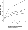
Weight loss of membranes during different soaking intervals. Notes: The image shows the difference in the degradation rates among the four groups. The cross-linked collagen membranes degraded slower than the collagen membranes (n=3).
Figure 5
Figure 5
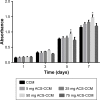
CCK-8 assay results of BMSCs distributed on different membranes after culturing for 1, 3, 5, and 7 days. Note: There was a statistically significant difference in cell proliferation between the 50 mg ACS-CCM group and the other four groups on day 5 (*P<0.05, n=3). Abbreviations: CCK-8, Cell Counting Kit-8; BMSCs, bone mesenchymal stem cells; CCM, collagen-chitosan membranes; ACS-CCM, aspirin-loaded chitosan nanoparticles contained in collagen-chitosan membranes.
Figure 6
Figure 6
ALP activities of BMSCs growing in different membranes after culturing for 7 and 14 days. Note: The difference between the ALP activity of the 50 mg ACS-CCM group and the other groups was statistically significant at each testing time point (*P<0.05, n=3). Abbreviations: BMSCs, bone mesenchymal stem cells; CCM, collagen-chitosan membranes; ACS-CCM, aspirin-loaded chitosan nanoparticles contained in collagen-chitosan membranes.
Figure 7
Figure 7
LSCM images of BMSCs attached to the collagen surface after culturing for 3 and 5 days (AO/EB staining). Note: The LSCM images showed no differences in cell morphology between the CCM group (3 days [A] and 5 days [C]) and the 50 mg ACS-CCM group (3 days [B] and 5 days [D]). Abbreviations: BMSCs, bone mesenchymal stem cells; LSCM, laser scanning confocal microscopy; AO/EB, acridine orange/ethidium bromide; ACS-CCM, aspirin-loaded chitosan nanoparticles contained in collagen-chitosan membranes; CCM, collagen-chitosan membranes.
Figure 8
Figure 8
Skull-penetrating defect model of SD rats. Notes: (A) The parietal calvarium defect of SD rat. (B) 50 mg ACS-CCM implanted in the defect. Abbreviations: SD, Sprague-Dawley; ACS-CCM, aspirin-loaded chitosan nanoparticles contained in collagen-chitosan membranes.
Figure 9
Figure 9
3D reconstruction and micro-CT images of new bone regeneration after 4 and 8 weeks. Note: The 3D reconstructions and the coronary sections of rat craniums in (A) the control and (B) the 50 mg ACS-CCM group after being implanted for 4 and 8 weeks. Abbreviations: micro-CT, micro-computed tomography; ACS-CCM, aspirin-loaded chitosan nanoparticles contained in collagen-chitosan membranes.
Figure 10
Figure 10
H&E staining of SD rat cranial defects at different time points. Notes: (A) Control group and (B) 50 mg ACS-CCM at 4 weeks. (C) Control group and (D) 50 mg ACS-CCM group at 8 weeks. Abbreviations: H&E, hematoxylin and eosin; SD, Sprague-Dawley; ACS-CCM, aspirin-loaded chitosan nanoparticles contained in collagen-chitosan membranes.

References

    1. Liu Y, Zheng Y, Ding G, et al. Periodontal ligament stem cell-mediated treatment for periodontitis in miniature swine. Stem Cells. 2008;26(4):1065–1073. - PMC - PubMed
    1. Chen FM, Zhang J, Zhang M, An Y, Chen F, Wu ZF. A review on endogenous regenerative technology in periodontal regenerative medicine. Biomaterials. 2010;31(31):7892–7927. - PubMed
    1. Pihlstrom BL, Michalowicz BS, Johnson NW. Periodontal diseases. Lancet. 2005;366(9499):1809–1820. - PubMed
    1. Bashutski JD, Wang HL. Periodontal and endodontic regeneration. J Endod. 2009;35(3):321–328. - PubMed
    1. Zhang K, Zhao M, Cai L, Wang Zk, Sun Yf, Hu QL. Preparation of chitosan/hydroxyapatite guided membrane used for periodontal tissue regeneration. Chinese Journal of Polymer Science. 2010;28(4):555–561.

MeSH terms

LinkOut - more resources