. 2017 Nov 30:8:1667.
doi: 10.3389/fimmu.2017.01667. eCollection 2017.

Generation and Functional In Vitro Analysis of Semliki Forest Virus Vectors Encoding TNF-α and IFN-γ

Affiliations

Generation and Functional In Vitro Analysis of Semliki Forest Virus Vectors Encoding TNF-α and IFN-γ

Baiba Kurena et al. Front Immunol. .

Abstract

Cytokine gene delivery by viral vectors is a promising novel strategy for cancer immunotherapy. Semliki Forest virus (SFV) has many advantages as a delivery vector, including the ability to (i) induce p53-independent killing of tumor cells via apoptosis, (ii) elicit a type-I interferon (IFN) response, and (iii) express high levels of the transgene. SFV vectors encoding cytokines such as interleukin (IL)-12 have shown promising therapeutic responses in experimental tumor models. Here, we developed two new recombinant SFV vectors encoding either murine tumor necrosis factor-α (TNF-α) or murine interferon-γ (IFN-γ), two cytokines with documented immunostimulatory and antitumor activity. The SFV vector showed high infection rate and cytotoxicity in mouse and human lung carcinoma cells in vitro. By contrast, mouse and human macrophages were resistant to infection with SFV. The recombinant SFV vectors directly inhibited mouse lung carcinoma cell growth in vitro, while exploiting the cancer cells for production of SFV vector-encoded cytokines. The functionality of SFV vector-derived TNF-α was confirmed through successful induction of cell death in TNF-α-sensitive fibroblasts in a concentration-dependent manner. SFV vector-derived IFN-γ activated macrophages toward a tumoricidal phenotype leading to suppressed Lewis lung carcinoma cell growth in vitro in a concentration-dependent manner. The ability of SFV to provide functional cytokines and infect tumor cells but not macrophages suggests that SFV may be very useful for cancer immunotherapy employing tumor-infiltrating macrophages.

Keywords: Lewis lung carcinoma; Semliki Forest virus; cancer immunotherapy; cytokines; gene delivery; macrophage activation.


Figures

Figure 1
Figure 1
Semliki Forest virus (SFV) vectors used in this study for replication-deficient expression system. All vectors comprised the prokaryotic SP6 RNA polymerase promoter for in vitro transcription of the recombinant SFV replicons. (A) Replication-deficient vectors encoding SFV non-structural protein genes (nsP1–4) and the heterologous genes (HGs) Tnfa or Ifng downstream of the SFV 26S subgenomic promoter. The mouse TNF-α gene (Tnfa) was followed by a FLAG-tag-encoding sequence. The mouse IFN-γ gene (Ifng) was inserted in frame with the minimal capsid translation enhancer sequence (Enh) using the 2A auto-protease sequence (2A) from foot and mouth disease virus as a linker. (B) Replication-deficient reporter construct consisting of SFV nsP1–4 and the fluorescent protein-encoding gene Ds-Red downstream of an SFV 26S subgenomic promoter. (C) The SFV helper vector SFV-Helper1, which encodes the full length of the structural protein ORF (i.e., C, capsid protein; p62, 6K and E1, and envelope proteins) downstream of an SFV 26S subgenomic promoter. (D) Production of recombinant particles and proteins by the replication-deficient SFV vector system: (1) in vitro transcription of RNA encoding the HG and the nsP1–4, and the transcription of helper RNA carrying SFV structural genes. The packaging signal (PS) for the selective encapsidation of the RNA encoding nsP1-4 and the HG is present in the nsP2 region of SFV-HG RNA. Both RNAs are capped at the 5-prime end and polyadenylated at the 3-prime end; (2) BHK-21 cells are transfected with SFV-HG and SFV-Helper RNAs by electroporation; (3) BHK-21 cells produce replication-deficient SFV particles carrying HGs; (4) target cells are infected with replication-deficient SFV particles; and (5) the heterologous protein is produced at high levels without production of new viral particles.
Figure 2
Figure 2
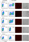
Susceptibility to Semliki Forest virus (SFV) infection varies between cell types. Cells were plated and infected with SFV-DsRed particles at MOI = 15 [determined using baby hamster kidney (BHK-21) cells] the next day. DsRed expression was evaluated at 24 h post-infection using flow cytometry and fluorescence microscopy. Non-infected cells were used as a negative control. From left to right: flow cytometry data for DsRed expression in non-infected and infected samples with the numbers representing the percentage of DsRed-positive cells as the mean of duplicates; and fluorescence microscopy images are shown with the corresponding bright-field microscopy images (scale bars, 50 µm). (A) The hamster fibroblast cell line BHK-21 was used as a positive control; (B) mouse lung carcinoma cell line Lewis lung carcinoma (LLC), (C) human lung carcinoma cell line A549, (D) mouse bone marrow-derived macrophages (BMDMs), and (E) mouse macrophage cell line J774A.1. The experiment was repeated three or more times with the SE not exceeding 10% between independent experiments. The average of duplicates with SEM <10% from one representative experiment is shown.
Figure 3
Figure 3
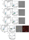
Human macrophages are resistant to Semliki Forest virus (SFV) infection. Human monocyte-derived macrophages (HMDMs), and the human lung carcinoma cell line A549 were infected with either SFV-DsRed or SeV-Gfp virus particles. The resulting cell monolayers were analyzed using phase-contrast microscopy, and the expression of DsRed and GFP was evaluated using fluorescence microscopy 24 h post-infection (scale bars, 20 µm). The numbers in the images indicate the percentages of infected cells. Non-infected cells were used as negative controls. HMDMs were not susceptible to infection with SFV-DsRed, whereas A549 cells were infected with SFV, as expected (shown in the second row). Sendai virus (SeV)-Gfp virus particles were used as a positive control because they are capable of infecting both HMDMs and A549 cells, as shown in the third row. Total HMDM resistance to SFV infection was confirmed in three independent experiments using HMDMs from different donors. Data from one representative experiment are shown where the percentages represent average fluorescent protein-expressing cell population with SEM <5%.
Figure 4
Figure 4
Macrophages remain viable, whereas cancer cells undergo cell death after challenge with Semliki Forest virus (SFV). J774A.1 mouse macrophages and Lewis lung carcinoma (LLC) carcinoma cells were infected with SFV-DsRed particles at MOI = 10 [determined using baby hamster kidney (BHK-21) cells]. DsRed expression (first column, x-axis) was evaluated at 48 h post-infection using flow cytometry and fluorescence microscopy (scale bar 100 µM). Cell morphology was observed using bright-field microscopy (scale bar 100 µM) and cell death was quantified 48 h post-infection using flow cytometry analysis after cell staining with annexin V-FITC (second column, x-axis) and DAPI (second column, y-axis). Annexin V-positive/DAPI-negative cells were regarded as early apoptotic, whereas annexin V-positive/PI-positive cells were regarded as late apoptotic and necrotic. (A) Non-infected J774A.1 macrophages were used as a negative control. (B) J774A.1 macrophages were resistant to SFV infection, stayed viable but changed their morphology 48 h after challenge with SFV. (C) Non-infected LLC cells were used as a negative control. (D) LLC cells were susceptible to SFV infection and underwent cell death. DsRed expression was observed using microscopy in both apoptotic (white arrows) and viable (yellow arrows) cells, whereas some apoptotic cells lacked DsRed expression (red arrows). (E) The infected DsRed-positive cells were both viable and undergoing cell death. The average of duplicates with SEM <10% from one experiment is shown.
Figure 5
Figure 5
Cancer cell growth is inhibited by infection with Semliki Forest virus (SFV) particles. (A) Schematic overview of the experiment. Baby hamster kidney (BHK-21) or Lewis lung carcinoma (LLC) cells were plated and incubated for 22 h before they were infected with SFV-Ifng or SFV-Tnfa particles at MOI = 15. The cells were incubated for 47 h after infection, and [3H]-thymidine was then added to detect proliferating cells. After 24 h, the cells were harvested, and cell growth was determined by measuring the incorporated [3H]-thymidine as counts per minute (cpm, depicted on the y-axis). (B) As expected, infecting BHK-21 cells with SFV resulted in substantial growth inhibition. (C) BHK-21 cells underwent cell death 47 h post-infection as observed using bright-field microscopy (scale bars, 50 µm). (D) LLC cell growth was inhibited by infection with different SFV particles. (E) LLC cells underwent cell death 47 h post-infection as observed using bright-field microscopy (scale bars, 50 µm). Two experiments were performed in triplicates with SE not exceeding 10% between independent experiments. The bars represent the mean values of triplicates ± SEM from one representative experiment.
Figure 6
Figure 6
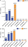
Semliki Forest virus (SFV)-encoded cytokines are produced and secreted by cancer cells. Baby hamster kidney (BHK-21) and Lewis lung carcinoma (LLC) cells were infected with either SFV-Ifng or SFV-Tnfa at MOI = 1, MOI = 10, or MOI = 40 (determined using BHK-21 cells). The level of secreted (A) TNF-α or (B) interferon (IFN)-γ in the cell culture medium was determined using Luminex bead-based assay at 24 h after infection and is shown on the y-axis. The cytokines were undetectable in both the negative controls of non-infected cells and the cultures that were infected with a mismatched SFV that did not encode the cytokine of interest. N.D.—not detectable. Two independent experiments were performed in duplicates. Data from one representative experiment is shown. The bars represent the mean values of duplicates ± SEM.
Figure 7
Figure 7
Vector-derived TNF-α induces cell death in L929 fibroblasts. L929 cells were cultured for 24 h until they reached 90% confluency. The cells were then (A) left untreated for 24 h and used as a negative control or (B) treated with 1 µM staurosporine for 24 h at 37°C and used as a positive control. The remaining cells were treated with rTNF-α or vdTNF-α for 24 h at the following concentrations: (C,D) 2.2 ng/mL, (E,F) 6.7 ng/mL, (G,H) 20 ng/mL or (I,J) 60 ng/mL. The resulting cell monolayers were analyzed using bright-field microscopy (scale bar, 50 µM). Cell death was determined using flow cytometry analysis after cell staining with annexin V-FITC (depicted on the x-axis) and propidium iodide (PI, depicted on the y-axis). Annexin V-positive/PI-negative cells were regarded as apoptotic, whereas annexin V-positive/PI-positive cells were regarded as necrotic. Two independent experiments were performed in duplicates with standard error not exceeding 10% between independent experiments. Data from one representative experiment are shown, where the percentages of the four distinct cell populations represent the averages of duplicates with SEM <10%.
Figure 8
Figure 8
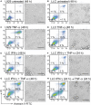
Interferon (IFN)-γ in combination with TNF-α induces cell death in mouse lung carcinoma cells. L929 fibroblasts and Lewis lung carcinoma (LLC) cancer cells were cultured for 24 h before starting treatment with recombinant cytokines. Both L929 and LLC cells were (A,B) left untreated for 48 h and used as negative controls or (C) L929 cells were treated with 50 ng/mL TNF-α for 48 h and used as a positive control. LLC cell viability was retained after treatment with (D) 50 ng/mL TNF-α or (E) 100 ng/mL IFN-γ and after (F) simultaneous treatment with both 50 ng/mL TNF-α and 100 ng/mL IFN-γ for 24 h. Cell death was induced in LLC cells after (G) simultaneous treatment with 100 ng/mL of IFN-γ in combination with 50 ng/mL of TNF-α for 48 h and after (H) LLC pretreatment with 100 ng/mL of IFN-γ for 24 h followed by treatment with TNF-α for 24 h. The resulting cell monolayers were analyzed using bright-field microscopy (scale bar, 50 µM). Cell death was determined using flow cytometry analysis after cell staining with annexin V-FITC (depicted on the x-axis) and propidium iodide (PI, depicted on the y-axis). Annexin V-positive/PI-negative cells were regarded as apoptotic, whereas annexin V-positive/PI-positive cells were regarded as necrotic. Experiment was performed in duplicates and repeated two times with standard error not exceeding 10% between independent experiments. One representative experiment is shown, where the percentages of the four distinct cell populations are averages of duplicates with SEM <5%.
Figure 9
Figure 9
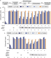
The effect of rIFN-γ or vdIFN-γ in combination with a TLR2/1 ligand on macrophage activation was detected using growth inhibition assay in Lewis lung carcinoma (LLC) cells. (A) Schematic timeline of the experiment. Mitomycin C-treated bone marrow-derived macrophages (BMDMs) were plated at three different densities and incubated for 24 h before macrophage-stimulating factors were added. After an additional 24 h, 100 µL of cell culture media was obtained from the wells with the highest macrophage density to measure NO2 levels. LLC cells were added to the activated BMDMs, and the co-cultures were incubated for an additional 20 h. [3H]-thymidine was then added to the co-cultures, and after 24 h, the LLC cells were harvested to analyze cell growth by measuring the amount of incorporated [3H]-thymidine as counts per minute (cpm, depicted on the y-axis). (B) LLC cell growth after co-cultivation with BMDMs that were activated with Pam3 (100 ng/mL) in combination with different concentrations of rIFN-γ ranging from 100 to 0.0015 ng/mL in fourfold dilutions. Controls, from left to right: BMDMs cultivated without LLC cells, LLC cells co-cultivated with unstimulated macrophages, and LLC cells co-cultivated with macrophages that were stimulated with either rIFN-γ alone or Pam3 alone. (C) LLC cell growth after co-cultivation with BMDMs that were activated with Pam3 (100 ng/mL) in combination with vdIFN-γ ranging from 100 to 0.0015 ng/mL in 16-fold dilutions. Controls, from left to right: BMDMs cultivated without LLC cells, LLC cells co-cultivated with unstimulated macrophages, LLC cells co-cultivated with macrophages that were treated with either rIFN-γ, vdIFN-γ, or Pam3 at a concentration of 100 ng/mL, and LLC cells co-cultivated with BMDMs that were treated with 100 ng/mL of both rIFN-γ and Pam3. Experiments were performed in triplicates and were repeated two times with similar results. The bars represent the mean values of triplicates ± SEM from one representative experiment.
Figure 10
Figure 10
The production of nitric oxide (NO) by macrophages at 24 h after activation was determined by analyzing NO2 levels in the cell culture medium. Mitomycin C-treated bone marrow-derived macrophages (BMDMs) were cultivated for 24 h before they were activated with the TLR2/1 agonist Pam3 (100 ng/mL) in combination with either (A) rIFN-γ at various concentrations ranging from 100 to 0.0015 ng/mL in 4-fold dilutions or (B) vdIFN-γ at concentratios ranging from 100 to 0.0015 ng/mL in 16-fold dilutions. Non-stimulated BMDMs and BMDMs that were stimulated with Pam3, rIFN-γ, or vdIFN-γ were used as the negative controls. Nitrite (NO2) levels were measured in the macrophage culture medium at 24 h post-stimulation using the Griess test. Experiments were performed in triplicates and were repeated two times with similar results. The bars represent the mean values of triplicates ± SEM from one representative experiment.

Similar articles

Cited by

References

    1. Lundstrom K. Viral vector-based cancer immunotherapy. Austin Immunol (2016) 1(2):1008.
    1. Colmenero P, Chen M, Castaños-Velez E, Liljeström P, Jondal M. Immunotherapy with recombinant SFV-replicons expressing the P815A tumor antigen or IL-12 induces tumor regression. Int J Cancer (2002) 98(4):554–60.10.1002/ijc.10184 - DOI - PubMed
    1. Melero I, Quetglas JI, Reboredo M, Dubrot J, Rodriguez-Madoz JR, Mancheño U, et al. Strict requirement for vector-induced type I interferon in efficacious antitumor responses to virally encoded IL12. Cancer Res (2015) 75(3):497–507.10.1158/0008-5472.CAN-13-3356 - DOI - PubMed
    1. Asselin-Paturel C, Lassau N, Guinebretière JM, Zhang J, Gay F, Bex F, et al. Transfer of the murine interleukin-12 gene in vivo by a Semliki Forest virus vector induces B16 tumor regression through inhibition of tumor blood vessel formation monitored by Doppler ultrasonography. Gene Ther (1999) 6(4):606–15.10.1038/sj.gt.3300841 - DOI - PubMed
    1. Yamanaka R, Zullo SA, Tanaka R, Ramsey J, Blaese M, Xanthopoulos KG. Induction of a therapeutic antitumor immunological response by intratumoral injection of genetically engineered Semliki Forest virus to produce interleukin-12. Neurosurg Focus (2000) 9(6):e7.10.3171/foc.2000.9.6.8 - DOI - PubMed