. 2017 Dec 8:7:509.
doi: 10.3389/fcimb.2017.00509. eCollection 2017.

Characterization of Ixodes ricinus Fibrinogen-Related Proteins (Ixoderins) Discloses Their Function in the Tick Innate Immunity

Affiliations

Characterization of Ixodes ricinus Fibrinogen-Related Proteins (Ixoderins) Discloses Their Function in the Tick Innate Immunity

Helena Honig Mondekova et al. Front Cell Infect Microbiol. .

Abstract

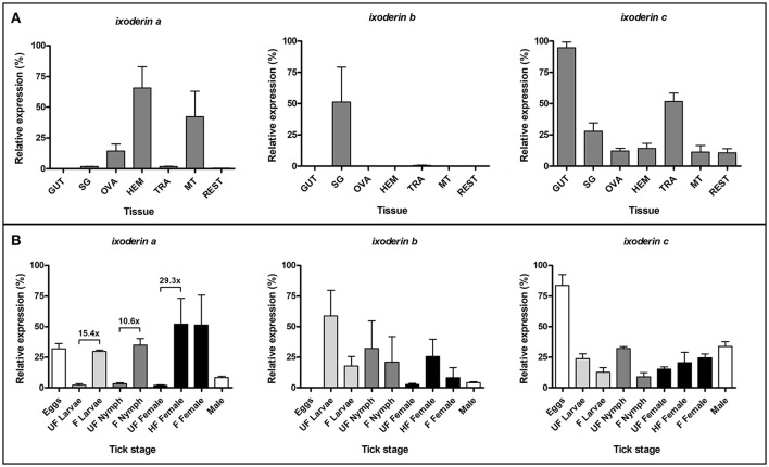
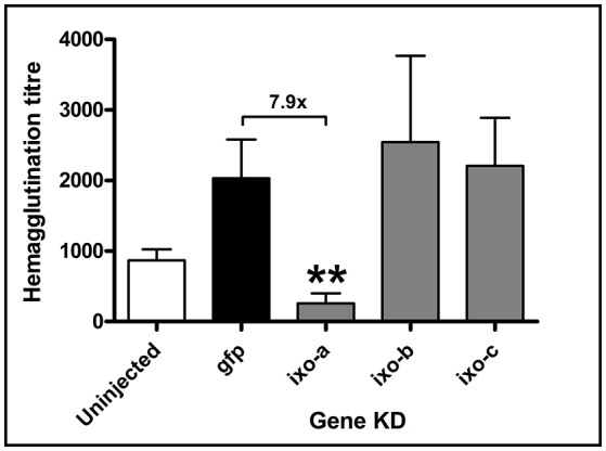
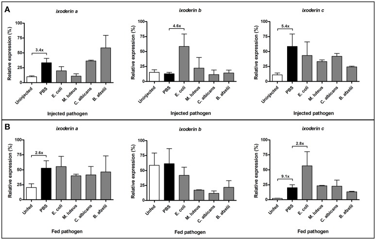
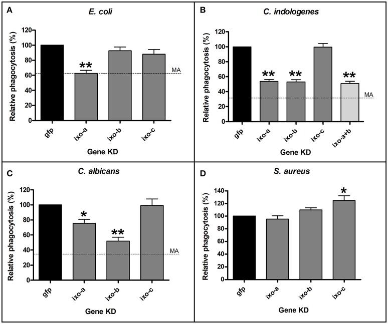
Ticks are important vectors of serious human and animal disease-causing organisms, but their innate immunity can fight invading pathogens and may have the ability to reduce or block transmission to mammalian hosts. Lectins, sugar-binding proteins, can distinguish between self and non-self-oligosaccharide motifs on pathogen surfaces. Although tick hemolymph possesses strong lectin activity, and several lectins have already been isolated and characterized, little is known about the implementation of these molecules in tick immunity. Here, we have described and functionally characterized fibrinogen-related protein (FReP) lectins in Ixodes ticks. We have shown that the FReP family contains at least 27 genes (ixoderins, ixo) that could, based on phylogenetic and expression analyses, be divided into three groups with differing degrees of expansion. By using RNA interference-mediated gene silencing (RNAi) we demonstrated that IXO-A was the main lectin in tick hemolymph. Further, we found that ixoderins were important for phagocytosis of Gram-negative bacteria and yeasts by tick hemocytes and that their expression was upregulated upon injection of microbes, wounding, or after blood feeding. However, although the tick hemocytes could swiftly phagocytose Borrelia afzelii spirochetes, their transmission and burst of infection in mice was not altered. Our results demonstrate that tick ixoderins are crucial immune proteins that work as opsonins in the tick hemolymph, targeting microbes for phagocytosis or lysis.

Keywords: Borrelia; Ixodes; RNAi; complement; fibrinogen-related protein; ixoderin; lectin; tick.


Figures

Figure 1
Figure 1
Tick FRePs cluster into three groups designated as Ixoderin A, B, and C. An unrooted phylogenetic tree of the tick and related invertebrate FReP amino acid sequences, reconstructed using the Neighbor Joining (NJ) method and based on alignment using ClustalX. Full circles indicate genomic ixoderin sequences of I. scapularis. Numbers at branches represent bootstrap support using NJ criterion with 1,000 replicates each. Bar: 0.1 substitutions per site. Ixoderin A I. ricinus: AAQ93650 (AY341424), Ixoderin B I. ricinus: AAV41827 (AY643518), Ixoderin C I. ricinus: GCJO01000224. Alignment and full sequence descriptions are provided as Supplementary Data Sheet 1 and 2.
Figure 2
Figure 2
Ixoderins a, b, and c show distinct tissue and stage-specific expression profiles. qRT-PCR analysis was performed on tissues of semi-engorged I. ricinus females (A) and a mixture of ticks from each developmental stage before and after blood feeding (B). The samples were prepared in three biological replicates. In each graph, cDNA with the highest expression was set as 100% (relative expression). Tick actin and elongation factor were used as housekeeping genes for the tissue and stage-specific profiling, respectively. (GUT) midgut, (SG) salivary glands, (OVA) ovaries, (HEM) hemolymph, (TRA) trachea and fat body, (MT) Malpighian tubules, (REST) rest of the body, (UF) unfed ticks, (HF) half-fed (semi-engorged) adult females fed for 5 days, (F) fully-fed ticks.
Figure 3
Figure 3
Silencing of ixoderin a abolishes lectin activity of the tick hemolymph. The effect of injection (wounding) and ixoderin KDs on the haemagglutination activity of semi-engorged tick hemolymph was measured using 2% mouse RBC. The hemagglutination titer was calculated as hemagglutination activity per 1 μl of tick hemolymph. Hemagglutination titer in the dsGFP group served as an injection control. Two asterisks indicate p-value < 0.001.
Figure 4
Figure 4
Wounding and exposure to microbes alter expression of ixoderins. Gene expression was measured by qRT-PCR 12 hrs after pathogen injection or feeding. The analysis was performed using unfed adult females injected (A) or capillary fed (B) with pathogens. The samples were prepared in three biological replicates. In each graph, cDNA with the highest expression was set as 100% (relative expression). Tick actin was used as a housekeeping gene.
Figure 5
Figure 5
Silencing of ixoderins impairs phagocytosis of microbes by tick hemocytes. Tick hemocytes acquired from the adult semi-engorged females after ixoderin KDs were incubated in vitro with E. coli (A), C. indologenes (B), C. albicans (C), and S. aureus (D). At least five independent slides with hemocytes were analyzed for each of biological triplicates. The level of phagocytosis in the dsGFP control was set as 100%. Dashed lines indicate phagocytosis after methylamine (MA) pre-treatment (no effect on S. aureus). One and two asterisks indicate p-value < 0.05 and <0.001, respectively.
Figure 6
Figure 6
Dual staining is necessary for proper interpretation of the Borrelia phagocytic assay. Hemocytes of adult semi-engorged females were mixed in vitro with B. afzelii CB43. The slides were then incubated with anti-Borrelia primary antibody and stained with Alexa 488 (A). Finally, cell membranes of hemocytes were permeabilized, incubated again with the anti-Borrelia primary antibody, and stained with Alexa 594 and DAPI (B). Borrelia spirochetes localized outside or on the surface of hemocytes are stained green and red, engulfed spirochetes are stained only red. The 488/594 (FITC/Texas red) dual filter can be used for rapid analysis of the slides (C) and can distinguish between phagocytosed (red; black arrow) and non-phagocytosed spirochetes (yellow; white arrow). Full and dashed lines indicate the hemocyte surface before and after permeabilization, respectively.
Figure 7
Figure 7
Pre-treatment of hemolymph with methylamine (MA), but not ixoderin KDs affects phagocytosis of Borrelia. Tick hemocytes after MA pre-treatment (A) or ixoderin KD (B) were incubated in vitro with B. afzelii CB43. At least five independent slides with hemocytes were analyzed for each of three biological replicates. The phagocytic index in (A) was determined as the ratio of phagocytic vs. non-phagocytic hemocytes, and the level of phagocytosis in (B) was set as 100% in the dsGFP control. (Uninjected) untreated hemolymph, (GLY) glycine pre-treatment (control), (MA) MA pre-treatment. Dashed line in (B) indicates phagocytosis level after MA treatment. Two asterisks indicate p-value < 0.001.
Figure 8
Figure 8
Silencing of ixoderins does not affect Borrelia transmission. Transmission of B. afzelii CB43 from naturally infected nymphs to mice was tested after ixoderins triple KD. Five mice were infested with 10 nymphs each in individual groups. (A) Percent of nymphs replete from each mouse. (B) Weights of individual replete nymphs. Dashed line indicates suggested border between female and male nymphs. (C) Number of bacteria (universal primers) in the individual nymphs measured by qRT-PCR (log10 scale). (D) Number of Borrelia in the individual nymphs measured by qRT-PCR (log10 scale). (E) Number of Borrelia in the mice ear biopsies during 4 weeks of infection and (F) number of Borrelia in the destination tissues 4 weeks after infestation measured by qRT-PCR. The number of Borrelia was normalized to 105 mouse genomes.

References

    1. Burešová V., Franta Z., Kopáček P. (2006). A comparison of Chryseobacterium indologenes pathogenicity to the soft tick Ornithodoros moubata and hard tick Ixodes ricinus. J. Invertebr. Pathol. 93, 96–104. 10.1016/j.jip.2006.05.006 - DOI - PubMed
    1. Buresova V., Hajdusek O., Franta Z., Loosova G., Grunclova L., Levashina E. A., et al. . (2011). Functional genomics of tick thioester-containing proteins reveal the ancient origin of the complement system. J. Innate Immun. 3, 623–630. 10.1159/000328851 - DOI - PubMed
    1. Burgdorfer W. (1984). Discovery of the lyme disease spirochete and its relation to tick vectors. Yale J. Biol. Med. 57, 515–520. - PMC - PubMed
    1. Cerenius L., Soderhall K. (2013). Variable immune molecules in invertebrates. J. Exp. Biol. 216, 4313–4319. 10.1242/jeb.085191 - DOI - PubMed
    1. DeFranco A., Locksley R. M., Robertson M. (2007). Immunity: The Immune Response in Infectious and Inflammatory Disease. London: New Science Press.

Publication types

LinkOut - more resources