

Two-dimensional digital photography for child body posture evaluation: standardized technique, reliable parameters and normative data for age 7-10 years
- PMID: 29276784
- PMCID: PMC5738151
- DOI: 10.1186/s13013-017-0146-7
Two-dimensional digital photography for child body posture evaluation: standardized technique, reliable parameters and normative data for age 7-10 years
Abstract
Background: Digital photogrammetry provides measurements of body angles or distances which allow for quantitative posture assessment with or without the use of external markers. It is becoming an increasingly popular tool for the assessment of the musculoskeletal system. The aim of this paper is to present a structured method for the analysis of posture and its changes using a standardized digital photography technique.
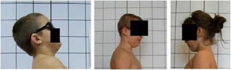
Material and methods: The purpose of the study was twofold. The first one comprised 91 children (44 girls and 47 boys) aged 7-10 (8.2 ± 1.0), i.e., students of primary school, and its aim was to develop the photographic method, choose the quantitative parameters, and determine the intraobserver reliability (repeatability) along with the interobserver reliability (reproducibility) measurements in sagittal plane using digital photography, as well as to compare the Rippstein plurimeter and digital photography measurements. The second one involved 7782 children (3804 girls, 3978 boys) aged 7-10 (8.4 ± 0.5), who underwent digital photography postural screening. The methods consisted in measuring and calculating selected parameters, establishing the normal ranges of photographic parameters, presenting percentile charts, as well as noticing common pitfalls and possible sources of errors in digital photography.
Results: A standardized procedure for the photographic evaluation of child body posture was presented. The photographic measurements revealed very good intra- and inter-rater reliability regarding the five sagittal parameters and good reliability performed against Rippstein plurimeter measurements. The parameters displayed insignificant variability over time. Normative data were calculated based on photographic assessment, while the percentile charts were provided to serve as reference values. The technical errors observed during photogrammetry are carefully discussed in this article.
Conclusions: Technical developments are allowed for the regular use of digital photogrammetry in body posture assessment. Specific child positioning (described above) enables us to avoid incidentally modified posture. Image registration is simple, quick, harmless, and cost-effective. The semi-automatic image analysis, together with the normal values and percentile charts, makes the technique reliable in terms of child's posture documentation and corrective therapy effects' monitoring.
Keywords: Digital photography; Normative data; Percentile charts; Photogrammetry; Primary school children; Standardization.
Conflict of interest statement
All study participants gave written consent, and the study was approved by the Institutional Review Board of Poznan University of Medical Sciences (832/11, date 6/10/2011).Not applicable.The authors declare that they have no competing interests.Springer Nature remains neutral with regard to jurisdictional claims in published maps and institutional affiliations.
Figures


Similar articles
-
Optimization of the Static Posture Evaluation Process Through Digital Processing of Photographic Images.Annu Int Conf IEEE Eng Med Biol Soc. 2021 Nov;2021:1801-1805. doi: 10.1109/EMBC46164.2021.9630704. Annu Int Conf IEEE Eng Med Biol Soc. 2021. PMID: 34891636
-
Intrarater and interrater reliability of photographic measurement of upper-body standing posture of adolescents.J Manipulative Physiol Ther. 2015 Jan;38(1):74-80. doi: 10.1016/j.jmpt.2014.10.009. Epub 2014 Nov 12. J Manipulative Physiol Ther. 2015. PMID: 25467608
-
Reliability of photographic posture analysis of adolescents.J Phys Ther Sci. 2015 Oct;27(10):3123-6. doi: 10.1589/jpts.27.3123. Epub 2015 Oct 30. J Phys Ther Sci. 2015. PMID: 26644658 Free PMC article.
-
Radiography and photogrammetry-based methods of assessing cervical spine posture in the sagittal plane: A systematic review with meta-analysis.Gait Posture. 2021 Feb;84:357-367. doi: 10.1016/j.gaitpost.2020.12.033. Epub 2021 Jan 7. Gait Posture. 2021. PMID: 33465736
-
Photographic analysis of human posture: a literature review.J Bodyw Mov Ther. 2014 Jan;18(1):56-61. doi: 10.1016/j.jbmt.2013.05.008. Epub 2013 Jun 14. J Bodyw Mov Ther. 2014. PMID: 24411150 Review.
Cited by
-
Clinical photography in severe idiopathic scoliosis candidate for surgery: is it a useful tool to differentiate among Lenke patterns?Eur Spine J. 2019 Dec;28(12):3018-3025. doi: 10.1007/s00586-019-06096-w. Epub 2019 Aug 8. Eur Spine J. 2019. PMID: 31396690
-
Regular School Sport versus Dedicated Physical Activities for Body Posture-A Prospective Controlled Study Assessing the Sagittal Plane in 7-10-Year-Old Children.J Clin Med. 2022 Feb 25;11(5):1255. doi: 10.3390/jcm11051255. J Clin Med. 2022. PMID: 35268346 Free PMC article.
-
Validity and Reliability of an Artificial Intelligence-Based Posture Estimation Software for Measuring Cervical and Lower-Limb Alignment Versus Radiographic Imaging.Diagnostics (Basel). 2025 May 26;15(11):1340. doi: 10.3390/diagnostics15111340. Diagnostics (Basel). 2025. PMID: 40506912 Free PMC article.
-
Body Posture and Low Back Pain: Differences between Folk and Ballroom Dancers.Healthcare (Basel). 2024 Jan 8;12(2):137. doi: 10.3390/healthcare12020137. Healthcare (Basel). 2024. PMID: 38255027 Free PMC article.
-
Reliability of a New Digital Tool for Photographic Analysis in Quantifying Body Asymmetry in Scoliosis.J Clin Med. 2024 Apr 5;13(7):2114. doi: 10.3390/jcm13072114. J Clin Med. 2024. PMID: 38610880 Free PMC article.
References
-
- Kendall FP, McCreary EK, Provance PG. Muscles testing and function with posture and pain. 4. USA: Lippincott Williams and Wilkins; 2005.
-
- Kiebzak W, Szmigiel C, Kowalski I, Sliwinski Z. Importance of risk factors in detecting psychomotor development disorders in children during their first year of life. Advances in Rehabilitation. 2008;22:29–33.
-
- Kowalski IM, Protasiewicz-Falowska H. Trunk measurements in the standing and sitting posture according to evidence based medicine (EBM) J Spine Surg. 2013;1:66–79.
-
- Kowalski IM, Protasiewicz-Faldowska H, Siwik P, Zaborowska-Sapeta K, Dabrowska A, Kluszczynski M, Raistenskis J. Analysis of the sagittal plane in standing and sitting position in girls with left lumbar idiopathic scoliosis. Pol Ann Med. 2013;20:30–34. doi: 10.1016/j.poamed.2013.07.001. - DOI
Publication types
LinkOut - more resources
Full Text Sources
Other Literature Sources