. 2018 Mar:195:498-507.
doi: 10.1016/j.chemosphere.2017.12.094. Epub 2017 Dec 15.

Embryonic exposure to Mono(2-ethylhexyl) phthalate (MEHP) disrupts pancreatic organogenesis in zebrafish (Danio rerio)

Affiliations

Embryonic exposure to Mono(2-ethylhexyl) phthalate (MEHP) disrupts pancreatic organogenesis in zebrafish (Danio rerio)

Haydee M Jacobs et al. Chemosphere. 2018 Mar.

Abstract

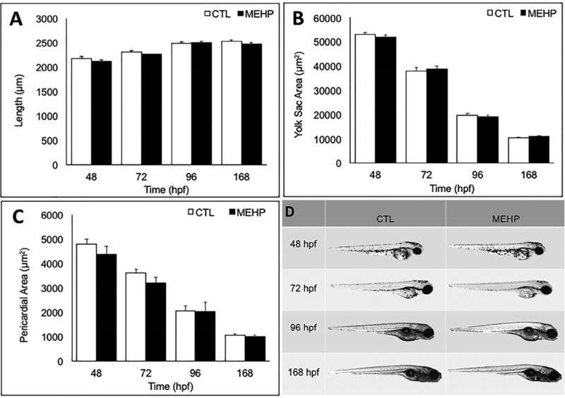
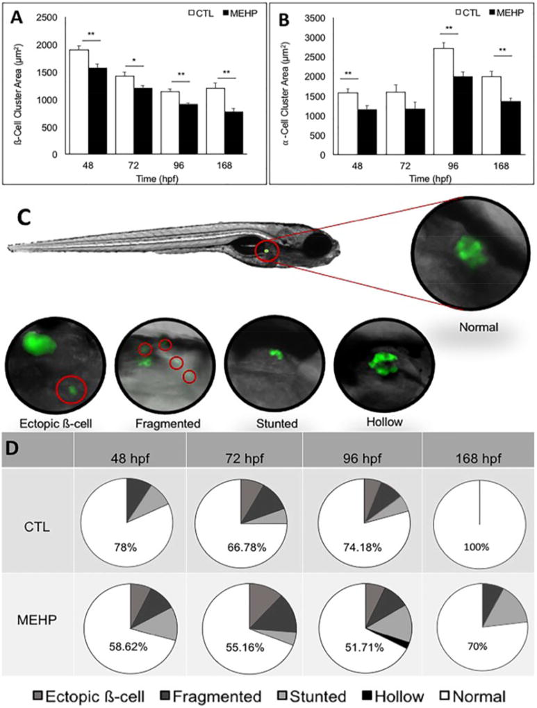
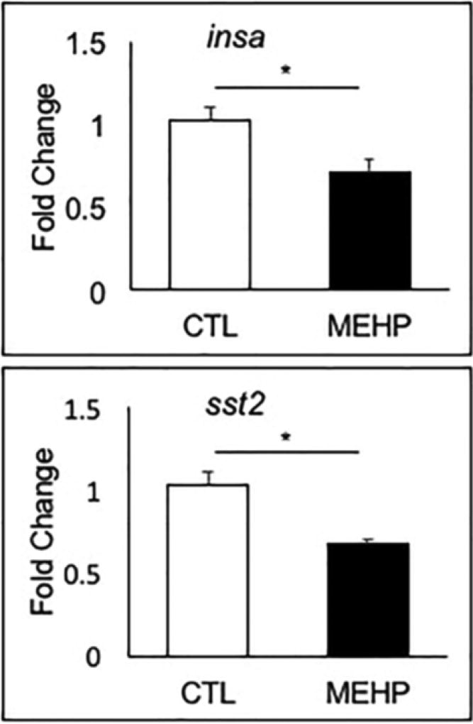
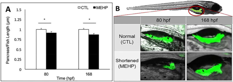
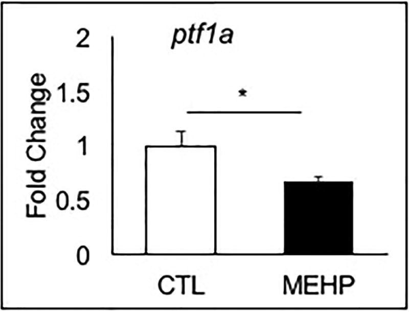
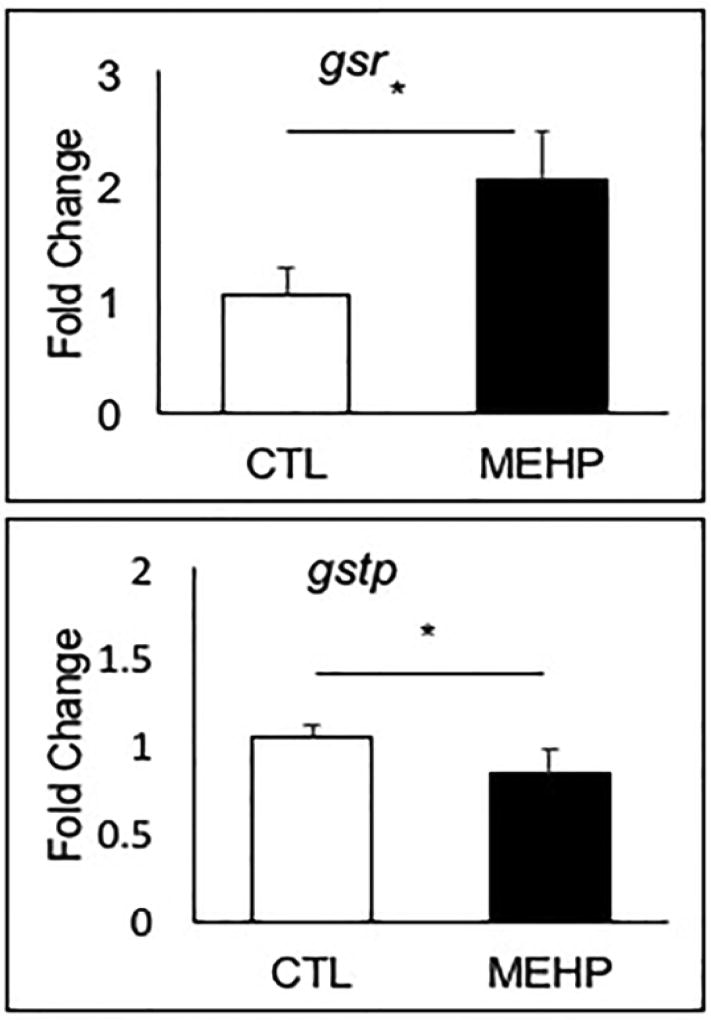
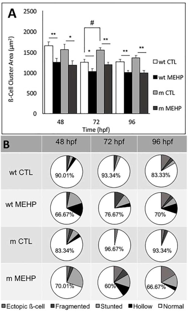
Mono(2-ethylhexyl) phthalate (MEHP) is the bioactive metabolite of di(2-ethylhexyl) phthalate, a plasticizing agent and persistent environmental contaminant associated with obesity, developmental abnormalities, and oxidative stress. Nrf2 (Nfe2l2) is a transcription factor that regulates cytoprotective genes as part of the adaptive antioxidant response. We previously identified the pancreas as a sensitive target of oxidative stress during embryonic development. The goals of this study were to 1) characterize the effects of MEHP exposure on pancreatic development, and 2) determine whether oxidative stress contributes to MEHP embryotoxicity. Zebrafish (Danio rerio) embryos from AB wildtype and Tg(ins:GFP;nrf2afh318/fh318) were exposed to 0 or 200 μg/L MEHP at 3 h post fertilization (hpf) through 168 hpf to assess pancreatic organogenesis. MEHP exposure significantly decreased β-cell area at all timepoints (48, 72, 96, 168 hpf), but Nrf2a did not significantly protect against islet hypomorphism. Tg(gcga:GFP) embryos exposed to MEHP showed a decrease in α-cell area in the islet across the same timepoints. Tg(ptf1a:GFP) embryos were assessed at 80 and 168 hpf for exocrine pancreas length. MEHP exposure decreased growth of the exocrine pancreas. Expression of pancreas genes insa, sst2 and ptf1a was significantly reduced by MEHP exposure compared to controls. Glutathione (GSH) concentrations and redox potentials were quantified at 72 hpf by HPLC, but no significant changes were observed. However, expression of the GSH-related genes gstp1 and gsr were significantly altered by MEHP exposure. These data indicate that the developing pancreas is a sensitive target tissue of embryonic exposure to MEHP.

Keywords: Endocrine; Exocrine; Islet; Nfe2l2; Redox; Toxicology.


Figures

Figure 1
Figure 1
Embryonic exposure to 200 µg/L MEHP does not alter gross morphology from 48–168 hpf. A) Length (rostral to caudal), B) yolk sac area, and C) pericardial sac area were measured at 48, 72, 96, and 168 hpf using Zen analysis software with a 2× objective. No significant differences in length, yolk sac area, pericardial area, or gross deformities were detected between exposed or control eleutheroembryos and larvae at any time point examined (n=30 fish; p>0.05).
Figure 2
Figure 2
Embryonic MEHP exposure significantly decreases pancreatic islet area compared to controls at 48, 72, 96, and 168 hpf, and significantly increases the incidence of pancreatic islet variants. A) Pancreatic ß-cell cluster area was visualized using Tg(ins:GFP) zebrafish eleutheroembryos and larvae following treatment with MEHP or DMSO, and islet area was measured using Zen analysis software. B) Pancreatic α-cell cluster area was visualized in Tg(gcga:GFP) zebrafish eleutheroembryos and larvae following treatment with MEHP or DMSO and measured using Zen analysis software. Islet architechture was assessed using Tg(ins:GFP) zebrafish eleutheroembryos and larvae. Morphologies that deviated from the normal spherical islet shape were categorized as fragmented, stunted, hollow, or having ectopic ß-cells. Variants were quantified across three study replicates. C) The position of the endocrine islet is shown on a 96 hpf eleutheroembryo, as well as representative images of the islet variants observed. D) Pie charts show the distribution of each variant, and numbers shown are the percentage of normal islets at each timepoint (n=40 for 48, 72, 96; n=20 for 168 hpf; *p<0.05; **p<0.005).
Figure 3
Figure 3
Embryonic MEHP exposure reduced expression of pancreatic hormone genes insa and sst2. AB zebrafish were exposed to MEHP from 3–96 hpf, and gene expression was assessed at 96 hpf. Data are presented as mean fold changes normalized to the biweight mean center of bactin and b2m expression ± SEM (n=6; *p<0.05; **p<0.005).
Figure 4
Figure 4
Embryonic MEHP exposure shortens the ratio of exocrine pancreas to total fish length at 80 and 168 hpf. Exocrine pancreas morphology was assessed using Tg(ptf1a:GFP) zebrafish eleutheroembryos and larvae. A) The position of the exocrine pancreas is shown, as well as representative images of shortened pancreata observed. B) Pancreas length was measured using Zen analysis software, from the center of the primary endocrine islet to the tail of the pancreas. Exocrine pancreas length was normalized to total fish length. Data presented are mean ratios of pancreas to fish length ± SEM (n=30; *p<0.05).
Figure 5
Figure 5
Embryonic MEHP exposure significantly reduces expression of ptf1a at 96 hpf. AB zebrafish were exposed to MEHP from 3–96 hpf, and gene expression was assessed at 96 hpf. Data are presented as mean fold change normalized to the biweight mean center of bactin and b2m expression ± SEM (n=6; *p<0.05).
Figure 6
Figure 6
Embryonic MEHP exposure significantly altered the expression of gstp1 and gsr. AB zebrafish were exposed to MEHP from 3–96 hpf, and gene expression was assessed at 96 hpf. Data are presented as mean fold changes normalized to the biweight mean center of bactin and b2m expression ± SEM (n=6; *p<0.05).
Figure 7
Figure 7
Embryonic MEHP exposure alters pancreatic structure in wild type (wt) and Nrf2afh318/fh318 mutant (m) zebrafish embryos. results in A) significantly reduced ß-cell cluster area, and B) significantly increased incidence of pancreatic islet variants at all time points observed. Data are presented as the mean ± SEM. Asterisks (*) indicate statistical significance between control and exposed within genotype; hash marks (#) indicate significant differences between controls across genotype (n=30; *p<0.05; **p<0.005; #p<0.05).

Similar articles

Cited by

References

    1. Agabi JO, Akhigbe AO. Comparative sonographic evaluation of the anteroposterior dimensions of the pancreas in diabetics and nondiabetics. Niger J Clin Pract. 2016;19:175–81. - PubMed
    1. Akirav E, Kushner JA, Herold KC. Beta-cell mass and type 1 diabetes: going, going, gone? Diabetes. 2008;57:2883–8. - PMC - PubMed
    1. Benjamin S, Masai E, Kamimura N, Takahashi K, Anderson RC, Faisal PA. Phthalates impact human health: Epidemiological evidences and plausible mechanism of action. J Hazard Mater. 2017;340:360–383. - PubMed
    1. Bento A, Baptista H, Oliveira F. Congenital pancreas malformations: a clinical case report. Rev Assoc Med Bras (1992) 2013;59:35–9. - PubMed
    1. Corbasson I, Hankinson SE, Stanek EJ, Reeves KW., 3 Urinary bisphenol-A, phthalate metabolites and body composition in US adults, NHANES 1999–2006. Int J Environ Health Res. 2016;26:606–17. - PubMed

MeSH terms