

Development of fluorescence imaging probes for nicotinic acetylcholine α4β2∗ receptors
- PMID: 29277457
- PMCID: PMC6004331
- DOI: 10.1016/j.bmcl.2017.12.036
Development of fluorescence imaging probes for nicotinic acetylcholine α4β2∗ receptors
Abstract
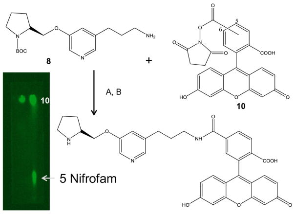
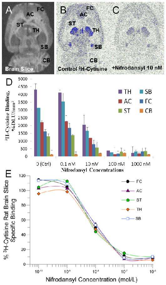
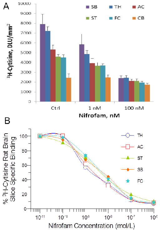
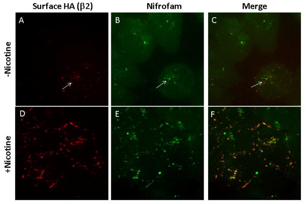
Nicotinic acetylcholine α4β2∗ receptors (nAChRs) are implicated in various neurodegenerative diseases and smoking addiction. Imaging of brain high-affinity α4β2∗ nAChRs at the cellular and subcellular levels would greatly enhance our understanding of their functional role. Since better resolution could be achieved with fluorescent probes, using our previously developed positron emission tomography (PET) imaging agent [18F]nifrolidine, we report here design, synthesis and evaluation of two fluorescent probes, nifrodansyl and nifrofam for imaging α4β2∗ nAChRs. The nifrodansyl and nifrofam exhibited nanomolar affinities for the α4β2∗ nAChRs in [3H]cytisine-radiolabeled rat brain slices. Nifrofam labeling was observed in α4β2∗ nAChR-expressing HEK cells and was upregulated by nicotine exposure. Nifrofam co-labeled cell-surface α4β2∗ nAChRs, labeled with antibodies specific for a β2 subunit extracellular epitope indicating that nifrofam labels α4β2∗ nAChR high-affinity binding sites. Mouse brain slices exhibited discrete binding of nifrofam in the auditory cortex showing promise for examining cellular distribution of α4β2∗ nAChRs in brain regions.
Keywords: Fluorescence imaging; Nicotine; Nifene; Nifrodansyl; Nifrofam; Nifrolidine.
Copyright © 2017 Elsevier Ltd. All rights reserved.
Conflict of interest statement
The authors have no conflict of interest.
Figures


References
-
- Pichika R, Easwaramoorthy B, Collins D, Christian BT, Shi B, Narayanan TK, Potkin SG, Mukherjee J. Nucl Med Biol. 2006;33:295–304. - PubMed
Publication types
MeSH terms
Substances
Grants and funding
LinkOut - more resources
Full Text Sources
Other Literature Sources
Miscellaneous
