Review
. 2018 Oct:71-72:28-39.
doi: 10.1016/j.matbio.2017.12.011. Epub 2017 Dec 24.

The pathogenic roles of heparan sulfate deficiency in hereditary multiple exostoses

Affiliations
Review

The pathogenic roles of heparan sulfate deficiency in hereditary multiple exostoses

Maurizio Pacifici. Matrix Biol. 2018 Oct.

Abstract

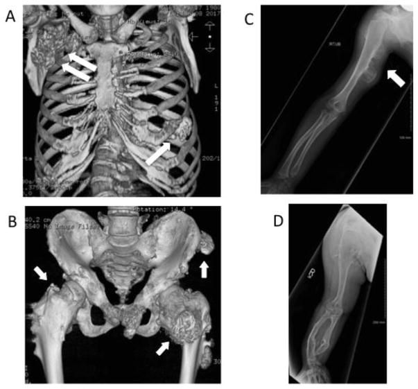
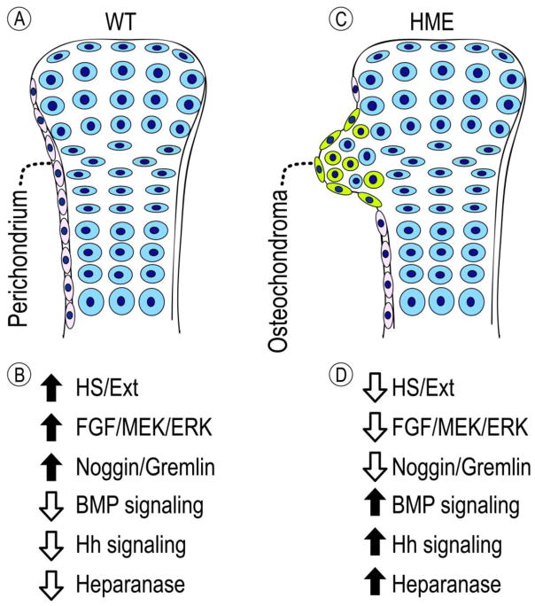
Heparan sulfate (HS) is an essential component of cell surface and matrix proteoglycans (HS-PGs) that include syndecans and perlecan. Because of their unique structural features, the HS chains are able to specifically interact with signaling proteins -including bone morphogenetic proteins (BMPs)- via their HS-binding domain, regulating protein availability, distribution and action on target cells. Hereditary Multiple Exostoses (HME) is a rare pediatric disorder linked to germline heterozygous loss-of-function mutations in EXT1 or EXT2 that encode Golgi-resident glycosyltransferases responsible for HS synthesis, resulting in a systemic HS deficiency. HME is characterized by cartilaginous/bony tumors -called osteochondromas or exostoses- that form within perichondrium in long bones, ribs and other elements. This review examines most recent studies in HME, framing them in the context of classic studies. New findings show that the spectrum of EXT mutations is larger than previously realized and the clinical complications of HME extend beyond the skeleton. Osteochondroma development requires a somatic "second hit" that would complement the germline EXT mutation to further decrease HS production and/levels at perichondrial sites of osteochondroma induction. Cellular studies have shown that the steep decreases in local HS levels: derange the normal homeostatic signaling pathways keeping perichondrium mesenchymal; cause excessive BMP signaling; and provoke ectopic chondrogenesis and osteochondroma formation. Data from HME mouse models have revealed that systemic treatment with a BMP signaling antagonist markedly reduces osteochondroma formation. In sum, recent studies have provided major new insights into the molecular and cellular pathogenesis of HME and the roles played by HS deficiency. These new insights have led to the first ever proof-of-principle demonstration that osteochondroma formation is a druggable process, paving the way toward the creation of a clinically-relevant treatment.

Keywords: Drug treatment; EXT1; EXT2; Heparan sulfate; Heparan sulfate proteoglycans; Hereditary multiple Exostoses; Signaling proteins and pathways.


Figures

Fig. 1
Fig. 1
Diagnostic CT and X-ray images obtained from HME patients. (A) This CT scan depicts the rib cage of a 37 yr-old HME patient exhibiting multiple osteochondromas along the ribs (arrow) and a large malignant chondrosarcoma Grade 1 on the upper right area near the clavicle (double arrow). The osteochondromas likely impinged on mechanical rib function, and the malignant tumor was eventually resected by surgery. (B) This CT scan shows the pelvis of a 23 yr-old patient exhibiting multiple osteochondromas along the upper and lower iliac crest (arrows), pubic ramus and femurs. The osteochondromas appear to fuse the pubic symphysis and their aggressive presence and size affected the left hip joint (arrow). (C) This X-ray image is from an 8 yr-old patient and reveals two large osteochondromas in posterior medial portion of humerus (arrow), a small osteochondroma in distal radius, and bending of radius toward ulna. (D) This X-ray image illustrates the right arm from a 20 yr-old patient displaying a marked degree of bending of radius and ulna. The severe shortening of distal ulna created an ulna-minus wrist. The bulkiest osteochondromas are located in the distal shaft of radius and ulna and are present also in scapula and humerus. All images were generously provided by the MHE Center at the Paley Orthopedic & Spine Institute, West Palm Beach, Florida.
Fig. 2
Fig. 2
Schematic illustrating possible changes in regulatory steps that underlie osteochondroma formation. (A) In healthy wild-type (WT) circumstances, perichondrium (in off-white) would define the boundary of growth plate (in blue) in skeletal elements such as long bones. Resident perichondrial cells would be characterized by a normal mesenchymal phenotype and express traits including: a flat cell morphology; normal EXT expression and HS levels; strong anti-chondrogenic mechanisms, including FGF and ERK/MEK signaling and Noggin and Gremlin expression [98]; and low activity/expression of pro-chondrogenic mechanisms including BMP and hedgehog signaling and heparanase. (B) Up and down arrows depict the high and low levels of respective traits in normal perichondrium. (C) During the course of HME, LOH or other “second hits” would cause a steep and nearly complete loss of EXT expression and/or HS levels in local cells (in green) within perichondrium bordering the heterozygous EXT mutant growth plate. This would cause concerted decreases in anti-chondrogenic pathways and reciprocal increases in pro-chondrogenic pathways and heparanase expression in mutant cells (summarized in D), thus altering homeostatic signaling mechanisms and triggering differentiation of perichondria progenitors into round-shaped chondrocytes (in green). The growing osteochondromas would contain a mixture of mutant (green) and heterozygous (blue) cells, the latter being recruited into the osteochondroma forming process by mutant cells. The changes occurring in BMP, hedgehog and FGF signaling pathways and in expression of heparanase could each offer plausible therapeutic targets to inhibit osteochondroma inception and/or growth.

Similar articles

Cited by

References

    1. Bishop JR, Schuksz M, Esko JD. Heparan sulphate proteoglycans fine-tune mammalian physiology. nature. 2007;446:1030–1037. - PubMed
    1. Lamanna WC, Kalus I, Pavda M, Baldwin RJ, Merry CLR, Dierks T. The heparanome - The enigma of encoding and decoding heparan sulfate sulfation. J Biotechnology. 2007;129:290–307. - PubMed
    1. Sarrazin S, Lamanna WC, Esko JD. Heparan sulfate proteoglycans. Cold Spring Harb Prospect Biol. 2011;3:a004952. - PMC - PubMed
    1. Iozzo RV, Schaefer L. Proteoglycan form and function: a comprehensive nomenclature for proteoglycans. Matrix Biol. 2015;42:11–55. - PMC - PubMed
    1. Dhoot GK, Gustafsson MK, Ai X, Sun w, Standiford DM, Emerson CP. Regulation of Wnt signaling and embryo patterning by an extracellular sulfatase. Science. 2001;293:1663–1666. - PubMed

Publication types

LinkOut - more resources