

The Emerging Relationship Between Interstitial Fluid-Cerebrospinal Fluid Exchange, Amyloid-β, and Sleep
- PMID: 29279202
- PMCID: PMC5767516
- DOI: 10.1016/j.biopsych.2017.11.031
The Emerging Relationship Between Interstitial Fluid-Cerebrospinal Fluid Exchange, Amyloid-β, and Sleep
Abstract
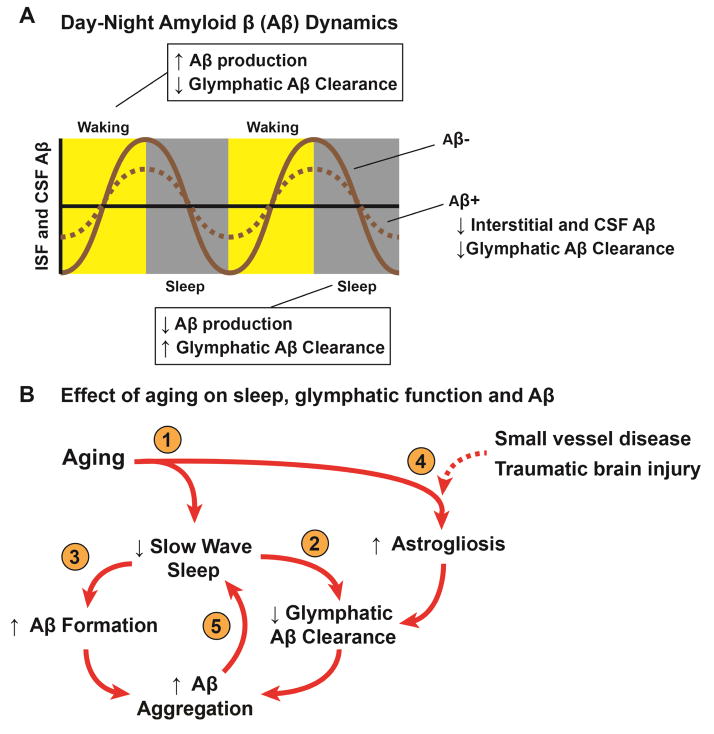
Amyloid-β (Aβ) plaques are a key histopathological hallmark of Alzheimer's disease (AD), and soluble Aβ species are believed to play an important role in the clinical development of this disease. Emerging biomarker data demonstrate that Aβ plaque deposition begins decades before the onset of clinical symptoms, suggesting that understanding the biological determinants of the earliest steps in the development of AD pathology may provide key opportunities for AD treatment and prevention. Although a clinical association between sleep disruption and AD has long been appreciated, emerging clinical studies and insights from the basic neurosciences have shed important new light on how sleep and Aβ homeostasis may be connected in the setting of AD. Aβ, like many interstitial solutes, is cleared in part through the exchange of brain interstitial fluid and cerebrospinal fluid along a brain-wide network of perivascular pathways recently termed the glymphatic system. Glymphatic function is primarily a feature of the sleeping brain, rather than the waking brain, and is slowed in the aging and posttraumatic brain. These changes may underlie the diurnal fluctuations in interstitial and cerebrospinal fluid Aβ levels observed in both the rodent and the human. These and other emerging studies suggest that age-related sleep disruption may be one key factor that renders the aging brain vulnerable to Aβ deposition and the development of AD. If this is true, sleep may represent a key modifiable risk factor or therapeutic target in the preclinical phases of AD.
Keywords: Alzheimer's; Aquaporin-4; Astrocytes; CSF; Cerebrospinal fluid; Glymphatic; Interstitial fluid; Perivascular; Sleep.
Copyright © 2017 Society of Biological Psychiatry. Published by Elsevier Inc. All rights reserved.
Figures
References
-
- Sperling RA, Aisen PS, Beckett LA, Bennett DA, Craft S, Fagan AM, et al. Toward defining the preclinical stages of Alzheimer’s disease: Recommendations from the National Institute on Aging-Alzheimer’s Association workgroups on diagnostic guidelines for Alzheimer’s disease. Alzheimer’s & Dementia. 2011;7:280–292. - PMC - PubMed
Publication types
MeSH terms
Substances
Grants and funding
LinkOut - more resources
Full Text Sources
Other Literature Sources
Medical
