. 2018 Feb 2;122(3):457-472.
doi: 10.1161/CIRCRESAHA.117.311983. Epub 2017 Dec 26.

Inositol Polyphosphate Multikinase Inhibits Angiogenesis via Inositol Pentakisphosphate-Induced HIF-1α Degradation

Affiliations

Inositol Polyphosphate Multikinase Inhibits Angiogenesis via Inositol Pentakisphosphate-Induced HIF-1α Degradation

Chenglai Fu et al. Circ Res. .

Abstract

Rationale: Inositol polyphosphate multikinase (IPMK) and its major product inositol pentakisphosphate (IP5) regulate a variety of cellular functions, but their role in vascular biology remains unexplored.

Objective: We have investigated the role of IPMK in regulating angiogenesis.

Methods and results: Deletion of IPMK in fibroblasts induces angiogenesis in both in vitro and in vivo models. IPMK deletion elicits a substantial increase of VEGF (vascular endothelial growth factor), which mediates the regulation of angiogenesis by IPMK. The regulation of VEGF by IPMK requires its catalytic activity. IPMK is predominantly nuclear and regulates gene transcription. However, IPMK does not apparently serve as a transcription factor for VEGF. HIF (hypoxia-inducible factor)-1α is a major determinant of angiogenesis and induces VEGF transcription. IPMK deletion elicits a major enrichment of HIF-1α protein and thus VEGF. HIF-1α is constitutively ubiquitinated by pVHL (von Hippel-Lindau protein) followed by proteasomal degradation under normal conditions. However, HIF-1α is not recognized and ubiquitinated by pVHL in IPMK KO (knockout) cells. IP5 reinstates the interaction of HIF-1α and pVHL. HIF-1α prolyl hydroxylation, which is prerequisite for pVHL recognition, is interrupted in IPMK-deleted cells. IP5 promotes HIF-1α prolyl hydroxylation and thus pVHL-dependent degradation of HIF-1α. Deletion of IPMK in mouse brain increases HIF-1α/VEGF levels and vascularization. The increased VEGF in IPMK KO disrupts blood-brain barrier and enhances brain blood vessel permeability.

Conclusions: IPMK, via its product IP5, negatively regulates angiogenesis by inhibiting VEGF expression. IP5 acts by enhancing HIF-1α hydroxylation and thus pVHL-dependent degradation of HIF-1α.

Keywords: Egln1 protein; blood–brain barrier; hydroxylation; inositol phosphates; vascular endothelial growth factor A; von Hippel-Lindau protein.


Figures

Figure 1
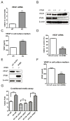
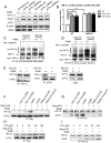
Figure 1. IPMK physiologically inhibits VEGF gene expression
A, The gene expression level of VEGF is significantly increased in IPMK deleted MEFs, analyzed by real time PCR. ***P < 0.001 by unpaired Student’s t test (n = 4, mean ± s.e.m.). B, Western blot shows VEGF protein level is much higher in IPMK KO cells. C, VEGF protein level is substantial increased in the cell culture medium of IPMK KO MEFs, assessed by ELISA. ***P < 0.001 by unpaired Student’s t test (n = 4, mean ± s.e.m.). D, Effect of IPMK rescue in IPMK KO MEFs on VEGF mRNA levels. ***P < 0.001 by unpaired Student’s t test (n = 4, mean ± s.e.m.). E, IPMK rescue in IPMK KO MEFs reduces the expression of VEGF protein, evaluated by Western blot. F, Re-expression of IPMK in IPMK deleted MEFs decreases VEGF protein in cell-culture medium, monitored by ELISA. ***P < 0.001 by unpaired Student’s t test (n=4, means±s.e.m.). G, Antibody neutralization of VEGF but not PDGF abolishes the IPMK deletion-induced angiogenesis in the in vitro Matrigel assay. ***P < 0.001 by unpaired Student’s t test; ns, no significant difference (n = 4, mean ± s.e.m.).
Figure 2
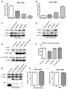
Figure 2. Negative regulation of VEGF expression requires IPMK kinase activity
A, IPMK knockdown in HEK293 cells by shRNA was analyzed by RT-qPCR. ***P < 0.001 by unpaired Student’s t test (n = 4, mean ± s.e.m.). B, Depletion of IPMK by shRNA increases VEGF mRNA levels in HEK293 cells as determined by RT-qPCR. ***P < 0.001 by unpaired Student’s t test (n = 4, mean ± s.e.m.). C, Depletion of IPMK by shRNA in HEK293 cells increases VEGF protein levels. D, Depletion of IPMK by shRNA in human fibroblasts increases VEGF protein levels. E, Re-expression of wild type IPMK but not kinase deficient mutants (IPMK KA or IPMK KA/SA) decreases VEGF protein levels in IPMK KO (-/-) cells. F, Re-expression of WT IPMK but not catalytically inactive IPMK (IPMK KA or IPMK KA/SA) reduces VEGF protein levels in the cell culture medium, as determined by ELISA. ***P < 0.001 by unpaired Student’s t test (n = 4, mean ± s.e.m.). G, Overexpressing WT or IPMK lacking nuclear localization signal (IPMK ΔNLS) or kinase activity (KA) in HEK293 cells does not decrease VEGF protein levels. H, Overexpression of IPMK in wild type human fibroblasts does not decrease VEGF mRNA levels. I, Overexpression of IPMK in wild type MEFs does not decrease VEGF mRNA levels.
Figure 3
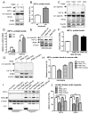
Figure 3. IPMK deletion causes HIF-1α protein accumulation
A and B, Western blot shows increased HIF-1α protein levels in IPMK KO MEFs under normoxic conditions. ***P < 0.001 by unpaired Student’s t test (n = 3, mean ± s.e.m.). C and D, Western blot reveals increased HIF-1α levels in IPMK KO MEFs both under normoxic (norm) and hypoxic (hypo) conditions. E and F, Knockdown of IPMK expression in HEK293 cells increases HIF-1α protein levels. G and H, Re-expression of wild type IPMK but not kinase deficient mutants (IPMK KA or IPMK KA/SA) in IPMK KO MEFs decreases HIF-1α protein levels. ***P < 0.001 by unpaired Student’s t test (n = 3, mean ± s.e.m.). I and J, Overexpressing WT IPMK or IPMK lacking the nuclear localization signal (ΔNLS), but not kinase deficient IPMK (KA), in HEK293 cells decreases hypoxia-induced HIF-1α and HIF-2α protein levels.
Figure 4
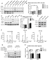
Figure 4. IP5 decreases HIF-1α protein levels
A and B, Depletion of HIF-1α by shRNA abrogates the upregulation of VEGF in IPMK KO MEFs. C and D, Inhibition of HIF-1α by 10 μM 2-methoxyestradiol (2ME2) or PX-478 abolishes the upregulation of VEGF in IPMK KO MEFs. E, Knockdown of IP5K expression in HEK293 cells by shRNA does not increase HIF-1α protein levels. F, Knockdown of IP5K expression in HEK293 cells decreases IP6 and increases IP5 levels. G and H, Depletion of IP5K in HEK293 cells decreases hypoxia-induced HIF-1α and HIF-2α protein levels.
Figure 5
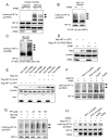
Figure 5. IP5 is critical for HIF-1α ubiquitination
A and B, Depletion of pVHL by shRNA increases HIF-1α protein levels in WT MEFs but fails to augment HIF-1α protein levels in IPMK KO cells. C, HIF-1α was immunoprecipitated from the lysates of whole cells that were exposed to either hypoxia (1% O2) or MG132 (10 μM) for 4 hours. Ubiquitination of HIF-1α is markedly diminished in IPMK KO MEFs. D, HIF-1α was immunoprecipitated from the nuclear fraction of MEFs that were exposed to either hypoxia (1% O2) or MG132 (10 μM) for 4 hours. HIF-1α ubiquitination is substantially decreased in IPMK KO MEFs. E, MEFs were treated with 10 μM MG132 for 4 hours; HIF-1α and pVHL were then immunoprecipitated. The physical association of pVHL and HIF-1α is disrupted in IPMK KO cells. F, Flag-pVHL was immunoprecipitated from lysates of IPMK KO cells that were treated with 50 μM Ins(1,3,4,5)P4, Ins(1,4,5,6)P4, IP5, IP6, PtdIns(4,5)P2 or PtdIns(3,4,5)P3. The interaction of pVHL and HIF-1α is rescued by IP5. G, HIF-1α was immunoprecipited from the same lysates described in (F). IP5 increases the binding of pVHL with HIF-1α. H, Flag-pVHL was immunoprecipitated from IPMK KO cell lysates to which were added different concentrations of IP5. IP5 restores the association of HIF-1α with pVHL. I, HIF-1α was immunoprecipitated from the same lysates described in (H). IP5 restores the association of HIF-1α with pVHL.
Figure 6
Figure 6. IP5 promotes HIF-1α prolyl hydroxylation
A, WT and IPMK KO MEFs were exposed to either hypoxia (1% O2) or MG132 (10 μM) for 4 hours and nuclear fractions were isolated. IPMK deletion elicits less hydroxylation of HIF-1α at proline-564 (arrows). B, WT and IPMK KO MEFs were treated with MG132 (10 μM for 4 hours), HIF-1α was then immunoprecipitated and blotted with anti-hydroxy-HIF-1α (Pro-564) antibody. IPMK deletion reveals diminished HIF-1α hydroxylation (arrows). C, IPMK or GFP was overexpressed in HEK293 cells and the cells were treated with MG132 (10 μM for 4 hours). HIF-1α was immunoprecipitated and blotted with anti-hydroxy-HIF-1α (pro564) antibody. Overexpression of IPMK increases HIF-1α prolyl hydroxylation (arrows). D, WT or hydroxylation site mutant (P402/564A) HIF-1α was overexpressed in MEFs. A substantial amount of WT HIF-1α is accumulated in IPMK deleted but not wild type cells. The difference of HIF-1α protein levels in WT and IPMK KO cells is abolished when HIF-1α prolyl hydroxylation sites are mutated. E, IPMK or GFP was overexpressed in HEK293 cells, which co-expressed either WT or hydroxylation site mutant (P402/564A) HIF-1α. The cells were exposed to hypoxia (1% O2 for 4 hours). Western blot shows overexpressing IPMK decreases levels of hypoxia-induced WT but not mutant HIF-1α. F, IPMK KO cells were treated with 10 μM MG132 together with 50 μM Ins(1,3,4,5)P4, Ins(1,4,5,6)P4, IP5 or IP6 for 4 hours. Western blot shows IP5 increases HIF-1α hydroxylation at proline-564 (arrows). G, IPMK KO cells were treated with 10 μM MG132 together with different concentrations of IP5 for 4 hours. Western blot shows IP5 increases HIF-1α hydroxylation (pro564) at 20–50 μM (arrows). H, PHD2 and flag-HIF-1α were overexpressed and purified from IPMK KO MEFs, then applied to an in vitro hydroxylation assay. Twenty μM of Ins(1,3,4,5)P4, Ins(1,4,5,6)P4, IP5 or IP6 were added to reactions. IP5 substantially increases HIF-1α hydroxylation.
Figure 7
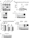
Figure 7. Hypoxia decreases IPMK/IP5 levels
A, PHD2 was knocked down by shRNA transduction in IPMK KO and WT MEFs. Knocking down PHD2 increases HIF-1α levels in WT but not IPMK KO cells. B,GST-PHD2 or GST-GFP was overexpressed in HEK293 cells. The cells were then labeled with [3H]-inositol. PHD2 or GFP was pulled down by glutathione agarose beads. [3H]-inositol phosphates that bind with PHD2 or GFP were detected by HPLC. C, HEK293 cells were exposed to hypoxia (1% for 4 hours). Western blot shows a substantial loss of IPMK protein levels in hypoxia treated cells. D, HEK293 cells were exposed to hypoxia (1%) for different times. Western blot shows that hypoxia decreases IPMK protein levels. E, HEK293 cells were labeled with [3H]-inositol and exposed to hypoxia (1% for 4 hours). The intracellular inositol phosphates were measured by HPLC. Hypoxia treatment significantly reduces intracellular IP5 levels. F, HIF-1α or HIF-2α was knocked down in HEK 293 cells. The cells were then exposed to hypoxia (1% for 4 hours). Western blot shows that knocking down HIF-1α or HIF-2α does not prevent hypoxia-induced IPMK degradation. G, HEK 293 cells were treated with cycloheximide for different times. Western blots show that IPMK protein level does not decrease with 8 hours of cycloheximide treatment.
Figure 8
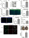
Figure 8. Increased HIF-1α /VEGF expression and blood vessel densities in IPMK KO mouse brain
A, A significantly increased amount of VEGF is present in the blood of IPMK KO mice compared to controls. ** P<0.01 by unpaired Student’s t test (n = 4, male, 6-week old). B, HIF-1α and VEGF protein levels are increased in cerebral cortex of IPMK KO mice (male, 6-week old). C, The gene expression level of angiopoietin 2 (ANGPT2) is substantially increased in IPMK KO brains. D, The gene expression level of erythropoietin (EPO) is markedly increased in IPMK KO brains. E and F, Immunostaining of blood vessels in cerebral cortex with anti-CD31 antibody. Vascular density increases two-fold in IPMK KO mice. Scale bar 100 μm. *** P<0.001 by unpaired Student’s t test (n = 4, male, 6-week old). G and H, Immunostaining of blood vessels in cerebellum with anti-CD31 antibody. Dramatically higher vascular densities are seen in IPMK deleted cerebellum. Scale bar 50μm. Twelve randomly chosen fields (360 × 360 μm) from 4 non-adjacent sections, were analyzed per mouse.** P<0.01 by unpaired Student’s t test (n = 4, male, 6-week old). I and J, Immunostaining of CD31 (blood vessels) and PDGFRβ (pericytes) on IPMK KO and WT mouse brain. The pericyte coverage of brain blood vessels is substantially decreased in IPMK KO mice. (n=3, male, 6-week old). K, Evans blue (2% in saline, 80 μL) was injected intraperitoneally to IPMK KO and WT mice. The animals were euthanized and perfused 2 hours later. IPMK KO brains were stained by Evans blue, indicating leaky blood vessels. (n=3, male, 6-week old). Evans Blue was measured in the cortex. ** P<0.01 by unpaired Student’s t test.

Similar articles

Cited by

References

    1. Monserrate JP, York JD. Inositol phosphate synthesis and the nuclear processes they affect. Curr Opin Cell Biol. 2010;22(3):365–373. - PubMed
    1. Frederick JP, Mattiske D, Wofford JA, et al. An essential role for an inositol polyphosphate multikinase, Ipk2, in mouse embryogenesis and second messenger production. Proc Natl Acad Sci U S A. 2005;102(24):8454–8459. - PMC - PubMed
    1. Seeds AM, Tsui MM, Sunu C, Spana EP, York JD. Inositol phosphate kinase 2 is required for imaginal disc development in Drosophila. Proc Natl Acad Sci U S A. 2015;112(51):15660–15665. - PMC - PubMed
    1. Nalaskowski MM, Deschermeier C, Fanick W, Mayr GW. The human homologue of yeast ArgRIII protein is an inositol phosphate multikinase with predominantly nuclear localization. Biochem J. 2002;366(Pt 2):549–556. - PMC - PubMed
    1. Resnick AC, Snowman AM, Kang BN, Hurt KJ, Snyder SH, Saiardi A. Inositol polyphosphate multikinase is a nuclear PI3-kinase with transcriptional regulatory activity. Proc Natl Acad Sci U S A. 2005;102(36):12783–12788. - PMC - PubMed

Publication types

MeSH terms

Substances