. 2018 Jan 31;38(5):1218-1231.
doi: 10.1523/JNEUROSCI.1896-17.2017. Epub 2017 Dec 26.

Conditional Deletion of Hippocampal CA2/CA3a Oxytocin Receptors Impairs the Persistence of Long-Term Social Recognition Memory in Mice

Affiliations

Conditional Deletion of Hippocampal CA2/CA3a Oxytocin Receptors Impairs the Persistence of Long-Term Social Recognition Memory in Mice

Yu-Ting Lin et al. J Neurosci. .

Abstract

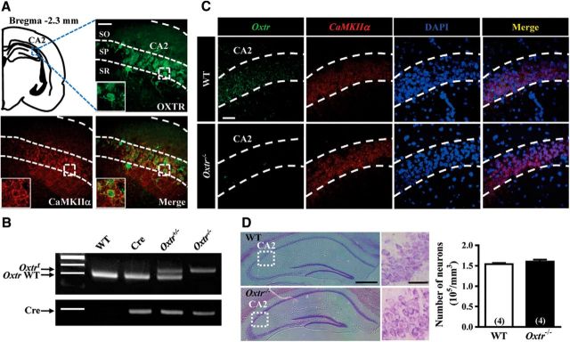
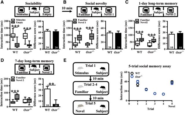
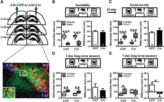
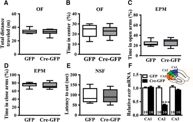
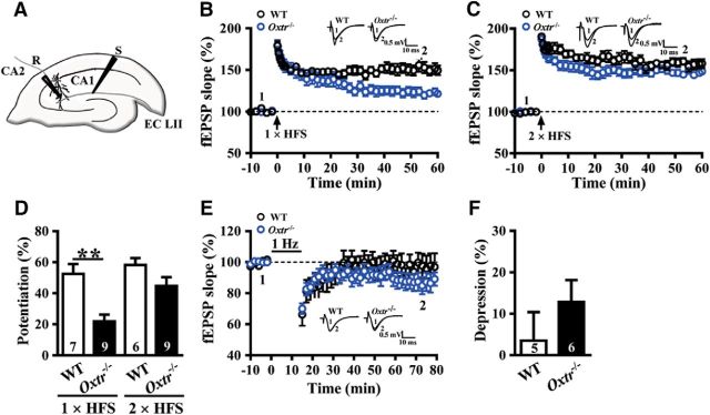
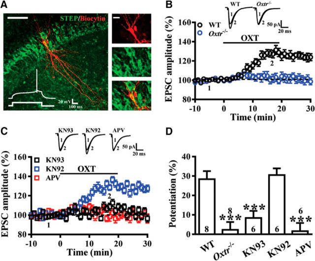
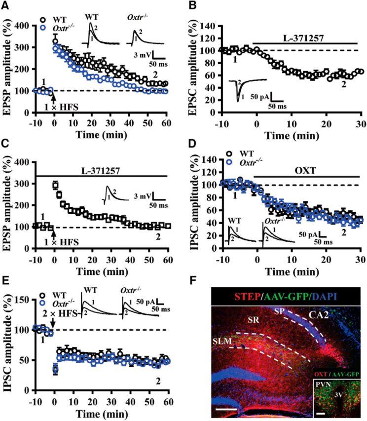
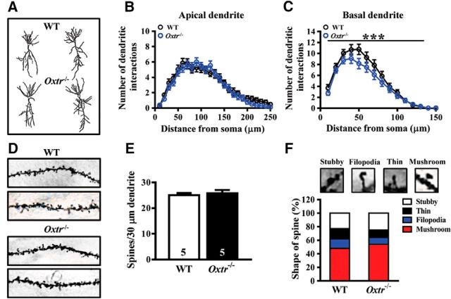
Oxytocin (OXT) receptors (OXTRs) are prominently expressed in hippocampal CA2 and CA3 pyramidal neurons, but little is known about its physiological function. As the functional necessity of hippocampal CA2 for social memory processing, we tested whether CA2 OXTRs may contribute to long-term social recognition memory (SRM) formation. Here, we found that conditional deletion of Oxtr from forebrain (Oxtr-/-) or CA2/CA3a-restricted excitatory neurons in adult male mice impaired the persistence of long-term SRM but had no effect on sociability and preference for social novelty. Conditional deletion of CA2/CA3a Oxtr showed no changes in anxiety-like behavior assessed using the open-field, elevated plus maze and novelty-suppressed feeding tests. Application of a highly selective OXTR agonist [Thr4,Gly7]-OXT to hippocampal slices resulted in an acute and lasting potentiation of excitatory synaptic responses in CA2 pyramidal neurons that relied on N-methyl-d-aspartate receptor activation and calcium/calmodulin-dependent protein kinase II activity. In addition, Oxtr-/- mice displayed a defect in the induction of long-term potentiation, but not long-term depression, at the synapses between the entorhinal cortex and CA2 pyramidal neurons. Furthermore, Oxtr deletion led to a reduced complexity of basal dendritic arbors of CA2 pyramidal neurons, but caused no alteration in the density of apical dendritic spines. Considering that the methodologies we have used to delete Oxtr do not rule out targeting the neighboring CA3a region, these findings suggest that OXTR signaling in the CA2/CA3a is crucial for the persistence of long-term SRM.SIGNIFICANCE STATEMENT Oxytocin receptors (OXTRs) are abundantly expressed in hippocampal CA2 and CA3 regions, but there are little known about their physiological function. Taking advantage of the conditional Oxtr knock-out mice, the present study highlights the importance of OXTR signaling in the induction of long-term potentiation at the synapses between the entorhinal cortex and CA2 pyramidal neurons and the persistence of long-term social recognition memory. Thus, OXTRs in the CA2/CA3a may provide a new target for therapeutic approaches to the treatment of social cognition deficits, which are often observed in patients with neuropsychiatric disorders.

Keywords: CA2/CA3a; hippocampus; long-term potentiation; oxytocin; oxytocin receptor; social recognition memory.


Figures

Figure 1.
Figure 1.
Conditional deletion of Oxtr in CA2 excitatory neurons of the mouse hippocampus. A, Doubled-labeled confocal immunofluorescent images showing the colocalization of OXTRs (green) and CaMKIIα (red) in the CA2 of the mouse hippocampus. Insets, High magnification of the boxed area. Scale bar, 10 μm. SO, Stratum oriens; SP, stratum pyramidale; SR, stratum radiatum. B, PCR screening of tail-derived genomic DNA for selection of Oxtr−/− mice. C, Dual-probe FISH images showing the expression of Oxtr mRNA and CaMKIIα mRNA in the CA2 of WT and Oxtr−/− mice (counterstained with DAPI, blue). Scale bar, 50 μm. Data were replicated in four mice. D, Representative photographs with cresyl violet staining of the CA2 showing that the number of pyramidal neurons was not significantly affected by conditional deletion of Oxtr compared with age-matched WT mice. Group data showing the summary results from four mice of each group at 10 weeks old. Scale bars: left, 10 μm; right, 200 μm. The total number of animal examined is indicated by n in parenthesis. Data represent the mean ± standard error of the mean (SEM).
Figure 2.
Figure 2.
Conditional deletion of Oxtr in forebrain excitatory neurons leads to impair the persistence of long-term SRM. A, Top, Schematic diagram of the three-chamber sociability test. Bottom left, Time spent in each side chamber containing the juvenile stimulus mouse or empty wire cage. Both WT and Oxtr−/− subject mice spent significantly more time interacting with the cage containing the juvenile stimulus mouse than with the empty wire cage. Bottom right, difference scores (stimulus minus empty) were similar between WT and Oxtr−/− mice in the sociability test. B, Top, Schematic diagram of the three-chamber social novelty test. Bottom left, Time spent in each side chamber containing a familiar mouse or a Novel 1 mouse, 10 min after the first exposure. Both WT and Oxtr−/− subject mice spent significantly more time interacting with the cage containing the novel mouse than with the familiar mouse. Bottom right, Difference scores (Novel 1 minus familiar) were similar between WT and Oxtr−/− mice in the social novelty test. C, Top, Schematic diagram of the three-chamber long-term SRM test. Bottom left, Time spent in each side chamber containing a familiar mouse or a Novel 2 mouse, 1 d after the exposure. Both WT and Oxtr−/− subject mice spent significantly more time interacting with the cage containing the novel mouse than with the familiar mouse. Bottom right, Difference scores (Novel 2 minus familiar) were similar between WT and Oxtr−/− mice in 1-d long-term SRM test. D, Top, Schematic diagram of the three-chamber long-term SRM test. Bottom, Time spent in each side chamber containing a familiar mouse or a Novel 2 mouse, 7 d after the exposure. WT, but not Oxtr−/−, subject mice spent significantly more time interacting with the cage containing the novel mouse than with the familiar mouse. Bottom right, Difference score (Novel 3 minus familiar) of Oxtr−/− mice was significantly less than that of WT mice in 7-d long-term SRM test. E, Left, Schematic diagram of the 5-trial social memory assay. Right, Both WT and Oxtr−/− subject mice showed a similar decline in the investigation time over trials one to four when they were repetitively presented the familiar ovariectomized female CD-1 mouse and increased in the investigation time on Trial 5 when they were presented with a novel ovariectomized female CD-1 mouse. The total number of animal examined is indicated by n in parenthesis. Data represent the mean ± SEM. **p < 0.01, ***p < 0.001.
Figure 3.
Figure 3.
Conditional deletion of CA2/CA3a Oxtr impairs the persistence of long-term SRM. A, Top, Schematic illustration of coronal sections illustrating the microinjection sites in the bilateral CA2. Bottom, Representative photograph showing the expression of AAV-Cre-GFP (green) in the CA2 and immunolabeled with STEP (red) and DAPI (blue). Scale bar, 50 μm. The inset represents high magnification of the boxed area. Scale bar, 20 μm. B, Top, Schematic diagram of the three-chamber sociability test. Bottom left, Time spent in each side chamber containing the juvenile stimulus mouse or empty wire cage. Both AAV-GFP- and AAV-Cre-GFP-treated subject mice spent significantly more time interacting with the cage containing the juvenile stimulus mouse than with the empty wire cage. Bottom right, Difference scores (stimulus minus empty) were similar between AAV-GFP and AAV-Cre-GFP mice in sociability test. C, Top, Schematic diagram of the three-chamber social novelty test. Bottom, Time spent in each side chamber containing a familiar mouse or a Novel 1 mouse, 10 min after the first exposure. Both AAV-GFP- and AAV-Cre-GFP-treated subject mice spent significantly more time interacting with the cage containing the novel mouse than with the familiar mouse. Bottom right, Difference scores (Novel 1 minus familiar) were similar between AAV-GFP and AAV-Cre-GFP mice in social novelty test. D, Top, Schematic diagram of the three-chamber long-term SRM test. Bottom, Time spent in each side chamber containing a familiar mouse or a Novel 2 mouse, 1 d after the exposure. Both AAV-GFP- and AAV-Cre-GFP-treated subject mice spent significantly more time interacting with the cage containing the novel mouse than with the familiar mouse. Bottom right, Difference scores (Novel 2 minus familiar) were similar between AAV-GFP and AAV-Cre-GFP mice in 1-d long-term SRM test. E, Top, Schematic diagram of the three-chamber long-term SRM test. Bottom, Time spent in each side chamber containing a familiar mouse or a Novel 2 mouse, 7 d after the exposure. AAV-GFP-, but not AAV-Cre-GFP-treated, subject mice spent significantly more time interacting with the cage containing the novel mouse than with the familiar mouse. Bottom right, Difference score (Novel 3 minus familiar) of AAV-Cre-GFP mice was significantly less than that of AAV-GFP mice in 7-d long-term SRM test. The total number of animal examined is indicated by n in parenthesis. Data represent the mean ± SEM. *p < 0.05, **p < 0.01, ***p < 0.001.
Figure 4.
Figure 4.
CA2/CA3a Oxtr deletion does not affect anxiety-like behavior. A, B, Bar graph comparing the performance of AAV-GFP- and AAV-Cre-GFP-treated mice on the total distance traveled (A) and the time spent in the center (B) in the OF test (open field [OF]). C, D, Bar graph comparing the performance of AAV-GFP- and AAV-Cre-GFP-treated mice on the percentage of time spent in the open (C) and closed arms (D) in the elevated plus maze (EPM) test. E, Bar graph comparing the performance of AAV-GFP- and AAV-Cre-GFP-treated mice on the latency to eat in the novelty suppressed feeding (NSF) test. F, Inset, Schematic of a hippocampal slice depicting the CA1, CA2, and CA3 hippocampal regions dissected for Oxtr mRNA analysis. Summary bar graphs showing qRT-PCR of Oxtr mRNA in the CA1, CA2, and CA3 regions from AAV-GFP- and AAV-Cre-GFP-treated mice. Data represent the mean ± SEM. ***p < 0.001.
Figure 5.
Figure 5.
Conditional deletion of CA2/CA3a Oxtr impairs the induction of LTP at EC→CA2 synapses. A, Schematic of experimental setup showing stimulation electrode placed in the SLM to stimulate EC inputs and field-potential recording electrode placed in the CA2 distal apical dendritic fields. B, Summary of experiments showing one train of 1 s HFS at 100 Hz induced LTP of fEPSPs at EC→CA2 synapses in slices from WT and Oxtr−/− mice. C, Summary of experiments showing two trains of 1 s HFS at 100 Hz induced LTP of fEPSPs at EC→CA2 synapses in slices from WT and Oxtr−/− mice. D, Summary bar graphs depicting levels of potentiation measured 50–60 min after one- or two-train of HFS at EC→CA2 synapses in slices from WT and Oxtr−/− mice. E, Summary graphs of LTD induced with a prolonged LFS (900 stimuli delivered at 1 Hz) at EC→CA2 synapses in slices from WT and Oxtr−/− mice. F, Summary bar graphs depicting levels of depression measured 50–60 min after LFS at EC→CA2 synapses in slices from WT and Oxtr−/− mice. Representative traces of fEPSPs were taken at the time indicated by number. Dashed lines show level of baseline. The total number of animals examined is indicated by n in parenthesis. Data are represented as mean ± SEM. **p < 0.01.
Figure 6.
Figure 6.
The selective OXTR agonist [Thr4,Gly7]-OXT induces a synaptic potentiation at EC→CA2 synapses. A, Left, Representative confocal image of a biocytin-stained CA2 pyramidal cell. Scale bar, 100 μm. Right, Single confocal attacks showing colocalization of biocytin (red) and STEP (green). Scale bar, 20 μm. Inset, Whole-cell patch-clamp recording from a CA2 pyramidal cell in current-clamp mode showing the response to a positive step current injection (100 pA, 500 ms). Note that the late-onset firing was initiated by a slow depolarizing ramp. B, Bath application of [Thr4,Gly7]-OXT (0.1 μm) for 20 min induced potentiation of EPSCs recorded in CA2 neurons from WT, but not Oxtr−/− mice. C, Summary of experiments showing [Thr4,Gly7]-OXT-induced synaptic potentiation was blocked by pretreatment of the hippocampal slices with the NMDAR antagonist APV (50 μm) or the broad spectrum CaMKII inhibitor KN93 (1 μm), but not by not its inactive structural analog KN92 (1 μm). D, Summary bar graphs depicting levels of potentiation measured 20 min after [Thr4,Gly7]-OXT treatment at EC→CA2 synapses in slices from different pretreatments. Representative traces of EPSCs were taken at the time indicated by number. Dashed lines show level of baseline. The total number of animals examined is indicated by n in parenthesis. Data are represented as mean ± SEM. ***p < 0.001.
Figure 7.
Figure 7.
OXTRs are involved in regulating synaptic transmission and plasticity in the CA2. A, Summary of experiments showing one train of 1 s HFS at 100 Hz induced LTP of EPSPs at EC→CA2 synapses in slices from WT and Oxtr−/− mice. B, Summary of experiments showing effect of L-371257 (1 μm) on EPSC amplitude at EC→CA2 synapses in slices from naive mice. C, Summary of experiments showing effect of L-371257 (1 μm) on the induction of LTP by one train of 1 s HFS at 100 Hz in slices from naive mice. D, Summary of experiments showing the effect of OXT (0.1 μm) on evoked IPSCs in CA2 pyramidal neurons in slices from WT and Oxtr−/− mice. E, Summary of experiments showing two train of 1 s HFS at 100 Hz induced LTD of IPSCs in CA2 pyramidal neurons in slices from WT and Oxtr−/− mice. F, A representative image showing the distribution of PVN projecting fibers in the SLM of the CA2. Sections were counterstained with DAPI (blue). Inset, OXT neurons in the PVN were labeled with anti-OXT antibody (red) and infected with AAV-Ubi-GFP. Scale bars: 200 μm; inset, 100 μm. SP, Stratum pyramidale; SR, stratum radiatum; 3V, third ventricle. Representative traces of EPSPs, EPSCs, or IPSCs were taken at the time indicated by number. Dashed lines show level of baseline. The total number of animals examined is indicated by n in parenthesis. Data are represented as mean ± SEM.
Figure 8.
Figure 8.
Effect of Oxtr deletion on dendritic morphology and spine density of pyramidal neurons in the hippocampal CA2 region. A, Representative camera lucida tracings of hippocampal CA2 pyramidal neurons from WT and Oxtr−/− mice. B, C, Sholl analysis of apical (B) basal (C) dendrites of CA2 pyramidal neurons from WT and Oxtr−/− mice. D, Representative images of the secondary branch of apical dendrites of CA2 pyramidal neurons from WT and Oxtr−/− mice. E, Summary bar graphs depicting the density of protrusions in apical dendrites of CA2 pyramidal cells from WT and Oxtr−/− mice. F, Representative images of labeled spines for analysis. Mushroom, filopodia, tine, and stubby spines are identified based on structural measures. There was no significant change in dendritic spine type proportion in Oxtr−/− mice compared with WT mice. Data represent the mean ± SEM. ***p < 0.001.

References

    1. Bekinschtein P, Cammarota M, Igaz LM, Bevilaqua LR, Izquierdo I, Medina JH (2007) Persistence of long-term memory storage requires a late protein synthesis- and BDNF-dependent phase in the hippocampus. Neuron 53:261–277. 10.1016/j.neuron.2006.11.025 - DOI - PubMed
    1. Bekinschtein P, Cammarota M, Katche C, Slipczuk L, Rossato JI, Goldin A, Izquierdo I, Medina JH (2008) BDNF is essential to promote persistence of long-term memory storage. Proc Natl Acad Sci U S A 105:2711–2716. 10.1073/pnas.0711863105 - DOI - PMC - PubMed
    1. Bird CM, Burgess N (2008) The hippocampus and memory: insights from spatial processing. Nat Rev Neurosci 9:182–194. 10.1038/nrn2335 - DOI - PubMed
    1. Buijs RM, Swaab DF (1979) Immuno-electron microscopical demonstration of vasopressin and oxytocin synapses in the limbic system of the rat. Cell Tissue Res 204:355–365. - PubMed
    1. Chafai M, Corbani M, Guillon G, Desarménien MG (2012) Vasopressin inhibits LTP in the CA2 mouse hippocampal area. PLoS One 7:e49708. 10.1371/journal.pone.0049708 - DOI - PMC - PubMed

Publication types

MeSH terms