. 2018 May;32(5):2574-2586.
doi: 10.1096/fj.201701008R. Epub 2018 Jan 8.

Angiogenic factor imbalance precedes complement deposition in placentae of the BPH/5 model of preeclampsia

Affiliations

Angiogenic factor imbalance precedes complement deposition in placentae of the BPH/5 model of preeclampsia

Jennifer L Sones et al. FASEB J. 2018 May.

Abstract

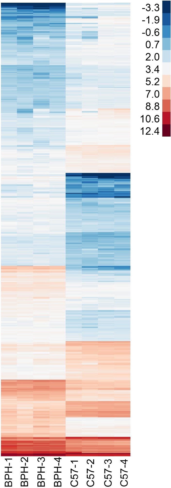
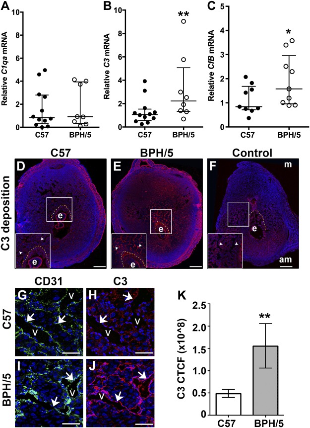
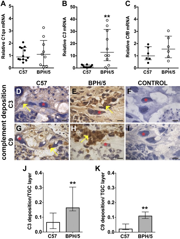
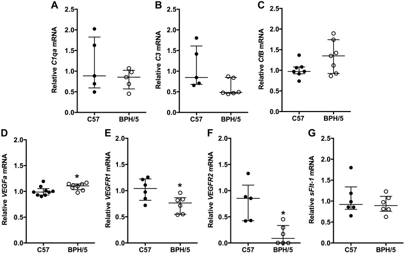
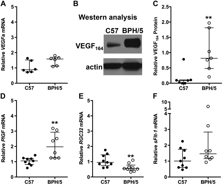
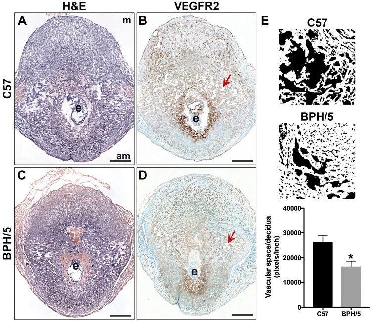
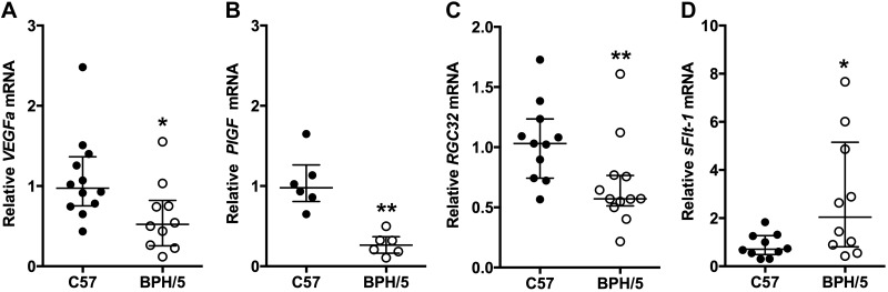
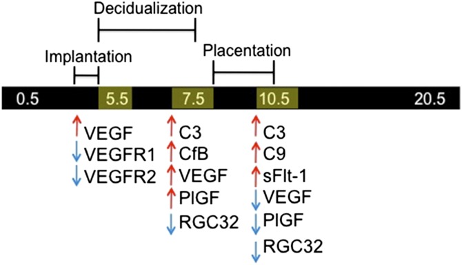
Preeclampsia (PE), a hypertensive disorder of pregnancy, is a leading cause of maternal and fetal morbidity and mortality. Although the etiology is unknown, PE is thought to be caused by defective implantation and decidualization in pregnancy. Pregnant blood pressure high (BPH)/5 mice spontaneously develop placentopathies and maternal features of human PE. We hypothesized that BPH/5 implantation sites have transcriptomic alterations. Next-generation RNA sequencing of implantation sites at peak decidualization, embryonic day (E)7.5, revealed complement gene up-regulation in BPH/5 vs. controls. In BPH/5, expression of complement factor 3 was increased around the decidual vasculature of E7.5 implantation sites and in the trophoblast giant cell layer of E10.5 placentae. Altered expression of VEGF pathway genes in E5.5 BPH/5 implantation sites preceded complement dysregulation, which correlated with abnormal vasculature and increased placental growth factor mRNA and VEGF164 expression at E7.5. By E10.5, proangiogenic genes were down-regulated, whereas antiangiogenic sFlt-1 was up-regulated in BPH/5 placentae. We found that early local misexpression of VEGF genes and abnormal decidual vasculature preceded sFlt-1 overexpression and increased complement deposition in BPH/5 placentae. Our findings suggest that abnormal decidual angiogenesis precedes complement activation, which in turn contributes to the aberrant trophoblast invasion and poor placentation that underlie PE.-Sones, J. L., Merriam, A. A., Seffens, A., Brown-Grant, D.-A., Butler, S. D., Zhao, A. M., Xu, X., Shawber, C. J., Grenier, J. K., Douglas, N. C. Angiogenic factor imbalance precedes complement deposition in placentae of the BPH/5 model of preeclampsia.

Keywords: RGC32; VEGF; decidual vasculature; implantation; sFlt-1; trophoblast.


Conflict of interest statement

The authors thank Robin L. Davisson for the generous gift of BPH/5 mice and additional support; Michelle A. Hoch for assistance with data analysis; Jan K. Kitajewski and Virginia E. Papaioannou for in-depth discussions and assistance with manuscript editing; and Theresa Swayne and Emilia Laura Munteanu for technical assistance with confocal microscopy. This work was supported by U.S. National Institutes of Health (NIH) National Heart Lung and Blood Institute Grant 1R01HL127013-01A1 (to N.C.D) and NIH Eunice Kennedy Shriver National Institute of Child Health and Human Development Grant P50-HD076210 (to J.L.S. and J.K.G.). Immunofluorescence images were collected in the Confocal and Specialized Microscopy Shared Resource of the Herbert Irving Comprehensive Cancer Center at Columbia University (New York, NY, USA), supported by NIH National Cancer Institute Grant P30 CA013696. The confocal microscope was purchased with NIH National Center for Research Resources Grant S10 RR025686. The authors declare no conflicts of interest.

Figures

Figure 1.
Figure 1.
Gene expression profile measured by RNA-Seq at E7.5. Heat map showing log2-FPKM values for 484 genes found to have differential expression between BPH/5 and C57 (q ≤ 0.05, minimum FPKM = 5 in at least 1 group, minimum 2-fold change between groups).
Figure 2.
Figure 2.
Increased decidual vascular complement deposition before placentation in BPH/5 pregnancies. AC) qRT-PCR analysis of C1qa, C3, and CfB mRNA expression in C57 and BPH/5 implantation sites at E7.5. A) C1qa mRNA expression was unchanged. B) C3 mRNA expression was increased in BPH/5 pregnancies. **P = 0.004. C) CfB mRNA expression was increased in BPH/5 pregnancies. *P = 0.05. DF) IF staining was performed on tissue sections of implantation sites at E7.5. Nuclei were stained with DAPI (blue). White boxes: areas magnified in the insets. Scale bars, 500 µm. C3 deposition was detected in the ectoplacental cone (area between the dashed yellow line and embryo) and around the decidual vasculature (arrowheads) of the C57 (D) and BPH/5 (E) implantation sites. Anti-goat secondary antibody alone (F) was used for C3 controls. GJ) Double staining IF was performed for CD31 and C3. Representative areas of the decidual vasculature are shown. Scale bars, 100 µm. CD31 identifies decidual vessels (G, I) C3 deposition around the vasculature was increased in BPH/5 pregnancies (J) as compared to C57 controls (H). To quantitatively assess the relative amount of C3 deposition in BPH/5 and C57 implantation sites, we calculated and compared CTCF. K) CTCF for C3 was significantly higher in BPH/5 pregnancies as compared to C57 controls. **P = 0.002. Results represent the median and IQR (n = 5–12 implantation sites/group). am, antimesometrial; e, embryo; m, mesometrial; v, vessels.
Figure 3.
Figure 3.
Midgestational BPH/5 placentae have excessive complement expression and deposition. AC) qRT-PCR analysis of C1qa, C3, and CfB mRNA expression in C57 and BPH/5 placentae at E10.5. A) C1qa mRNA expression was unchanged. B) C3 mRNA expression was increased in BPH/5 placentae. **P = 0.001. C) CfB mRNA expression was unchanged. DI) IHC was performed to detect C3 and C9 deposition in placental sections at E10.5. Red asterisks: TGCs; yellow arrows: positive staining, indicative of complement deposition. Scale bar, 500 µm. Representative sections of the TGC layer show C3 deposition (D, E) and C9 deposition (G, H). Anti-goat secondary antibody (F) or anti-rabbit secondary antibody (I) alone was used for C3 and C9 controls, respectively. To quantitatively assess the amount of C3 and C9 deposition in the TGC layer of BPH/5 and C57 placentae, we measured the total amount of complement deposition and normalized that to the total TGC area in a placenta section. J) Median C3 deposition in the TGC layer of BPH/5 placentae was significantly increased as compared to C57 controls. **P = 0.001. K) Median C9 deposition in the TGC layer of BPH/5 placentae was significantly increased as compared to C57 controls. **P = 0.009. Results represent the median and IQR (n = 4–12 placentas/group).
Figure 4.
Figure 4.
Angiogenic gene expression is altered in BPH/5 implantation sites during decidualization. mRNA expression of complement and angiogenic genes in C57 control and BPH/5 implantation sites at E5.5 was quantified by qRT-PCR. mRNA expression of C1qa (A), C3 (B), CfB (C), and sFlt-1(G) was similar in C57 control and BPH/5 implantation sites. Expression of VEGFa (D) was significantly increased in BPH/5 pregnancies. *P = 0.05. mRNA expression of both VEGFR1 (E) *P = 0.03 and VEGFR2 (F) *P = 0.02 was decreased in BPH/5 pregnancies. Results represent the median and IQR; n = 5–9 implantation sites/group.
Figure 5.
Figure 5.
Angiogenic gene expression remains altered in BPH/5 implantation sites before placentation. A) VEGFa mRNA expression in C57 and BPH/5 implantation sites at E7.5 was quantified using qRT-PCR. mRNA expression of VEGFa was not significantly increased in BPH/5 pregnancies. P = 0.06. B) Representative Western blots of VEGF164 and actin in BPH/5 and C57 implantation sites at E7.5. C) Quantification of VEGF164 protein expression relative to actin by Western blot analysis. **P = 0.004. DF) Quantification by qRT-PCR of PlGF, RGC32, and sFlt-1 transcripts in implantation sites at E7.5. D) mRNA expression of PlGF was increased in BPH/5 pregnancies. **P = 0.01. E) mRNA expression of RGC32 was decreased in BPH/5 pregnancies. **P = 0.01. F) mRNA expression of sFlt-1 showed no significant difference between BPH/5 and C57 pregnancies. Results represents the median and IQR; n = 6–10 implantation sites/group.
Figure 6.
Figure 6.
Development of the decidual vasculature was reduced in BPH/5 implantation sites. A, C) H&E staining was performed on tissue sections of implantation sites at E7.5. B, D) IHC was performed to detect VEGFR2 expression in the decidual vasculature. Nuclei were stained with methylene green. Scale bars, 500 µm. To quantitatively assess development of the decidual vasculature in BPH/5 and C57 pregnancies at E7.5, we measured the total amount of vascular space [red arrows in capillary lumens in (B, D)] and normalized that to the area of decidua measured in each section. E) Representative images used to quantify vascular space in ImageJ are shown. Mean vascular space/area of decidua in BPH/5 implantation sites was significantly decreased as compared to C57 controls. P = 0.03. Results represent the mean and sem (n = 4 implantation sites/group). am, antimesometrial; e, embryo; m, mesometrial.
Figure 7.
Figure 7.
Midgestational BPH/5 placentae have abnormal angiogenic gene expression. mRNA expression of angiogenic genes in C57 and BPH/5 placentae at E10.5 was quantified with qRT-PCR. mRNA expression of proangiogenic was significantly decreased in BPH/5 placentae: VEGFa (A) *P = 0.02; PlGF (B) **P = 0.002; and RGC32 (C), **P = 0.004. D) mRNA expression of antiangiogenic sFlt-1 was significantly increased in BPH/5 placentae. *P = 0.04. Results represent the median and IQR (n = 6–12 placentae/group).
Figure 8.
Figure 8.
Summary of dynamic complement and angiogenic factor expression in BPH/5 pregnancy at the maternal–fetal interface as compared controls.

References

    1. Ghulmiyyah L., Sibai B. (2012) Maternal mortality from preeclampsia/eclampsia. Semin. Perinatol. 36, 56–59 10.1053/j.semperi.2011.09.011 - DOI - PubMed
    1. American College of Obstetricians and Gynecologists ; Task Force on Hypertension in Pregnancy . (2013) Hypertension in pregnancy: report of the American College of Obstetricians and gynecologists’ task force on hypertension in pregnancy. Obstet. Gynecol. 122, 1122–1131 10.1097/01.AOG.0000437382.03963.88 - DOI - PubMed
    1. Tranquilli A. L., Dekker G., Magee L., Roberts J., Sibai B. M., Steyn W., Zeeman G. G., Brown M. A. (2014) The classification, diagnosis and management of the hypertensive disorders of pregnancy: a revised statement from the ISSHP. Pregnancy Hypertens. 4, 97–104 10.1016/j.preghy.2014.02.001 - DOI - PubMed
    1. Chen C. W., Jaffe I. Z., Karumanchi S. A. (2014) Pre-eclampsia and cardiovascular disease. Cardiovasc. Res. 101, 579–586 10.1093/cvr/cvu018 - DOI - PMC - PubMed
    1. Huppertz B. (2008) Placental origins of preeclampsia: challenging the current hypothesis. Hypertension 51, 970–975 10.1161/HYPERTENSIONAHA.107.107607 - DOI - PubMed

Publication types

MeSH terms

Substances