. 2018 Jan 9;115(2):E190-E199.
doi: 10.1073/pnas.1708584115. Epub 2017 Dec 26.

RSK2 drives cell motility by serine phosphorylation of LARG and activation of Rho GTPases

Affiliations

RSK2 drives cell motility by serine phosphorylation of LARG and activation of Rho GTPases

Geng-Xian Shi et al. Proc Natl Acad Sci U S A. .

Abstract

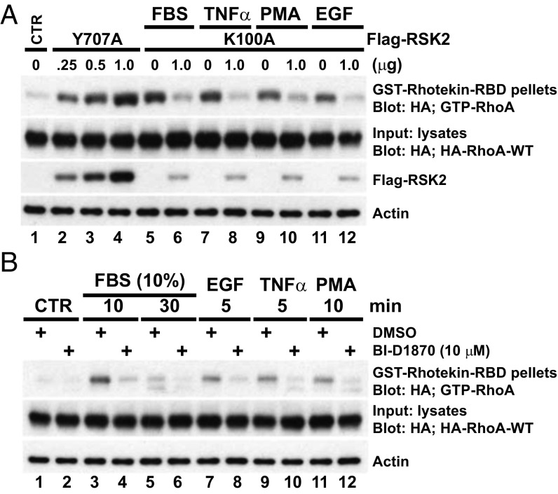
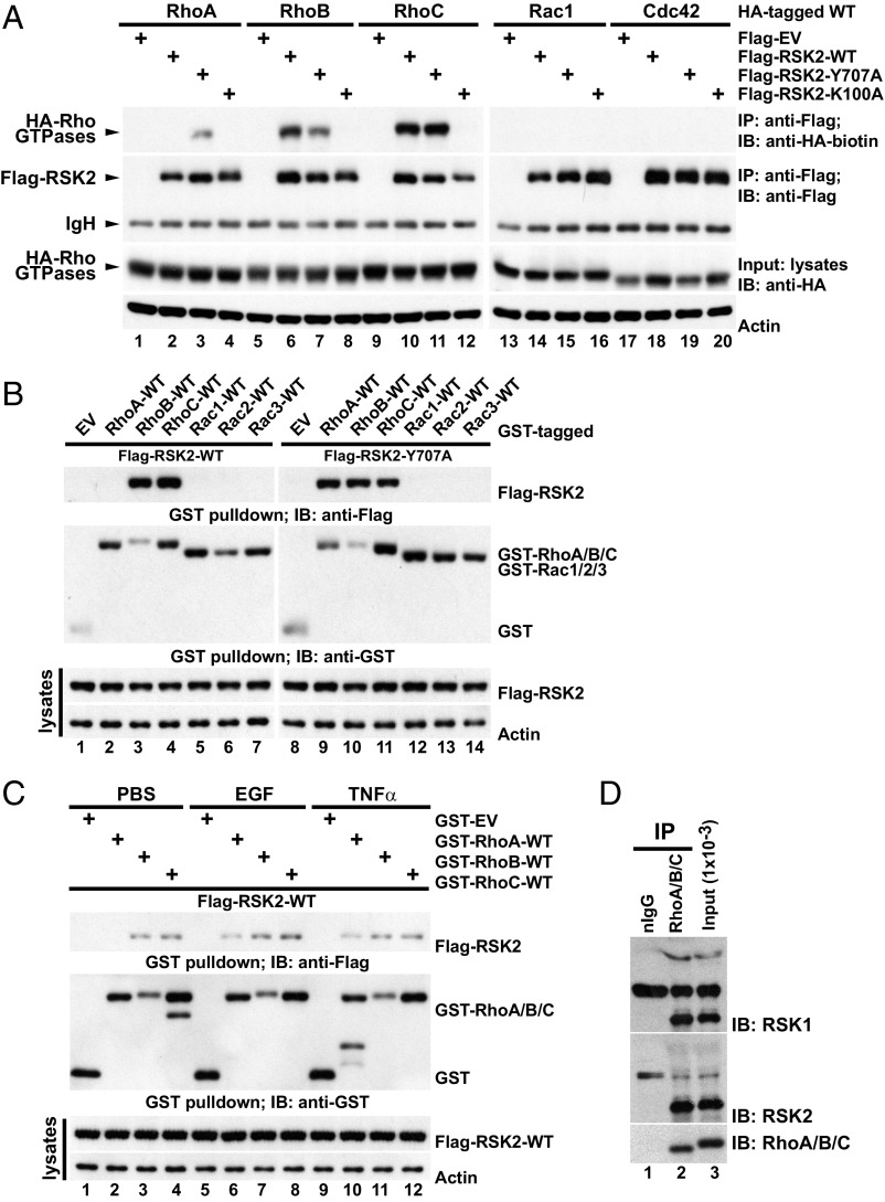
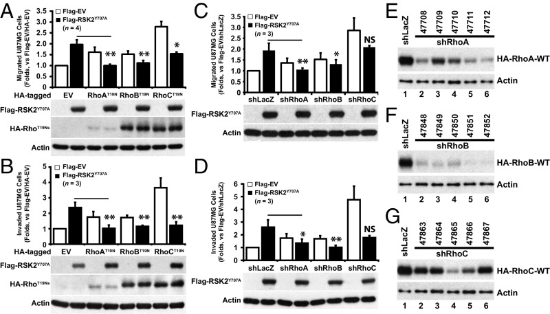
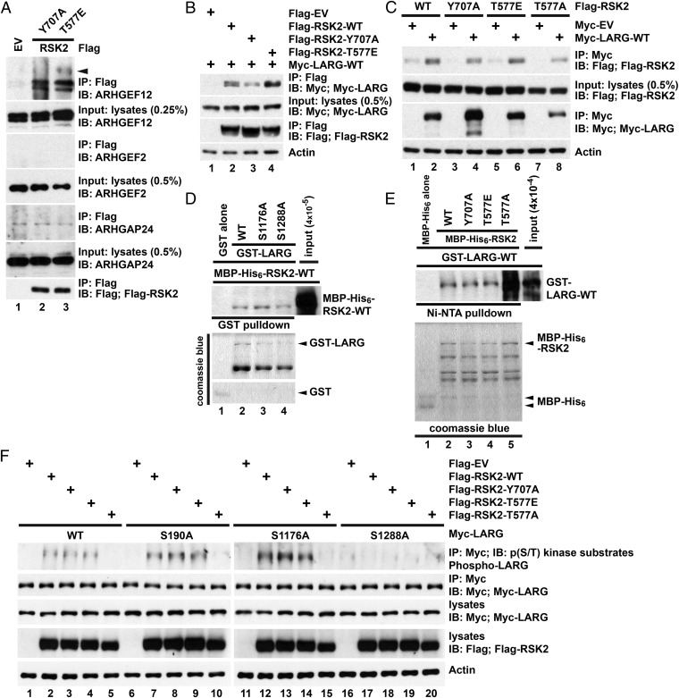
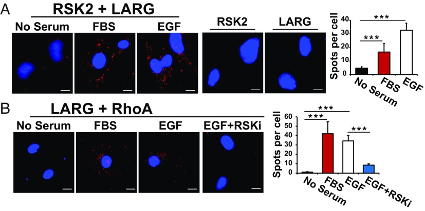
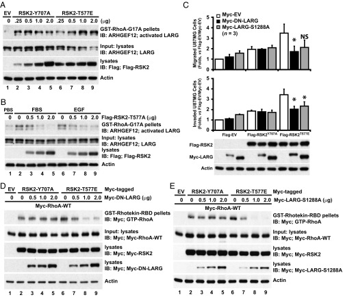
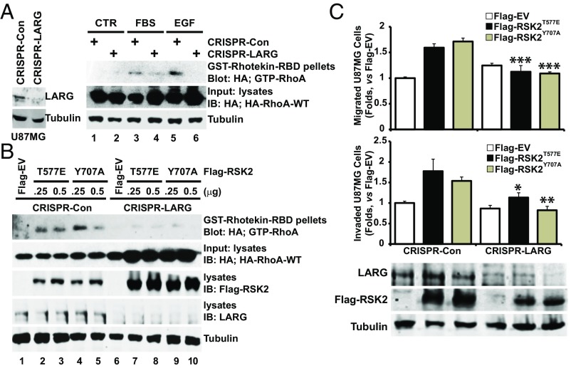
Directed migration is essential for cell motility in many processes, including development and cancer cell invasion. RSKs (p90 ribosomal S6 kinases) have emerged as central regulators of cell migration; however, the mechanisms mediating RSK-dependent motility remain incompletely understood. We have identified a unique signaling mechanism by which RSK2 promotes cell motility through leukemia-associated RhoGEF (LARG)-dependent Rho GTPase activation. RSK2 directly interacts with LARG and nucleotide-bound Rho isoforms, but not Rac1 or Cdc42. We further show that epidermal growth factor or FBS stimulation induces association of endogenous RSK2 with LARG and LARG with RhoA. In response to these stimuli, RSK2 phosphorylates LARG at Ser1288 and thereby activates RhoA. Phosphorylation of RSK2 at threonine 577 is essential for activation of LARG-RhoA. Moreover, RSK2-mediated motility signaling depends on RhoA and -B, but not RhoC. These results establish a unique RSK2-dependent LARG-RhoA signaling module as a central organizer of directed cell migration and invasion.

Keywords: ARHGEF12; LARG; RSK2; Rho GTPases; motility.


Conflict of interest statement

The authors declare no conflict of interest.

Figures

Fig. 1.
Fig. 1.
RSK2 activity is required for RhoA activation in response to diverse stimuli. (A) U87MG cells expressing HA-RhoA-WT were transfected with Flag-RSK2-Y707A vector or dominant negative (DN) Flag-RSK2-K100A. Flag-EV transfection was used as control. Cells were serum starved for 24 h before stimulation with FBS (10%, 10 min), TNFα (50 ng/mL, 5 min), PMA (100 ng/mL, 10 min), or EGF (100 ng/mL, 5 min). Five hundred micrograms of total cell lysates was subjected to GST-rhotekin-RBD precipitation. The levels of GTP-RhoA were determined by immunoblotting with anti-HA antibody. Equal loading of HA-RhoA proteins and expression levels of transfected Flag-RSK2 proteins are shown. Results are representative of seven independent experiments. (B) U87MG cells expressing HA-RhoA-WT were serum starved and then stimulated as indicated after pretreatment with BI-D1870 (10 μM). GTP-RhoA was detected by rhotekin assay as described above. Results are representative of four independent experiments.
Fig. 2.
Fig. 2.
RSK2 forms a complex with RhoA GTPases. (A) U87MG cells transfected with the indicated Flag-RSK2 and wild-type (WT) HA-Rho GTPase constructs and serum starved. Lysates were subjected to anti–Flag-RSK2 immunoprecipitation (IP) and the coprecipitated HA-tagged Rho GTPases were detected by immunoblotting with biotinylated anti-HA antibody. The IP efficiency of Flag-RSK2 proteins and the equal loading of HA-Rho GTPases were determined. A representative of seven independent experiments is shown. (B) U87MG cells expressing GST-tagged WT-Rho GTPases together with Flag-RSK2-WT or Flag-RSK2-Y707A and serum starved. GST-Rho GTPases were recovered by GST pulldown, and coprecipitated Flag-RSK2 proteins were detected by immunoblotting with anti-Flag antibody. The precipitated GST-fused protein and equal loading of the Flag-RSK2 proteins are shown. The results shown are representative of three independent experiments. (C) U87MG cells were transfected with Flag-RSK2-WT and the indicated GST-Rho constructs and serum starved before stimulation with EGF (100 ng/mL, 5 min) or TNFα (50 ng/mL, 15 min). Lysates were subjected to GST pulldown. Coprecipitated Flag-RSK2 proteins were detected by immunoblotting with anti-Flag monoclonal antibody, while recovered GST-Rho proteins were determined by anti-GST immunoblotting. A representative of four independent experiments is shown. (D) U87MG cell lysate was subjected to immunoprecipitation with anti-RhoA/B/C antibody and bound proteins were recovered by incubation with protein-G Sepharose. The presence of coprecipitated RSK1/2 was determined by immunoblotting with RSK1 or -2 antibodies. The input levels of RSK1/2 and the immunoprecipitated Rho proteins are shown. nIgG, normal rabbit control IgG. A representative of four independent experiments is shown.
Fig. 3.
Fig. 3.
RSK2-stimulated cell migration requires Rho GTPase activity. (A and B) U87MG cells were transfected with Flag-EV or Flag-RSK2-Y707A together with HA-tagged DN-Rho isoforms or HA-EV and subjected to G418 selection and serum starved (24 h) before Transwell migration (A) or Matrigel invasion (B) assays. Results are presented as mean ± SD of four independent experiments each performed in triplicate. (C and D) U87MG cells infected with indicated rLenti-shRNA against different Rho isoforms were transfected with Flag-RSK2-Y707A. shLacZ infection and Flag-EV transfection were used as controls. Cells were subjected to G418 and puromycin selection and serum starved before Transwell assays. The results are presented as mean ± SD of three independent experiments performed in triplicate. (EG) Lentivirus shRNA-mediated Rho silencing. U87MG cells expressing human WT-HA-Rho isoforms were transduced with the indicated lenti-shRNA against human RhoA (E), RhoB (F), or RhoC (G). Cells were subjected to puromycin selection before lysate preparation, and the levels of HA-Rho isoforms were detected by anti-HA immunoblotting. Actin was used as a loading control. For all figures, error bars represent SD. Statistical significance between the indicated sample versus EV+RSK2Y707A is *P < 0.05, **P < 0.01, or NS (not significant, P ≥ 0.05). Refer to Dataset S1 A and B for all P values.
Fig. 4.
Fig. 4.
RSK2 forms a signaling complex composed of Rho GTPases and the RhoGEF, LARG. (A) Active RSK2 precipitates endogenous LARG. A total of 10 mg of U87MG cell lysates with overexpressed Flag-RSK2-Y707A or Flag-RSK2-T577E was incubated with anti-Flag antibody at 4 °C overnight and bound proteins were detected by immunoblotting with indicated antibodies. The arrow indicates possible phosphorylated LARG. (B and C) Association of RSK2 and LARG. U87MG cells were transfected with indicated Flag- or Myc-tagged constructs and serum starved for 24 h before lysate preparation. Flag (C)- or Myc (D)-tagged proteins were immunoprecipitated by anti-Flag or -Myc antibodies, respectively. Bound proteins were detected by immunoblotting with anti-Flag (RSK2) or -Myc (LARG) antibodies. Results are representative of three independent experiments. (D and E) RSK2 interacts directly with LARG. GST-LARG proteins and MBP-His6-RSK2-WT were incubated with either glutathione Sepharose (D) or Ni-NTA His-Bind resin (E). Bound MBP-His6-RSK2-WT (D) or GST-LARG (E) was determined by immunoblotting. (F) RSK2 phosphorylates LARG on Ser1288 residue. U87MG cells were cotransfected with Flag-RSK2 constructs and the indicated Myc-LARG constructs. The Myc-LARG proteins were immunoprecipitated after serum starvation for 24 h. LARG phosphorylation was determined by immunoblotting anti-Myc immunoprecipitates with a phospho-AKT substrate-specific antibody. Results are representative of three independent experiments.
Fig. 5.
Fig. 5.
Activation of RSK2 by FBS or EGF stimulates endogenous RSK2 binding to LARG and LARG to RhoA. (A) U373MG cells were starved for 24 h and then stimulated with FBS (10%, 10 min) or EGF (100 ng/mL, 5 min). Endogenous RSK2 and LARG protein complex formation was determined by proximity ligation assay as described in Materials and Methods. RSK2 or LARG antibody alone was used as negative controls. Complex formation is indicated where there are distinct red spots. Complex formation in cells from each slide was quantified by ImageJ software and is presented as bar graph. (B) Starved overnight U373MG cells were treated with RSK inhibitor (RSKi, BI-D1870) for 30 min before stimulation. Complex formation is indicated where there are distinct red spots. Complex formation in cells from each slide was quantified by ImageJ software and is presented as bar graph. n = 5. (Scale bar, 5 μM.) ***P < 0.001.
Fig. 6.
Fig. 6.
RSK2 activation of RhoA signaling in cell migration and invasion requires LARG. (A) RSK2-T577E activates LARG. U87MG cells were transfected with varying amounts of Flag-RSK2-T577E. Flag-EV transfection was used as a negative control. Cells were serum starved before cell lysate preparation. Activated LARG (ARHGEF12) was captured by GST-RhoA-G17A pulldown and detected by immunoblotting with anti-LARG antibody. The levels of LARG and Flag-RSK2 are shown. Results are representative of four independent experiments. (B) U87MG cells transfected with Flag-RSK2-T577A and serum starved were stimulated by FBS (10%, 10 min) or EGF (100 ng/mL, 5 min). Activated LARG was retrieved by GST-RhoA-G17A pulldown and detected by immunoblotting with anti-LARG antibody. Expression of LARG and Flag-RSK2-T577A levels is shown. (C) RSK2-T577E–induced U87MG cell migration and invasion requires LARG activity. U87MG cells were cotransfected with Flag-RSK2-T577E and Myc-LARG-S1288A or -DN-LARG. Flag-EV and Myc-EV transfection was used as controls. Transfected cells were serum starved before migration and invasion assays. Cell migration (Upper) and invasion (Middle) were assessed by Transwell assays. Expression levels of exogenous RSK2 and LARG proteins are shown (Lower). Refer to Dataset S1C for all P values. *P < 0.05. (D and E) Inhibition of LARG activity disrupts RSK2-T577E–triggered RhoA activation. U87MG cells expressing Myc-RhoA-WT were transfected with Myc-RSK2-T577E (1 μg) in the presence or absence of Myc-tagged DN-LARG (D) or LARG-S1288A (E). Cells were starved before lysate preparation and GST-rhotekin-RBD pulldown analysis. GTP-RhoA levels were determined by immunoblotting pulldown precipitate with anti-Myc antibody. Expression levels of Myc-RhoA-WT, Myc-RSK2, and Myc-LARG are determined by immunoblot and shown. Results are representative of three independent experiments.
Fig. 7.
Fig. 7.
Deletion of LARG impairs RSK2 activation of RhoA and cell migration and invasion. (A and B) LARG was deleted in U87MG cells by CRISPR/Cas9-targeted deletion as described in Materials and Methods. LARG-deleted cells and nonspecific CRISPR/Cas9-treated control cells expressing HA-RhoA-WT were stimulated with FBS (10%, 10 min), EGF (100 ng/mL, 5 min) (A), or transfected with Flag-EV, Flag-RSK2-T577E, or Y707A (B). GTP-RhoA was retrieved using GST-rhotekin-RBD, and GTP-RhoA levels were determined by immunoblotting with anti-HA antibody as described above. Expression levels of HA-RhoA, LARG, and Flag-RSK2 were determined by immunoblotting. (C) RSK2-induced migration and invasion was impaired in LARG-deleted U87MG cells. CRISPR/Cas9-mediated nonspecific control or LARG-deleted cells were transfected with Flag-EV, RSK2-T577E, and Y707A. Transfected cells were enriched by G418 selection and serum starved for 24 h before migration and invasion assays. Cell migration (Upper) and invasion (Middle) were assessed by Transwell assays. Refer to Dataset S1D for all P values. Expression levels of RSK2 and LARG proteins are shown (Lower). *P < 0.05, **P < 0.01, ***P < 0.001.

References

    1. Huttenlocher A, Horwitz AR. Integrins in cell migration. Cold Spring Harb Perspect Biol. 2011;3:a005074. - PMC - PubMed
    1. Parsons JT, Horwitz AR, Schwartz MA. Cell adhesion: Integrating cytoskeletal dynamics and cellular tension. Nat Rev Mol Cell Biol. 2010;11:633–643. - PMC - PubMed
    1. Sulzmaier FJ, Ramos JW. RSK isoforms in cancer cell invasion and metastasis. Cancer Res. 2013;73:6099–6105. - PMC - PubMed
    1. Sulzmaier FJ, et al. RSK2 activity mediates glioblastoma invasiveness and is a potential target for new therapeutics. Oncotarget. 2016;7:79869–79884. - PMC - PubMed
    1. Hauge C, Frödin M. RSK and MSK in MAP kinase signalling. J Cell Sci. 2006;119:3021–3023. - PubMed

Publication types

Substances

LinkOut - more resources