. 2018 Jan 9;115(2):E210-E217.
doi: 10.1073/pnas.1716055115. Epub 2017 Dec 26.

Listeria monocytogenes triggers noncanonical autophagy upon phagocytosis, but avoids subsequent growth-restricting xenophagy

Affiliations

Listeria monocytogenes triggers noncanonical autophagy upon phagocytosis, but avoids subsequent growth-restricting xenophagy

Gabriel Mitchell et al. Proc Natl Acad Sci U S A. .

Abstract

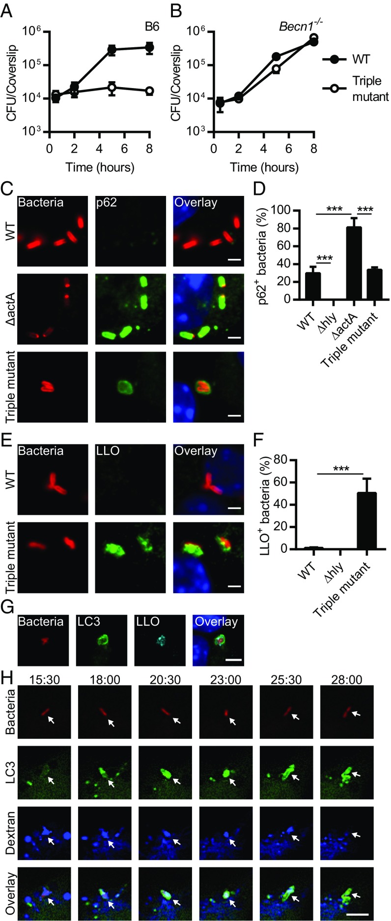
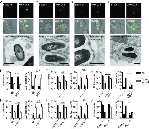
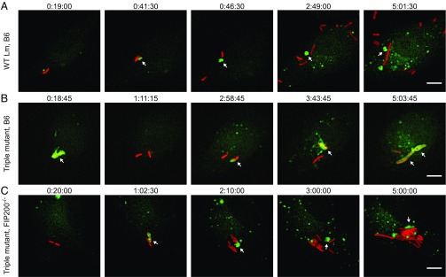
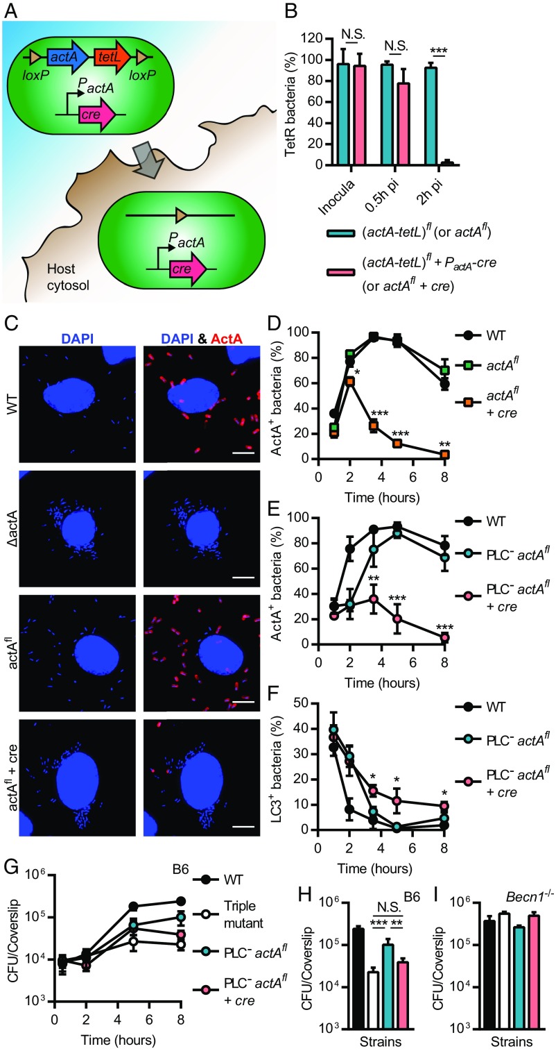
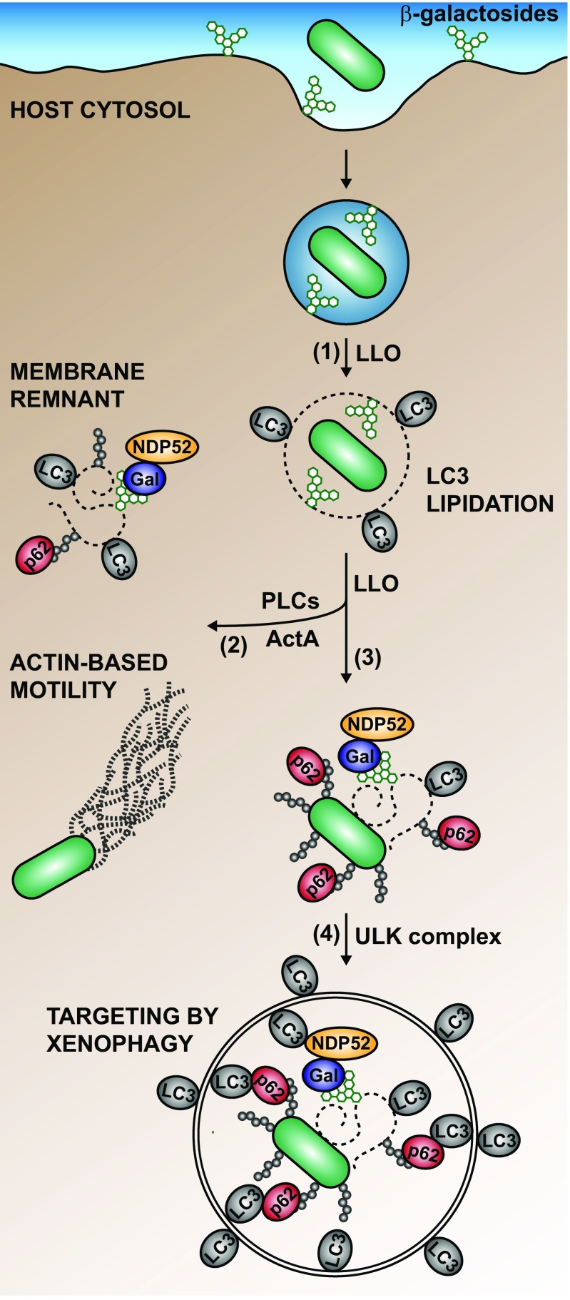
Xenophagy is a selective macroautophagic process that protects the host cytosol by entrapping and delivering microbes to a degradative compartment. Both noncanonical autophagic pathways and xenophagy are activated by microbes during infection, but the relative importance and function of these distinct processes are not clear. In this study, we used bacterial and host mutants to dissect the contribution of autophagic processes responsible for bacterial growth restriction of Listeria monocytogenesL. monocytogenes is a facultative intracellular pathogen that escapes from phagosomes, grows in the host cytosol, and avoids autophagy by expressing three determinants of pathogenesis: two secreted phospholipases C (PLCs; PlcA and PlcB) and a surface protein (ActA). We found that shortly after phagocytosis, wild-type (WT) L. monocytogenes escaped from a noncanonical autophagic process that targets damaged vacuoles. During this process, the autophagy marker LC3 localized to single-membrane phagosomes independently of the ULK complex, which is required for initiation of macroautophagy. However, growth restriction of bacteria lacking PlcA, PlcB, and ActA required FIP200 and TBK1, both involved in the engulfment of microbes by xenophagy. Time-lapse video microscopy revealed that deposition of LC3 on L. monocytogenes-containing vacuoles via noncanonical autophagy had no apparent role in restricting bacterial growth and that, upon access to the host cytosol, WT L. monocytogenes utilized PLCs and ActA to avoid subsequent xenophagy. In conclusion, although noncanonical autophagy targets phagosomes, xenophagy was required to restrict the growth of L. monocytogenes, an intracellular pathogen that damages the entry vacuole.

Keywords: ActA; LC3-associated phagocytosis; bacteria; macrophage; phospholipases.


Conflict of interest statement

Conflict of interest statement: D.A.P. has a consulting relationship with and a financial interest in Aduro Biotech. Both he and the company stand to benefit from the commercialization of the results of this research. G.M. and Ralph R. Isberg are coauthors on a 2017 review article.

Figures

Fig. 1.
Fig. 1.
L. monocytogenes escapes from an autophagic process that targets damaged vacuoles. Growth kinetics of WT L. monocytogenes and the triple mutant lacking ActA, PlcA, and PlcB in B6 (A) and Becn1−/− (B) BMMs (n = 2–4). (C) Representative micrographs of BMMs infected for 2 h with WT, ΔactA, and the triple mutant. Infected cells were stained for L. monocytogenes (red), p62 (green), and DNA (blue). (D) Colocalization of p62 with WT, Δhly, ΔactA, and the triple mutant in BMMs infected for 2 h. Relevant statistically significant differences are indicated [***P < 0.001 (ANOVA with Tukey’s post hoc test); n = 5]. (E) Representative micrographs of BMMs infected with WT and the triple mutant for 2 h and stained with L. monocytogenes (red), LLO (green), and DNA (blue). (F) Colocalization of LLO with WT, Δhly, and the triple mutant in BMMs infected for 2 h. Relevant statistically significant differences are indicated [***P < 0.001 (ANOVA with Tukey’s post hoc test); n = 3]. (G) Representative micrographs of GFP-LC3 BMMs infected with the triple mutant for 2 h and stained for L. monocytogenes (red), GFP-LC3 (green), LLO (cyan), and DNA (blue). (H) Selected Z-stacked micrographs from a time-lapse microscopy experiment performed with GFP-LC3 (green) BMMs infected with the triple mutant expressing mCherry (red) in presence of fluorescent dextran (blue). Arrowheads indicate the position of bacteria in the different channels and at different time points. Times are indicated (min:s) above the top row. Results are expressed as means and SDs. (Scale bars: C and E, 2 µm; G and H, 5 µm.)
Fig. 2.
Fig. 2.
L. monocytogenes is targeted by noncanonical autophagy. (AD) CLEM of GFP-LC3 BMMs infected with the triple mutant expressing mCherry for 1 h. Micrographs showing bacteria (red), GFP-LC3 (green), differential interference contrast (DIC) images, and transmission electron microscopy (TEM) images are shown. (Scale bars: fluorescence/DIC micrographs, 5 µm; TEM micrographs, 500 nm.) CFUs (at 5 h p.i.) and LC3 colocalization (at 2 h p.i.) in p62−/− (E), Ndp52−/− (F), Tbk1−/− Tnfr1−/− (G), Ulk1−/− (H), Fip200−/− (I), and Becn1−/− (J) BMMs infected with WT or the triple mutant. Tnfr1−/− (tumor necrosis factor receptor 1, TNFR1) BMMs are the control cells for the Tbk1−/− Tnfr1−/− BMMs. Results are expressed as means and SDs [N.S., nonsignificant; *P < 0.05; **P < 0.01; ***P < 0.001 (unpaired t test); n = 2–4].
Fig. 3.
Fig. 3.
Multiple autophagic processes sequentially target intracellular bacteria. Selected Z-stacked micrographs from time-lapse microscopy experiments of GFP-LC3 (green) BMMs infected with mCherry L. monocytogenes strains (red). (A) GFP-LC3 WT BMMs infected with WT bacteria. Arrowheads show a LC3+ vacuole that was removed from a bacterium and formed a membrane aggregate in the host cytosol. (B) GFP-LC3 WT BMMs infected with the triple mutant. Arrowheads show bacteria that colocalized with LC3. Note that the triple mutant colocalized with LC3 multiple distinct times. (C) GFP-LC3 Fip200−/− BMMs infected with the triple mutant. Arrowheads show a LC3+ vacuole that was removed from bacteria and formed a membrane aggregate in the host cytosol. Times are indicated (h:min:s) above each micrograph. (Scale bars: 5 µm.)
Fig. 4.
Fig. 4.
L. monocytogenes avoids growth-restricting xenophagy in the macrophage cytosol. (A) A Cre-lox system that deletes actA (and tetL) upon access to the host cytosol was engineered. The gene encoding the Cre recombinase was cloned downstream of the actA promoter, which is robustly up-regulated intracellularly. Upon access to the host cytosol, both actA and cre are expressed until Cre mediates recombination of loxP sites and deletion of the (actA-tetL) cassette (hereafter referred to as actAfl). (B) Proportion of tetracycline resistant (TetR) CFU in inocula and samples recovered from BMMs infected with actAfl and actAfl + PactA-cre (referred to as actAfl + cre) in the PlcAH86A PlcBH69G (referred to as PlcAB) background [N.S., nonsignificant; ***P < 0.001 (unpaired t test); n = 2–5]. (C) Micrographs of BMMs infected for 5 h with WT, ΔactA, and ΔactA bacteria carrying actAfl or actAfl + cre. Infected cells were strained for DNA (blue) and ActA (red). (D and E) Kinetics of ActA-positive bacteria in BMMs infected with WT and actAfl +/− cre (D), or WT and PlcAB actAfl +/− cre (E). (F) Colocalization kinetics of GFP-LC3 with L. monocytogenes in BMMs infected with WT and PlcAB actAfl +/− cre. (DF) Significant differences between actAfl and actAfl +/− cre strains are indicated for each time point [*P < 0.05; **P < 0.01; ***P < 0.001 (ANOVA with Tukey’s post hoc test performed for each time point); n = 2–3]. (G) Growth kinetics for WT, the triple mutant, and PlcAB actAfl +/− cre in BMMs. CFU recovered from the 8 h time point are represented in H. Relevant statistically significant differences are indicated [N.S., nonsignificant; **P < 0.01; ***P < 0.001 (ANOVA with Tukey’s post hoc test); n = 4]. (I) CFU recovered from Becn1−/− BMMs infected for 8 h with WT, the triple mutant, and PlcAB actAfl +/− cre [no significant differences (ANOVA with Tukey’s post hoc test); n = 2]. Results are expressed as means and SDs. (Scale bars: 5 µm.)
Fig. 5.
Fig. 5.
Model for the interaction of L. monocytogenes with the host autophagy machinery during macrophage infection. L. monocytogenes is targeted sequentially by distinct autophagic processes during its intracellular life cycle. (1) LLO triggers lipidation of LC3 on the Listeria-containing vacuole through a process independent of the ULK complex. (2) Bacteria that expressed ActA and PLCs interfere with autophagy and proliferate in the host cytosol. (3) During escape from the phagolysosomal pathway, L. monocytogenes might associate with a ruptured vacuole or membrane remnants, which are marked by galectins (Gal) and ubiquitin (Ub) chains, and recruit autophagy adaptors (e.g., p62 and NDP52). Once in the cytosol, L. monocytogenes can also directly associate with Ub and autophagy adaptors (e.g., p62). (4) Autophagy adaptors mediate the engulfment of bacteria through xenophagy, a process that requires the ULK complex. Not represented on this model is the possibility that L. monocytogene-containing autophagosomes are damaged by LLO and retargeted by the autophagy machinery.

References

    1. Galluzzi L, et al. Molecular definitions of autophagy and related processes. EMBO J. 2017;36:1811–1836. - PMC - PubMed
    1. Jiang P, Mizushima N. Autophagy and human diseases. Cell Res. 2014;24:69–79. - PMC - PubMed
    1. Huang J, Brumell JH. Bacteria-autophagy interplay: A battle for survival. Nat Rev Microbiol. 2014;12:101–114. - PMC - PubMed
    1. Randow F, Youle RJ. Self and nonself: How autophagy targets mitochondria and bacteria. Cell Host Microbe. 2014;15:403–411. - PMC - PubMed
    1. Martinez J, et al. Molecular characterization of LC3-associated phagocytosis reveals distinct roles for Rubicon, NOX2 and autophagy proteins. Nat Cell Biol. 2015;17:893–906. - PMC - PubMed

Publication types

MeSH terms