. 2018 Jan 9;115(2):E325-E333.
doi: 10.1073/pnas.1717794115. Epub 2017 Dec 26.

ALKBH5-dependent m6A demethylation controls splicing and stability of long 3'-UTR mRNAs in male germ cells

Affiliations

ALKBH5-dependent m6A demethylation controls splicing and stability of long 3'-UTR mRNAs in male germ cells

Chong Tang et al. Proc Natl Acad Sci U S A. .

Abstract

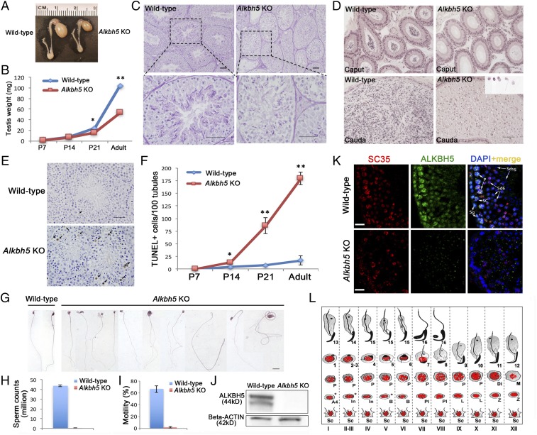
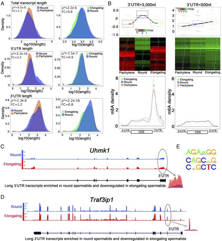
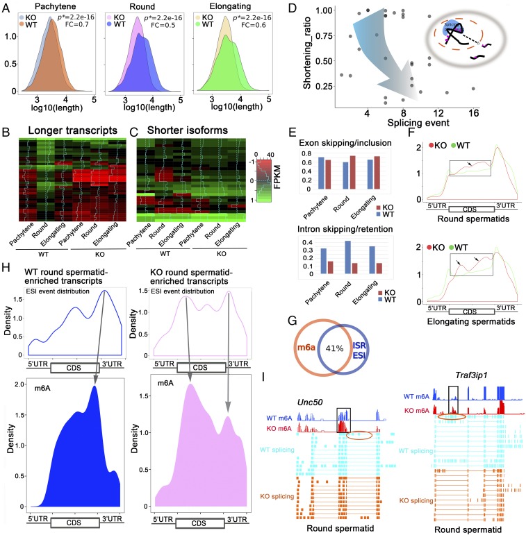
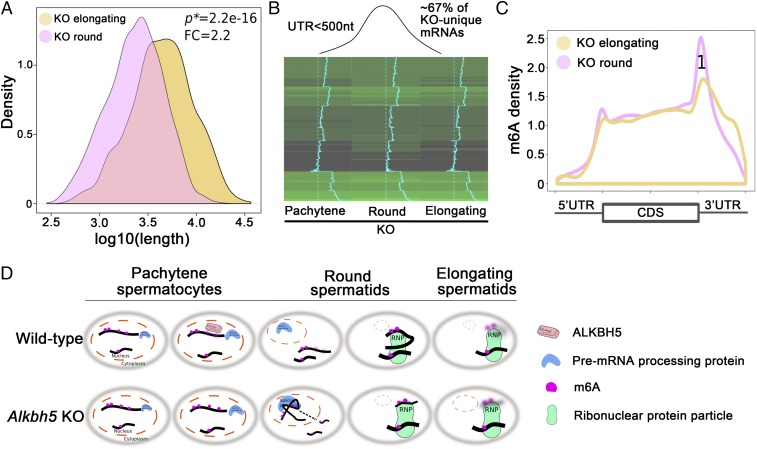
N6-methyladenosine (m6A) represents one of the most common RNA modifications in eukaryotes. Specific m6A writer, eraser, and reader proteins have been identified. As an m6A eraser, ALKBH5 specifically removes m6A from target mRNAs and inactivation of Alkbh5 leads to male infertility in mice. However, the underlying molecular mechanism remains unknown. Here, we report that ALKBH5-mediated m6A erasure in the nuclei of spermatocytes and round spermatids is essential for correct splicing and the production of longer 3'-UTR mRNAs, and failure to do so leads to aberrant splicing and production of shorter transcripts with elevated levels of m6A that are rapidly degraded. Our study identified reversible m6A modification as a critical mechanism of posttranscriptional control of mRNA fate in late meiotic and haploid spermatogenic cells.

Keywords: 3′-UTR shortening; RNA methylation; alternative splicing; fertility; mRNA stability.


Conflict of interest statement

The authors declare no conflict of interest.

Figures

Fig. 1.
Fig. 1.
Alkbh5 is essential for meiotic and haploid phases of spermatogenesis. (A) Alkbh5−/− (Alkbh5 KO) testes are much smaller than wild-type controls. (B) Significant decrease in testicular weight started at postnatal day 14 (P14) and persisted thereafter into adulthood. (C) Paraffin-embedded, periodic acid-Schiff (PAS)-stained testis sections showing robust spermatogenesis in wild-type mice, but disrupted spermatogenesis in Alkbh5 KO testes, characterized by drastically reduced number of meiotic (spermatocytes) and haploid (spermatids) male germ cells in the seminiferous epithelium and the presence of numerous vacuoles, a hallmark of active germ cell depletion. (Scale bars, 50 µm.) (D) Paraffin-embedded, HE-stained epididymal sections showing the presence of fully developed spermatozoa in wild-type caput and cauda epididymides, whereas the Alkbh5 KO caput and cauda epididymides contain no mature spermatozoa, but degenerated germ cells resembling round, elongating, or elongated spermatids, which were most likely those depleted from the seminiferous epithelium (Inset). (Scale bars, 50 µm). (E) Representative images showing TUNEL staining of apoptotic germ cells in 6-wk-old wild-type and Alkbh5 KO testes. (F) Quantitative analyses of apoptotic (TUNEL+) germ cells in developing wild-type and Alkbh5 KO testes. The number of TUNEL+ cells were counted per 100 cross-sections of seminiferous tubules. Data are presented as means ± SEM (n = 3). *P < 0.01, **P < 0.001; Student’s t test, two-tailed, homoscedasticity assumed. (G) HE-stained spermatozoa collected from wild-type and Alkbh5 KO epididymides (very rarely seen). All panels were at the same magnification. (Scale bar, 10 µm.) (H) Sperm counts in wild-type and Alkbh5 KO mice. Data are presented as means ± SEM (n = 10). (I) Sperm motility in wild-type and Alkbh5 KO mice. Data are presented as means ± SEM (n = 10). (J) Representative Western blots showing the detection of ALKBH5 protein in wild-type testes and the absence of ALKBH5 in Alkbh5 KO testes. β-Actin was used as a loading control. (K) Immunofluorescent localization of ALKBH5 (green) and SC35 (marker for nuclear speckles) in stage VI seminiferous tubules of wild-type and Alkbh5 KO testes. ALKBH5 is localized to the nuclei of Sertoli cells (SCs), spermatogonia (Sg), spermatocytes with higher levels in pachytene (P) spermatocytes, and step 6 round spermatids (Sd6). Step 15 spermatids (sd15) are devoid of ALKBH5. ALKBH5 is only partially colocalized with SC35. (Scale bars, 20 µm.) (L) Schematic illustration showing ALKBH5 localization in the murine seminiferous epithelia. A4, type A4 spermatogonia; B, type B spermatogonia; Di, diplotene spermatocytes; In, intermediate spermatogonia; L, leptotene spermatocytes; M, dividing spermatocytes; P, pachytene spermatocytes; PL, preleptotene spermatocytes; Sc, Sertoli cells; Z, zygotene spermatocytes; and 1–16, steps 1–16 spermatids.
Fig. 2.
Fig. 2.
m6A marks the longer 3′-UTR transcripts that are destined to be eliminated during spermiogenesis (from round to elongating/elongated spermatids). (A) Density plots showing that the total transcript, 5′-UTR, and 3′-UTR lengths all decreased when round spermatids developed into elongating spermatids although such a trend is less obvious during late meiotic and early haploid phases of spermatogenesis (i.e., from pachytene spermatocytes to round spermatids). Total transcript, 5′-UTR, and 3′-UTR lengths were determined based on RNA-Seq data using SpliceR. P values of statistical significance (P*) and fold changes (FC) are shown. (B) m6A sites and levels in longer vs. shorter 3′-UTR transcripts in pachytene spermatocytes and round and elongating spermatids. Two types of transcripts were analyzed: (i) Thirty longer 3′-UTR (>3,000 nt) mRNAs that were mainly synthesized in round spermatids, but were drastically down-regulated when round spermatids developed into elongating spermatids. (ii) Two hundred shorter 3′-UTR (<500 nt) mRNAs, whose levels continuously increased from round to elongating spermatids. These transcripts are most likely those required for the final several steps of sperm assembly and are subjected to delayed translation in pachytene spermatocytes and round spermatids. Density of m6A reads detected in m6A RIP-Seq datasets were plotted against the total mRNA length. Longer 3′-UTR mRNAs contain much higher levels of m6A, which is mainly enriched in 3′-UTRs proximal to the stop codon. In contrast, levels of m6A in shorter 3′-UTR transcripts are much lower and no significant enrichment was noticed. Note that m6A levels of the longer 3′-UTR mRNAs were noticeably higher in elongating spermatids than in pachytene spermatocytes and round spermatids (peak 1). (C and D) Two example genes (Uhmk1 and Traf3ip1) showed higher m6A levels in the 3′-UTRs close to the stop codon in elongating spermatids than in round spermatids. (E) Common motifs detected surrounding the m6A sites.
Fig. 3.
Fig. 3.
Proper m6A erasure is required for the production of longer 3′-UTR mRNAs in pachytene spermatocytes and spermatids. (A) Density plots showing that the total transcript length was significantly decreased in Alkbh5 KO (KO) pachytene spermatocytes and round and elongating spermatids compared with corresponding wild-type (WT) spermatogenic cells. P values of statistical significance (P*) and fold changes (FC) are shown. (B) Heat maps showing the 30 longer 3′-UTR (>3,000 nt) transcripts predominantly expressed in round spermatids in wild-type testes were significantly down-regulated in all three spermatogenic cell types in Alkbh5 KO testes. (C) Heat maps showing that the shorter 3′-UTR isoforms of these 30 longer transcripts were all up-regulated in three spermatogenic cell types in Alkbh5 KO testes compared with wild-type (WT) controls. (D) Dot plot showing the relationship between shortened transcripts and splicing events in the three Alkbh5 KO spermatogenic cell types (pachytene spermatocytes and round and elongating spermatids) analyzed. Shortening ratios (defined as the length of shorter isoform/the length of the longest transcript) were plotted against the number of total splicing events detected based on the RNA-Seq data. The shorter the transcript isoforms become, the more splicing events they tend to have, suggesting that those shorter transcript isoforms were derived from enhanced splicing of those longer transcripts in the three Alkbh5 KO spermatogenic cell types. (E) Histograms showing splicing events, including exon skipping/inclusion and intron skipping/retention, in the three spermatogenic cell types in WT and Alkbh5 KO testes. (F) Comparison of m6A density in transcripts enriched in Alkbh5 KO round (Upper) and elongating (Lower) spermatids with >3 exon skipping/inclusion (ESI) events and those enriched in wild-type (WT) round and elongating spermatids without ESI events. Note that frames indicate elevated m6A levels in the CDS region of the transcripts. (G) Venn diagram showing ∼41% of the m6A sites overlap with the sites with splicing events (±200 nt distance), including exon skipping/inclusion (ESI) and intron skipping/retention (ISR). (H) Density plots showing correlations between splicing and m6A sites in wild-type (WT) and Alkbh5 KO (KO) round spermatid-enriched transcripts. The transcripts enriched in KO round spermatids appear to contain more splicing sites proximal to the m6A sites, compared with those enriched in WT round spermatids, suggesting enhanced splicing events due to m6A accumulation in the KO cells. (I) Examples of two mRNAs (Unc50 and Traf3ip1) up-regulated in Alkbh5 KO round spermatids showing exon skipping events (circled) near the m6A accumulation sites.
Fig. 4.
Fig. 4.
Fate of the aberrantly spliced short transcripts in Alkbh5 KO (KO) round spermatids. (A) Density plot showing the average length of total transcripts increased from round to elongating spermatids in Alkbh5 KO testes; this expression pattern is opposite to the shortening trend in wild-type controls, as shown in Fig. 2A (Upper Right). P values of statistical significance (P*) and fold changes (FC) are shown. (B) Heat map showing ∼2/3 of the shorter 3′-UTR (<500 nt) transcripts up-regulated in Alkbh5 KO round spermatids were quickly down-regulated when round spermatids develop into elongating spermatids. Given that the up-regulated shorter 3′-UTR transcripts in Alkbh5 KO round spermatids were mostly those aberrantly spliced from the longer transcripts in the wild-type cells, this result suggests that these KO cell-specific shorter 3′-UTR transcripts are not stable in the Alkbh5 KO cells. (C) Elevated levels of m6A in the 3′-UTR close the stop codon in Alkbh5 KO cell-unique shorter 3′-UTR transcripts. Note that this m6A pattern is typical to the longer, but not the shorter 3′-UTR transcripts in WT round and elongating spermatids, as shown in Fig. 2B, Lower Right. (D) Schematic presentation showing the physiological functions of ALKBH5-dependent m6A erasure in late meiotic (pachytene spermatocytes) and haploid (round and elongating spermatids) phases of spermatogenesis in the wild-type testes, and the molecular consequences of m6A erasure failure due to Alkbh5 inactivation.

Similar articles

Cited by

References

    1. He C. Grand challenge commentary: RNA epigenetics? Nat Chem Biol. 2010;6:863–865. - PubMed
    1. Cantara WA, et al. The RNA modification database, RNAMDB: 2011 update. Nucleic Acids Res. 2011;39:D195–D201. - PMC - PubMed
    1. Machnicka MA, et al. MODOMICS: A database of RNA modification pathways–2013 update. Nucleic Acids Res. 2013;41:D262–D267. - PMC - PubMed
    1. Rottman F, Shatkin AJ, Perry RP. Sequences containing methylated nucleotides at the 5′ termini of messenger RNAs: Possible implications for processing. Cell. 1974;3:197–199. - PubMed
    1. Wei CM, Gershowitz A, Moss B. Methylated nucleotides block 5′ terminus of HeLa cell messenger RNA. Cell. 1975;4:379–386. - PubMed

Publication types