Review
. 2017 Dec 18:16:Doc07.
doi: 10.3205/cto000146. eCollection 2017.

Tympanoplasty - news and new perspectives

Affiliations
Review

Tympanoplasty - news and new perspectives

Marcus Neudert et al. GMS Curr Top Otorhinolaryngol Head Neck Surg. .

Abstract

Techniques and biomaterials for reconstructive middle ear surgery are continuously and steadily developing. At the same time, clinical post-surgery results are evaluated to determine success or failure of the therapy. Routine quality assessment and assurance is of growing importance in the medical field, and therefore also in middle ear surgery. The exact definition and acquisition of outcome parameters is essential for both a comprehensive and detailed quality assurance. These parameters are not the audiological results alone, but also additional individual parameters, which influence the postoperative outcome after tympanoplasty. Selection of patients and the preoperative clinical situation, the extent of the ossicular chain destruction, the chosen reconstruction technique and material, the audiometric frequency selection and the observational interval are only some of them. If these parameters are not well documented, the value of comparative analyses between different studies is very limited. The present overview aims at describing, comparing, and evaluating some of the existing assessment and scoring systems for middle ear surgery. Additionally, new methods for an intraoperative quality assessment in ossiculoplasty and the postoperative evaluation of suboptimal hearing results with imaging techniques are available. In the area of implant development, functional elements were integrated in prostheses to enable not only good sound transmission but also compensation of occurring atmospheric pressure changes. In combination with other components for ossicular repair, they can be used in a modular manner, which so far show experimentally and clinically promising results.

Keywords: PORP; TORP; middle ear mechanics; middle ear prosthesis; ossiculoplasty; outcome parameters; quality control; quality of life; tympanoplasty.


Conflict of interest statement

The authors declare that they were supported by Heinz Kurz Medical Technique Company during the last 3 years in the context of travelling grants and performed contract research.

Figures

Table 1
Table 1. Recommendations of the AAO-HNS for documentation of hearing results in studies [13] (1995).
Table 2
Table 2. Classification of the status of the defective ossicles according to Austin [39]. The fourfold table displays the combinations of the 4 possible conditions of manubrium (M) and stapes arch (S), i.e. type A–D, as well as the recommended reconstruction technique. To better describe the findings, the PTA-ABG values (average value and 95% CI interval) stratified according to the Austin types, are given by Stankovic [63]. The clearly lower ABG values of the Austin types A and B indicate the significance of the manubrium for the hearing outcome.
Table 3
Table 3. This table displays the middle ear risk (MER) index and the ossiculoplasty outcome parameter staging (OOPS) index. For each risk factor, corresponding scores were allotted that were summed up. For the MER index, the categories from 0–3 (light), 4–6 (moderate), and 7–12 (severe) disease were applied. The higher the total score was in the according index, the more difficult is it to determine the disease and the poorer is the prognosis for the reduction of the postoperative air-bone gap (M: malleus; I: incus:, S: stapes; +: present; –: missing).
Figure 1
Figure 1. Scheme of the assessment levels of tympanoplasty or ear surgery. By triangulation of assessment instruments (possible methods), a more exact statement can be made with an increasing number of methods. As of level 4, the results of single patients are considered in the context of studies. The more detailed the characterization was performed on the previous levels, the more exact and valid are the conclusions. (OP: surgery; MER: middle ear risk; OOPS: ossiculoplasty outcome parameter staging; HRQoL: health-related quality of life; COMOT-15: chronic otitis media outcome test 15; ZCMEI-21: Zurich chronic middle ear inventory; CES: chronic ear survey; COMQ-12: chronic otitis media questionnaire 12).
Figure 2
Figure 2. Diagnostics and assessment instruments are applied at different points and times in the treatment course. In comparison of the time course (pre- and postoperative) data related to audiology and the quality of life have a good assessment quality while imaging can be used with different questions throughout the whole process. Assessment instruments that integrate the patient’s history as well as intraoperative findings in order to evaluate the individual risk profile and the probable success, combine pre- and postoperative data. Classic instruments for the standardized, but merely intraoperative assessment and documentation are the classifications described by Wullstein [1] and Austin-Kartush [40]. The intraoperative real-time feedback is a new procedure to generate statements on the acoustic reconstruction quality.
Figure 3
Figure 3. MER-OOPS: correlations of the postoperative air-bone gap (ABG) and the scores of the middle ear risk (MER) index (upper y-axis: data of dB as average value and standard deviation) according to Felek et al. (2010) [57] and the ossiculoplasty outcome parameter staging (OOPS) index (lower y axis; data of dB as average value according to Dornhoffer and Gardner in 2001 [43]. For the OOPS-index, a correlation coefficient of 0.8 is given. The correlation between the increasing scores in the indices and the remaining ABG is clearly seen.
Figure 4
Figure 4. Display of the real-time feedback for ossiculoplasty. a schematic measurement setup; b magnet (*) on the umbo/eardrum; c surgeon with earphones; d improved middle ear transfer function by acoustic feedback. Via the coil that is placed under the patient’s head the magnet is set in vibration on the umbo/eardrum after performed ossiculoplasty. This is transmitted on the stapes and registered on the footplate by means of laser-Doppler-vibrometer (LDV). As stimulation signal of the coil, music can be played that is forwarded via the ossicular reconstruction to the footplate and registered by the surgeon via earphones. In real-time, changes of the prosthesis position and coupling can be “heard” and an optimal reconstruction result can be achieved [80].
Figure 5
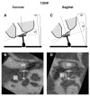
Figure 5. Imaging for postoperative quality control. In order to assess the position of the prosthesis in the middle ear by means of rotational tomography (here: total prosthesis, total ossicular replacement prosthesis (TORP)) the coupling angle a between the prosthesis head plate and the eardrum/manubrium and the angular deviation ß of the prosthesis stem from the vertical stapes axis is determined in coronal section (a; b) and sagittal section (courtesy of Zaoui et al., 2014 [95]).
Figure 6
Figure 6. Correlation of the coupling angle and hearing outcome. Correlations between the coupling angle a (a) and the inclination angle ß (b) (measured in °) and the postoperative air-bone gap (ABG) improvement. The figure displays all prostheses examined in the study, 52 PORP and 55 TORP. (TORP: total ossicular replacement prosthesis; PORP: partial ossicular replacement prosthesis) (courtesy of Zaoui et al., 2014 [95]).
Figure 7
Figure 7. MRP-TORP. Middle ear transmission function measured in fresh temporal bone specimens. The transmission function for the total prosthesis (TORP, green line) coupled to the normal manubrium and the total prosthesis coupled to the manubrium prosthesis (MRP + TORP, removed manubrium, red line) show nearly identical results as for the intact ossicular chain (black line); measurement by means of laser-Doppler-vibrometry on the footplate; stimulation of 94 dB SPL, intact eardrum.
Figure 8
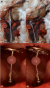
Figure 8. Bird’s columella and bended prosthesis. Direct comparison of an ostrich’s middle ear a and b with a bended TORP c and d under conditions of pressure balance a and c or a positive pressure in the auditory canal (negative pressure in the tympanic cavity) b and d. The movement of the prosthesis stem reduces the stress of ring ligament and footplate. When the pressure decreases, the original position is restored in both cases. So the bended prosthesis does not only represent the reconstruction of the sound conduction apparatus, but also to a certain extent the pressure balance of the intact ossicular chain (figures A and B: courtesy of Beleites et al. 2007 [108]).
Figure 9
Figure 9. Force-displacement relation for the stapes footplate (red) and the tympanic membrane (black) in case of axial displacement in direction of the vestibulum. Because of the visco-elastic properties, the behavior for loading and releasing is different which explains the hysteresis curves. It is obvious that the annular ligament stiffens already in case of low forces and allows only little deflection. The eardrum, however, shows a nearly linear course with constant stiffness in the examined area.
Figure 10
Figure 10. Axial loading of the stapes. Loss of the middle ear transmission function in dB, related to the neutral position of the stapes (zero line) with increasing deflection in direction of the vestibulum. The graph displays the deflection in µm and the resulting factor of annular ligament stiffening (referring to the neutral position). In case of deflection of 67 µm, stiffening of the annular ligament by factor 15 resulted in this measurement with a transmission loss of 25 dB at 2 kHz. Already low pretensions of the annular ligament (for example by longer prostheses) lead to measurable transmission losses (measurements performed in fresh temporal bone specimens, stimulation with 50 mV (corresponding to 94 dB SPL) via the floating mass transducer on the stapes head).
Figure 11
Figure 11. Loss of the middle ear transmission function in dB related to the neutral position of the stapes (zero line) and increasing deflection in direction of the promontorium (tilting around the longitudinal axis of the footplate). The deflection forces are measured in mN. Also tilting alone without additional displacement in direction of the vestibulum reveals transmission losses. They are clearly lower compared to axial forces (see figure 10) (measurement in fresh temporal bone specimens, stimulation with 50 mV (corresponding to 94 dB SPL) via the floating mass transducer on the stapes head).

References

    1. Wullstein H. Theory and practice of tympanoplasty. Laryngoscope. 1956 Aug;66(8):1076–1093. doi: 10.1288/00005537-195608000-00008. Available from: http://dx.doi.org/10.1288/00005537-195608000-00008. - DOI - DOI - PubMed
    1. Zollner F. Surgical technics for the improvement of sound conduction after radical operation. Arch Ital Otol Rinol Laringol. 1953 Jul-Aug;64(4):455–468. - PubMed
    1. Hüttenbrink KB. Die Funktion der Gehörknöchelchenkette und der Muskeln des Mittelohres. In: Feldmann H, Ganzer U, editors. Teil I: Referate. Verhandlungsbericht 1995 der Deutschen Gesellschaft für Hals-Nasen-Ohren-Heilkunde, Kopf- und Hals-Chirurgie. Berlin, Heidelberg: Springer; 1995. Available from: http://dx.doi.org/10.1007/978-3-642-79553-4_1. - DOI - DOI
    1. Hüttenbrink KB. Zur Rekonstruktion des Schallleitungsapparates unter biomechanischen Gesichtspunkten. [Biomechanics of Middle Ear Reconstruction]. Laryngo-Rhino-Otol. 2000;79(S2):S23–S51. doi: 10.1055/s-2000-15917. (Ger). Available from: http://dx.doi.org/10.1055/s-2000-15917. - DOI - DOI
    1. Zahnert T. Laser in der Ohrforschung. Laryngo-Rhino-Otol. 2003;82:157–180. doi: 10.1055/s-2003-38931. Available from: http://dx.doi.org/10.1055/s-2003-38931. - DOI - DOI - PubMed