. 2018 Apr;17(2):e12711.
doi: 10.1111/acel.12711. Epub 2017 Dec 27.

Senescence promotes in vivo reprogramming through p16INK4a and IL-6

Affiliations

Senescence promotes in vivo reprogramming through p16INK4a and IL-6

Lluc Mosteiro et al. Aging Cell. 2018 Apr.

Abstract

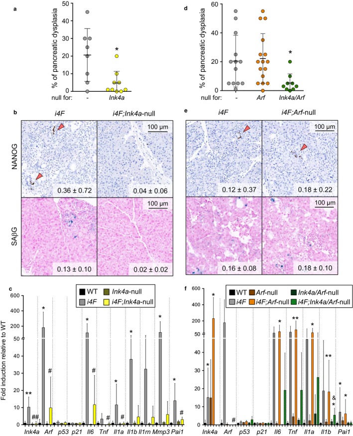
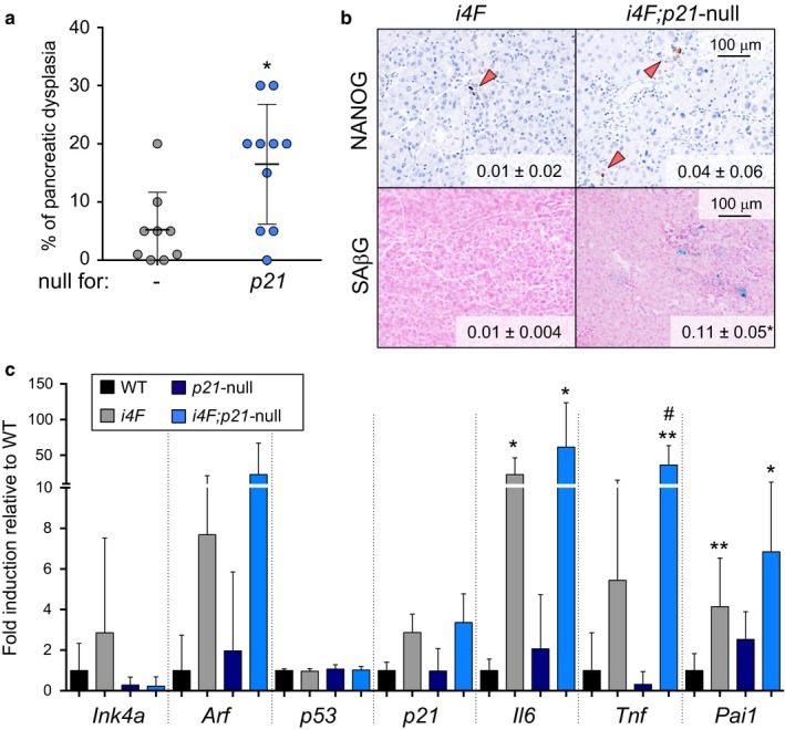
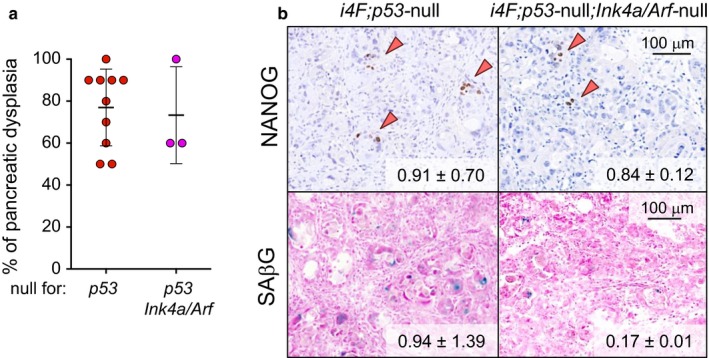
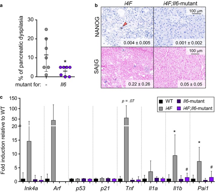
Cellular senescence is a damage response aimed to orchestrate tissue repair. We have recently reported that cellular senescence, through the paracrine release of interleukin-6 (IL6) and other soluble factors, strongly favors cellular reprogramming by Oct4, Sox2, Klf4, and c-Myc (OSKM) in nonsenescent cells. Indeed, activation of OSKM in mouse tissues triggers senescence in some cells and reprogramming in other cells, both processes occurring concomitantly and in close proximity. In this system, Ink4a/Arf-null tissues cannot undergo senescence, fail to produce IL6, and cannot reprogram efficiently; whereas p53-null tissues undergo extensive damage and senescence, produce high levels of IL6, and reprogram efficiently. Here, we have further explored the genetic determinants of in vivo reprogramming. We report that Ink4a, but not Arf, is necessary for OSKM-induced senescence and, thereby, for the paracrine stimulation of reprogramming. However, in the absence of p53, IL6 production and reprogramming become independent of Ink4a, as revealed by the analysis of Ink4a/Arf/p53 deficient mice. In the case of the cell cycle inhibitor p21, its protein levels are highly elevated upon OSKM activation in a p53-independent manner, and we show that p21-null tissues present increased levels of senescence, IL6, and reprogramming. We also report that Il6-mutant tissues are impaired in undergoing reprogramming, thus reinforcing the critical role of IL6 in reprogramming. Finally, young female mice present lower efficiency of in vivo reprogramming compared to male mice, and this gender difference disappears with aging, both observations being consistent with the known anti-inflammatory effect of estrogens. The current findings regarding the interplay between senescence and reprogramming may conceivably apply to other contexts of tissue damage.

Keywords: SASP; interleukin-6; p16Ink4a; plasticity; pluripotency; reprogramming; senescence.


Figures

Figure 1
Figure 1
Ink4a is required, and Arf is dispensable for in vivo OSKM‐induced senescence and reprogramming. a. Percentage of dysplasia in the pancreas of i4F and i4F;Ink4a‐null mice treated with 0.2 mg/ml doxycycline for 10 days and analyzed at the end of the treatment. Values correspond to the percentage (%) of pancreatic area affected (evaluated blindly). b. NANOG immunohistochemistry and SAβG staining in the pancreas of the same mice as in panel a. Images are representative for at least six mice (n ≥ 6) in NANOG and three mice (n ≥ 3) in SAβG. Values correspond to ‰ of NANOG+ cells and % of SAβG area quantified with an automated software. c. mRNA levels of the indicated genes in the pancreas of the same mice as in panel a. The number of mice tested is: n = 5 in WT and Ink4a‐null; n = 8 in i4F; n = 10 in i4F;Ink4a‐null. d. Percentage of dysplasia in the pancreas of i4F, i4F;Arf‐null, and i4F;Ink4a/Arf‐null mice treated with 0.2 mg/ml doxycycline for 7 days and analyzed at the end of the treatment. Values correspond to the percentage (%) of pancreatic area affected (evaluated blindly). e. NANOG immunohistochemistry and SAβG staining in the pancreas of the same mice as in panel d. Images are representative for at least 13 mice (n ≥ 13) in NANOG and three mice (n ≥ 3) in SAβG. Values correspond to ‰ of NANOG+ cells and % of SAβG area quantified with an automated software. f. mRNA levels of the indicated genes in the pancreas of the same mice as in panel d. The number of mice tested is: n = 3 in WT, Arf‐null and Ink4a/Arf‐null; n = 10 in i4F; n = 12 in i4F;Arf‐null and n = 6 in i4F;Ink4a/Arf‐null. All the mice tested were males of 8–10 weeks of age. All values are expressed as average ± SD Statistical significance compared to WT controls was assessed using the unpaired two‐tailed Student's t test with Welch's correction: < .05, *; < .01, **. Comparisons of each genotype to i4F controls are indicated in the same manner but using the symbol “#.” Comparisons between i4F;Arf‐null and i4F;Ink4a/Arf‐null are indicated in the same manner but using the symbol “&”
Figure 2
Figure 2
p21 limits in vivo OSKM‐induced senescence and reprogramming. a. Percentage of dysplasia in the pancreas of i4F and i4F; p21‐null mice treated with 0.2 mg/ml doxycycline for 7 days and analyzed at the end of the treatment. Values correspond to the percentage (%) of pancreatic area affected (evaluated blindly). b. NANOG immunohistochemistry and SAβG staining in the pancreas of the same mice as in panel a. Images are representative for at least 9 mice (n ≥ 9) in NANOG and 3 mice (n ≥ 3) in SAβG. Values correspond to ‰ of NANOG+ cells and % of SAβG area quantified with an automated software. c. mRNA levels of the indicated genes in the pancreas of the same mice as in panel a. The number of mice tested is: n = 6 in WT; n = 8 in p21‐null; n = 9 in i4F; and n = 10 in i4F;p21‐null. All the mice tested were females of 12–16 weeks of age. All values are expressed as average ± SD Statistical significance compared to WT controls was assessed using the unpaired two‐tailed Student's t test with Welch's correction: < .05, *; < .01, **. Comparisons between i4F and i4F;p21‐null are indicated in the same manner but using the symbol “#”
Figure 3
Figure 3
Deletion of p53 is dominant over the deletion of Ink4a/Arf during in vivo OSKM‐induced senescence and reprogramming. a. Percentage of dysplasia in the pancreas of i4F;p53‐null and i4F;p53‐null;Ink4a/Arf‐null mice treated with 0.2 mg/ml doxycycline for 7 days and analyzed at the end of the treatment. Values correspond to the percentage (%) of pancreatic area affected (evaluated blindly). b. NANOG immunohistochemistry and SAβG staining in the pancreas of the same mice as in panel a. Images are representative for at least four mice (n ≥ 4) except in i4F; p53‐null; Ink4a/Arf‐null (n = 2). Values correspond to ‰ of NANOG+ cells and % of SAβG area quantified with an automated software. All the mice tested are males of 10–14 weeks of age
Figure 4
Figure 4
Deletion of Il6 impairs in vivo OSKM‐induced senescence and reprogramming. a. Percentage of dysplasia in the pancreas of i4F and i4F;Il6‐mutant mice treated with 0.2 mg/ml doxycycline for 7 days and analyzed at the end of the treatment. Values correspond to the percentage (%) of pancreatic area affected (evaluated blindly). b. NANOG immunohistochemistry and SAβG staining in the pancreas of the same mice as in panel a. Images are representative for at least four mice (n ≥ 4) in NANOG and at least three mice (n ≥ 3) in SAβG. Values correspond to ‰ of NANOG+ cells and % of SAβG area quantified with an automated software. c. mRNA levels of the indicated genes in the pancreas of the same mice as in panel a. The number of mice tested is n = 5 in WT and Il6‐mutant and n = 7 in i4F and i4F;Il6‐null. All the mice tested were males of 9–18 weeks of age. All values are expressed as average ± SD Statistical significance compared to WT controls was assessed using the unpaired two‐tailed Student's t test with Welch's correction: < .05, *. Comparisons of each genotype to i4F controls are indicated in the same manner but using the symbol “#”
Figure 5
Figure 5
Female mice present reduced in vivo reprogramming. a. Percentage of dysplasia in the pancreas of i4F mice from young male (n = 28, from Figures 1a,d, and 4a), young female (n = 9, from Figure 2a), old male (n = 7) and old female (n = 5), treated with 0.2 mg/ml doxycycline for 7 days, and analyzed at the end of the treatment. Young male and female mice were 8–12 weeks of age; old male and female mice were ≥54 weeks of age. Values correspond to the percentage (%) of pancreatic area affected (evaluated blindly). b. mRNA levels of the indicated genes in the pancreas of the indicated mice, treated with 0.2 mg/ml doxycycline for 7 days, and analyzed at the end of the treatment. The number of mice tested is n = 8 in male WT; n = 9 in female WT; n = 8 in male i4F and n = 9 in female i4F. All values are expressed as average ± SD Statistical significance was assessed using the unpaired two‐tailed Student's t test with Welch's correction: < .001, ***. Some nonsignificant (n.s.) comparisons are also indicated
Figure 6
Figure 6
Genetic dissection of OSKM‐induced reprogramming and senescence. a. Compilation of senescence induction (SAβG+ cells) by OSKM in the pancreas of the indicated mice from Figures 1, 2, 3, 4. N values are: i4F, n = 10; i4F;Il6‐mutant, n = 3; i4F;Ink4a/Arf‐null, n = 3; i4F;Ink4a‐null, n = 3; i4F;Arf‐null, n = 5; i4F;p53‐null, n = 4; i4F;p53‐null;Ink4a/Arf‐null, n = 2; female i4F, n = 3; female i4F;p21‐null, n = 4. b. Compilation of dysplasia induction by OSKM in the pancreas of the indicated mice from Figures 1, 2, 3, 4. Compilation of data from Figures 1, 2, 3, 4. N values are: i4F, n = 28; i4F; Il6‐mutant, n = 7; i4F;Ink4a/Arf‐null, n = 9; i4F;Ink4a‐null, n = 9; i4F; Arf‐null, n = 16; i4F;p53‐null, n = 10; i4F;p53‐null;Ink4a/Arf‐null, n = 3; female i4F, n = 9; female i4F;p21‐null, n = 10. c. Compilation of reprogramming induction (NANOG+ cells) by OSKM in the pancreas of the indicated mice from Figures 1, 2, 3, 4. Compilation of data from Figures 1, 2, 3, 4. N values are: i4F, n = 23; i4F; Il6‐mutant, n = 4; i4F;Ink4a/Arf‐null, n = 9; i4F;Ink4a‐null, n = 9; i4F;Arf‐null, n = 15; i4F;p53‐null, n = 5; i4F;p53‐null;Ink4a/Arf‐null, n = 2; female i4F, n = 9; female i4F;p21‐null, n = 10. d. Correlation between the ‰ NANOG+ cells (assessed by IHC) and the mRNA levels of Il6 (relative to WT mice and assessed by qRTPCR). Compilation of all the paired values obtained in this study (n = 78). Statistical significance was evaluated by two‐tailed Student's t test (< .0001) and Pearson correlation (= .60). In a–c, all values are expressed as average ± SEM Statistical significance was assessed using the unpaired two‐tailed Student's t test with Welch's correction: < .05, *; < .01, **; < .001, ***; < .0001, ****

References

    1. Abad, M. , Mosteiro, L. , Pantoja, C. , Cañamero, M. , Rayon, T. , Ors, I. , … Serrano, M. (2013). Reprogramming in vivo produces teratomas and iPS cells with totipotency features. Nature, 502, 340–345. - PubMed
    1. Banito, A. , Rashid, S. T. , Acosta, J. C. , Li, S. , Pereira, C. F. , Geti, I. , … Gil, J. (2009). Senescence impairs successful reprogramming to pluripotent stem cells. Genes & Development, 23, 2134–2139. - PMC - PubMed
    1. Brady, J. J. , Li, M. , Suthram, S. , Jiang, H. , Wong, W. H. , & Blau, H. M. (2013). Early role for IL‐6 signalling during generation of induced pluripotent stem cells revealed by heterokaryon RNA‐Seq. Nature Cell Biology, 15, 1244–1252. - PMC - PubMed
    1. Brugarolas, J. , Chandrasekaran, C. , Gordon, J. I. , Beach, D. , Jacks, T. , & Hannon, G. J. (1995). Radiation‐induced cell cycle arrest compromised by p21 deficiency. Nature, 377, 552–557. - PubMed
    1. Cevenini, E. , Monti, D. , & Franceschi, C. (2013). Inflamm‐ageing. Current Opinion in Clinical Nutrition and Metabolic Care, 16, 14–20. - PubMed

Publication types

LinkOut - more resources