. 2017 Dec 27;13(12):e1005905.
doi: 10.1371/journal.pcbi.1005905. eCollection 2017 Dec.

High-resolution global peptide-protein docking using fragments-based PIPER-FlexPepDock

Affiliations

High-resolution global peptide-protein docking using fragments-based PIPER-FlexPepDock

Nawsad Alam et al. PLoS Comput Biol. .

Abstract

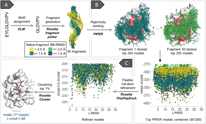
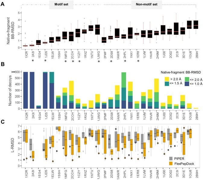
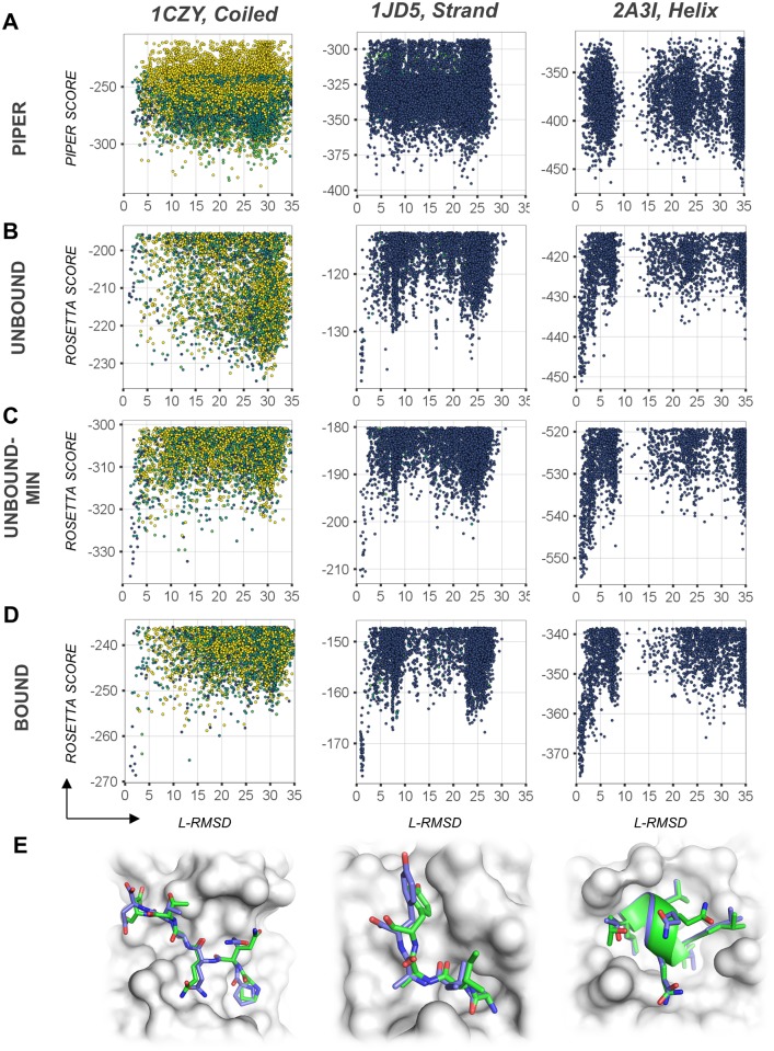
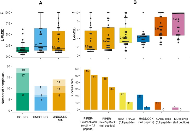
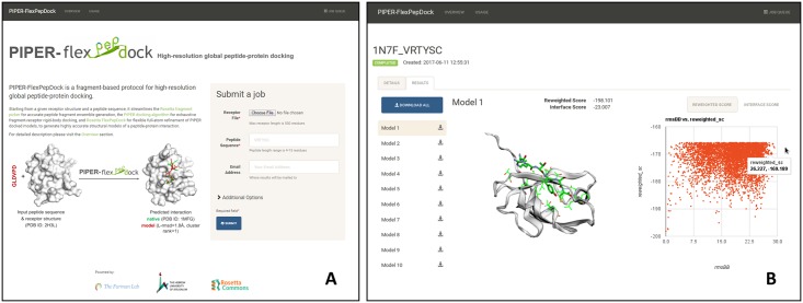
Peptide-protein interactions contribute a significant fraction of the protein-protein interactome. Accurate modeling of these interactions is challenging due to the vast conformational space associated with interactions of highly flexible peptides with large receptor surfaces. To address this challenge we developed a fragment based high-resolution peptide-protein docking protocol. By streamlining the Rosetta fragment picker for accurate peptide fragment ensemble generation, the PIPER docking algorithm for exhaustive fragment-receptor rigid-body docking and Rosetta FlexPepDock for flexible full-atom refinement of PIPER docked models, we successfully addressed the challenge of accurate and efficient global peptide-protein docking at high-resolution with remarkable accuracy, as validated on a small but representative set of peptide-protein complex structures well resolved by X-ray crystallography. Our approach opens up the way to high-resolution modeling of many more peptide-protein interactions and to the detailed study of peptide-protein association in general. PIPER-FlexPepDock is freely available to the academic community as a server at http://piperfpd.furmanlab.cs.huji.ac.il.


Conflict of interest statement

The authors have declared that no competing interests exist.

Figures

Fig 1
Fig 1. Overview of the PIPER-FlexPepDock peptide docking protocol.
Example shown: PDZ domain-peptide interaction [PDB IDs of receptor structure 1MFG (bound) and 2H3L (free)]. For a given receptor structure and peptide sequence, the divide and conquer strategy involves first the description of the peptide as an ensemble of fragments (A), their fast and exhaustive rigid body docking (using PIPER) onto the whole receptor (binding site region is shaded salmon) (B), and subsequent high-resolution refinement (using Rosetta FlexPepDock; the top 5000 models are included in the plot) (C), followed by clustering and selection of top ranking representatives. Fragments are colored according to their similarity to the native bound peptide conformation. L-RMSD: Ligand root mean square deviation from crystal structure; see text for more details.
Fig 2
Fig 2. Assessment of performance of the different steps of PIPER-FlexPepDock.
(A) Fragment quality: distribution of fragment backbone RMSDs relative to the native bound peptide conformation (defined as fragment quality). PDBs with and without motif information are grouped separately. The initial calibration set is marked with asterisks (*). (B) PIPER rigid body docking: distribution of the number of models within 5Å ligand (L)-RMSD from the native, colored according to fragment quality. (C) Improvement after FlexPepDock refinement: distribution of the L-RMSDs of the top 1% FlexPepDock refined models (in orange) and corresponding PIPER models (in gray). Shown are the results of runs starting from the unbound receptor structure and including receptor minimizations (see also Fig 3). The circles represent the L-RMSD values of the best model among the top 10 ranking clusters. The Y-axis has been trimmed to 7Å. Note that for the PIPER runs, circles represent the top-ranked model of a PIPER run (including density clustering, as described in Methods and Porter et al. [24]), while the distributions represent the subset of models that served as starting structures for the models selected after FlexPepDock refinement. The former allows the comparison of the final results from a PIPER run to a corresponding PIPER-FlexPepDock run, while the latter shows improvement due to FlexPepDock refinement for the finally selected models.
Fig 3
Fig 3. Examples of global peptide docking energy landscapes.
Left: PDB id 1CZY (coiled peptide); Center: 1JD5 (extended peptide); Right: 2A3I (helical peptide). (A) Energy landscape as sampled in the first docking step of the protocol by PIPER rigid body docking of peptide fragments onto the unbound receptor structure. (B-D) Energy landscapes for the PIPER-FPD scheme, starting from the unbound receptor structure (B), the unbound receptor structure including receptor flexibility (C), and the corresponding bound receptor for comparison (D). Models are colored according to fragment quality, as in previous Figures. (E) Comparison of the modeled to the native structure (shown in blue and green, respectively).
Fig 4
Fig 4. PIPER-FlexPepDock peptide docking performance.
(A) Overall performance on a non-redundant set of 27 peptide-protein complexes. Top: Distribution of best model L-RMSDs (among top 10 ranking clusters) for runs using the bound (BOUND) and free (UNBOUND & UNBOUND-MIN) receptor structures, the latter including also receptor flexibility in the final refinement step (only the motif region was modeled for the 12 complexes with known motif). Shown are both the L-RMSD values for each protein-peptide complex (grey circles, rounded values for improved visibility are provided), as well as the distribution (quartiles and medians, with median values printed alongside). Bottom: Distribution of the ranks of the first near-native cluster (defined as L-RMSD < = 2.0Å), shown using different shades (for corresponding results among the top1, top3 and top10 ranked predictions). (B) Comparison to performance by other algorithms. Top: Box plots of best L-RMSDs among top 10 ranking clusters, including results for the motif part where the motif is known (as in A), as well as for the full peptide, for comparison. Bottom: Performance is shown for different cutoffs (3.0Å and 2.0Å L-RMSD in left and right boxes, respectively) (See S1B Table for more details).
Fig 5
Fig 5. The PIPER-FlexPepDock server.
(A) Job submission page: the required input includes the structure of the receptor and the sequence of the peptide; advanced options are accessible via a button. The tabs at the top provide links to detailed descriptions of the server, as well as to the Queue (upper right). (B) Results of an example peptide docking run: The liprin C-terminal peptide sequence VRTYSC docked onto the PDZ domain of GRIP1 (free receptor PDB id 1N7E). The top10 ranking models can be downloaded, and links to the individual models are provided to the left for inspection using an interactive viewer. In this case, Model 1 is an accurate prediction (L-RMSD = 1.0Å from solved structure PDB id 1N7F). On the right side a scatter plot shows the sampled energy landscape (relative to the lowest energy structure of the simulation).

References

    1. Pawson T, Nash P. Assembly of cell regulatory systems through protein interaction domains. Science. 2003;300(5618):445–52. doi: 10.1126/science.1083653 - DOI - PubMed
    1. Petsalaki E, Russell RB. Peptide-mediated interactions in biological systems: new discoveries and applications. Curr Opin Biotechnol. 2008;19(4):344–50. doi: 10.1016/j.copbio.2008.06.004 - DOI - PubMed
    1. Neduva V, Linding R, Su-Angrand I, Stark A, de Masi F, Gibson TJ, et al. Systematic discovery of new recognition peptides mediating protein interaction networks. PLoS Biol. 2005;3(12):e405 doi: 10.1371/journal.pbio.0030405 - DOI - PMC - PubMed
    1. Vacic V, Oldfield CJ, Mohan A, Radivojac P, Cortese MS, Uversky VN, et al. Characterization of molecular recognition features, MoRFs, and their binding partners. J Proteome Res. 2007;6(6):2351–66. doi: 10.1021/pr0701411 - DOI - PMC - PubMed
    1. Gamble TR, Vajdos FF, Yoo S, Worthylake DK, Houseweart M, Sundquist WI, et al. Crystal structure of human cyclophilin A bound to the amino-terminal domain of HIV-1 capsid. Cell. 1996;87(7):1285–94. - PubMed

Publication types

Substances

LinkOut - more resources