Duodenal Adenocarcinoma of Brunner Gland Origin: A Case Report
- PMID: 29281782
- PMCID: PMC5964285
- DOI: 10.4132/jptm.2017.10.09
Duodenal Adenocarcinoma of Brunner Gland Origin: A Case Report
Abstract
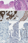
We report a case of adenocarcinoma originating from the duodenal Brunner glands in a 47-year-old female patient. The lesion was 0.8 cm in extent and located at the posterior wall of the first part of the duodenum. Histologically, the tumor showed transition from non-neoplastic Brunner glands through dysplastic epithelium into adenocarcinoma. The carcinoma cells were strongly positive for MUC6 protein, which is an epithelial marker for the Brunner glands. Tumor protein p53 was overexpressed in the carcinoma cells, but not in the non-neoplastic or dysplastic epithelium. Dystrophic calcification was predominant. This is the first case report of duodenal adenocarcinoma of Brunner gland origin in Korea.
Keywords: Adenocarcinoma; Brunner glands; Duodenum.
Conflict of interest statement
No potential conflict of interest relevant to this article was reported.
Figures


References
-
- Fenoglio-Preiser CM, Noffsinger AE, Stemmermann GN, Lantz PE, Isaacson PG. Gastrointestinal pathology: an atlas and text. Philadelphia: Lippincott Williams & Wilkins; 2008. p. 471.
-
- Chang HK, Yu E, Kim J, et al. Adenocarcinoma of the small intestine: a multi-institutional study of 197 surgically resected cases. Hum Pathol. 2010;41:1087–96. - PubMed
-
- Ohta Y, Saitoh K, Akai T, Uesato M, Ochiai T, Matsubara H. Early primary duodenal carcinoma arising from Brunner's glands synchronously occurring with sigmoid colon carcinoma: report of a case. Surg Today. 2008;38:756–60. - PubMed
LinkOut - more resources
Full Text Sources
Other Literature Sources
Research Materials
Miscellaneous

