. 2017 Dec 26;21(13):3708-3716.
doi: 10.1016/j.celrep.2017.12.006.

Identification of Oxa1 Homologs Operating in the Eukaryotic Endoplasmic Reticulum

Affiliations

Identification of Oxa1 Homologs Operating in the Eukaryotic Endoplasmic Reticulum

S Andrei Anghel et al. Cell Rep. .

Abstract

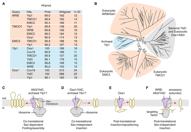
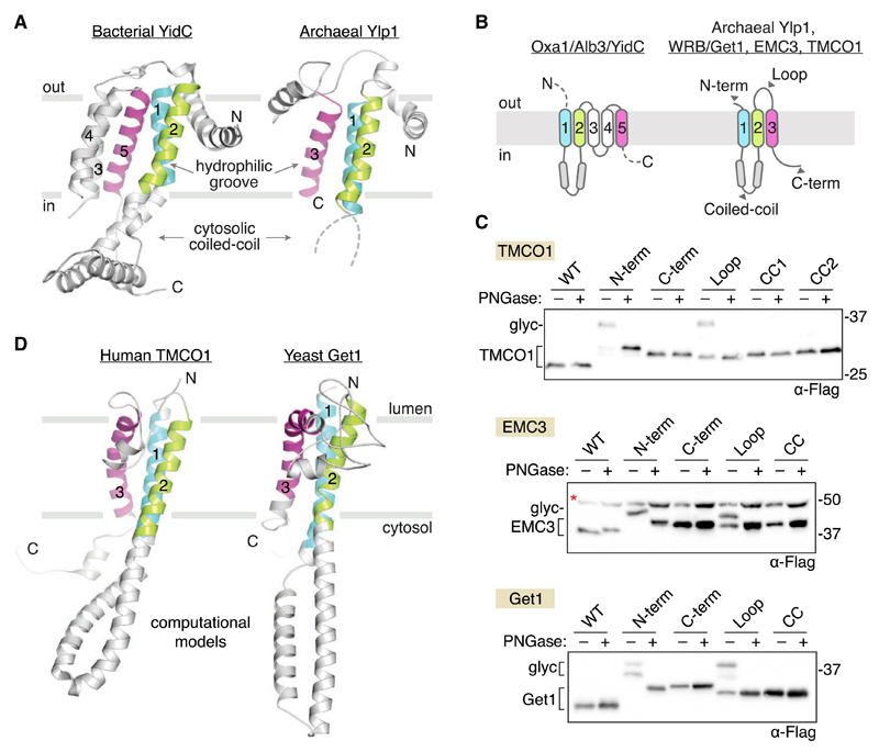
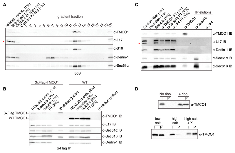
Members of the evolutionarily conserved Oxa1/Alb3/YidC family mediate membrane protein biogenesis at the mitochondrial inner membrane, chloroplast thylakoid membrane, and bacterial plasma membrane, respectively. Despite their broad phylogenetic distribution, no Oxa1/Alb3/YidC homologs are known to operate in eukaryotic cells outside the endosymbiotic organelles. Here, we present bioinformatic evidence that the tail-anchored protein insertion factor WRB/Get1, the "endoplasmic reticulum (ER) membrane complex" subunit EMC3, and TMCO1 are ER-resident homologs of the Oxa1/Alb3/YidC family. Topology mapping and co-evolution-based modeling demonstrate that Get1, EMC3, and TMCO1 share a conserved Oxa1-like architecture. Biochemical analysis of human TMCO1, the only homolog not previously linked to membrane protein biogenesis, shows that it associates with the Sec translocon and ribosomes. These findings suggest a specific biochemical function for TMCO1 and define a superfamily of proteins-the "Oxa1 superfamily"-whose shared function is to facilitate membrane protein biogenesis.


Conflict of interest statement

Declaration of Interests

The authors declare no competing interests.

Figures

Figure 1
Figure 1. Phylogenetic and Functional Comparison Defines the Oxa1 Superfamily
(A) Identification of remote DUF106 homologs using HHpred. Eukaryotic (human), bacterial (E. coli), and archaeal (M. jannaschii) proteomes were searched for each query (UniProt ID: WRB, O00258; Oxa1, Q15070; TMCO1, Q9UM00; EMC3, Q9P0I2; Ylp1, Q57904) using default settings in HHpred in “global” alignment mode. Top hits are listed, along with the HHpred probability score, the number of residues aligned, and the sequence identity. (B) Maximum-likelihood tree of representative sequences. Branch lengths for the five main clades are indicated. (C) During Sec-dependent, co-translational assembly and folding, substrates are delivered to the membrane by the ribosome; insertion requires participation of the Sec translocon. Substrates of this pathway typically contain multiple TMDs and/or large translocated regions. Superfamily members exemplifying this activity include bacterial YidC and chloroplast Alb3. (D) During Sec-independent, co-translational insertion, topologically “simple” substrates that lack large or highly charged translocated regions are delivered to the membrane by the ribosome. Superfamily members exemplifying this activity include Oxa1 and YidC; archaeal Ylp1 proteins function similarly in vitro. (E) Post-translational TMD repositioning, exemplified by Oxa1. (F) During Sec-independent, post-translational insertion, topologically simple substrates are delivered to the membrane by soluble targeting factors. Superfamily members exemplifying this activity include WRB/Get1, which inserts tail-anchored proteins delivered by TRC40/Get3; chloroplast Alb3, which inserts specific proteins delivered to the thylakoid membrane by cpSRP43; and bacterial YidC.
Figure 2
Figure 2. Oxa1 Superfamily Members Share a Conserved Membrane Topology and Core Structural Features
(A) Comparison of known structures from two clades: bacterial YidC (left; PDB 3WO6) and archaeal Ylp1 (right; PDB 5C8J). These proteins share a common N-out/C-in topology, a cytosolic-facing coiled coil between TM1 and TM2 (disordered in the archaeal structure), and a three-TMD core (colored) that harbors a lipid-exposed hydrophilic groove implicated in binding to nascent polypeptides during insertion. (B) Predicted topology of the Oxa1 superfamily members. (C) Experimentally defined topology of human TMCO1, EMC3, and yeast Get1. Glycosylation acceptor sequences were introduced at the indicated positions (gray arrowheads in B and Figure S2), and glycosylation was monitored by western blotting after treatment with or without PNGase F. All three proteins conform to the predicted Oxa1 superfamily topology. A non-specific, cross-reacting band visible in all EMC3 samples is marked (red asterisk). (D) Evolutionary covariation-based computational 3D models of human TMCO1 and yeast Get1 recapitulate the core structural features of bacterial YidC and archaeal Ylp1: lumenal N terminus; cytosolic-facing coiled coil and C terminus; and a three-TMD core. Here, the predicted coiled coil region of Get1 has been replaced with the experimentally determined structure of the Get1 coiled coil (PDB 3ZS8). The resulting hybrid model is in good agreement with the covariation-based 3D model calculated for human WRB (Figure S2). See also Figures S1 and S2.
Figure 3
Figure 3. TMCO1 Forms a Complex with the Sec61 Translocon and RNCs
(A) HEK293 membranes were solubilized with digitonin, fractionated by sucrose cushion, separated on a high-resolution sucrose gradient, and analyzed by western blotting. TMCO1 co-fractionates with intact 80S particles and the Sec61 translocon, but not the unrelated ER membrane protein Derlin-1, which does not bind to ribosomes. Blots for the large (L17) and small (S16) ribosomal subunits are also shown; a non-specific, cross-reacting band visible in the L17 blot is indicated with an asterisk. (B) Digitonin-solubilized membranes from wild-type (WT) HEK293 cells or a HEK293 cell line containing an N-terminal 3×Flag-tagged TMCO1 allele were analyzed by anti-Flag immunoprecipitation, sucrose cushion, and western blotting. (C) Digitonin-solubilized canine pancreatic rough microsomes were tested for interaction between TMCO1 and Sec61 by co-immunoprecipitation and western blotting. An anti-TMCO1, but not a control anti-3F4 antibody, pulls down two components of Sec61. The absence of TMCO1 in the reciprocal pull-down is consistent with the higher levels of Sec61 in these membranes. (D) Recombinant, purified TMCO1 co-sediments with unprogrammed ribosomes isolated from rabbit reticulocyte lysate (top panel). This interaction is salt sensitive and can be stabilized by chemical crosslinking (XL) (bottom panel). The pellet (P) fractions correspond to 5× volume equivalents of the input (I) fractions. See also Figure S3.

Comment in

References

    1. Alanay Y, Ergüner B, Utine E, Haçarız O, Kiper POS, Taşkıran EZ, Perçin F, Uz E, Sağıroğlu MŞ, Yuksel B, et al. TMCO1 deficiency causes autosomal recessive cerebrofaciothoracic dysplasia. Am J Med Genet A. 2014;164:291–304. - PubMed
    1. Altschul SF, Madden TL, Schäffer AA, Zhang J, Zhang Z, Miller W, Lipman DJ. Gapped BLAST and PSI-BLAST: a new generation of protein database search programs. Nucleic Acids Res. 1997;25:3389–3402. - PMC - PubMed
    1. Aschtgen MS, Zoued A, Lloubès R, Journet L, Cascales E. The C-tail anchored TssL subunit, an essential protein of the enteroaggregative Escherichia coli Sci-1 Type VI secretion system, is inserted by YidC. MicrobiologyOpen. 2012;1:71–82. - PMC - PubMed
    1. Aviram N, Schuldiner M. Embracing the void–how much do we really know about targeting and translocation to the endoplasmic reticulum? Curr Opin Cell Biol. 2014;29:8–17. - PubMed
    1. Aviram N, Ast T, Costa EA, Arakel EC, Chuartzman SG, Jan CH, Haβdenteufel S, Dudek J, Jung M, Schorr S, et al. The SND proteins constitute an alternative targeting route to the endoplasmic reticulum. Nature. 2016;540:134–138. - PMC - PubMed

Publication types

MeSH terms