. 2017 Dec 26;21(13):3833-3845.
doi: 10.1016/j.celrep.2017.11.104.

Degree of Tissue Differentiation Dictates Susceptibility to BRAF-Driven Colorectal Cancer

Affiliations

Degree of Tissue Differentiation Dictates Susceptibility to BRAF-Driven Colorectal Cancer

Kevin Tong et al. Cell Rep. .

Abstract

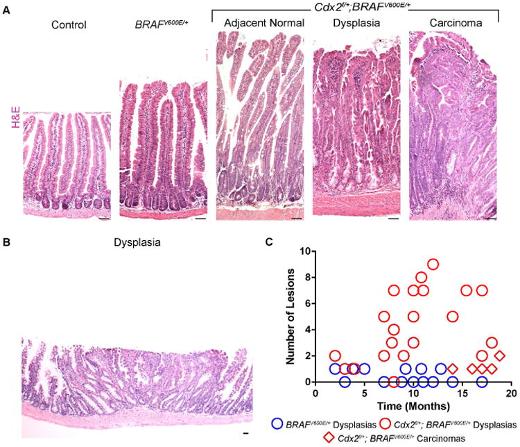
Oncogenic mutations in BRAF are believed to initiate serrated colorectal cancers; however, the mechanisms of BRAF-driven colon cancer are unclear. We find that oncogenic BRAF paradoxically suppresses stem cell renewal and instead promotes differentiation. Correspondingly, tumor formation is inefficient in BRAF-driven mouse models of colon cancer. By reducing levels of differentiation via genetic manipulation of either of two distinct differentiation-promoting factors (Smad4 or Cdx2), stem cell activity is restored in BRAFV600E intestines, and the oncogenic capacity of BRAFV600E is amplified. In human patients, we observe that reduced levels of differentiation in normal tissue is associated with increased susceptibility to serrated colon tumors. Together, these findings help resolve the conditions necessary for BRAF-driven colon cancer initiation. Additionally, our results predict that genetic and/or environmental factors that reduce tissue differentiation will increase susceptibility to serrated colon cancer. These findings offer an opportunity to identify susceptible individuals by assessing their tissue-differentiation status.

Keywords: BRAF; Cdx2; Smad4; intestinal homeostasis; serrated colorectal cancer.


Conflict of interest statement

The authors declare no conflicts of interest

Figures

Figure 1
Figure 1. BRAFV600E activation alters intestinal homeostasis, promoting differentiation
(A) GSEA analysis reveals that MAPK target genes (Rad et al., 2013), were elevated in the BRAFV600E/+ mutants, as expected (K-S test). (B) Assessment of morphological changes (H&E), proliferation (Ki67), differentiation (AP), stem cells (OLFM4), and senescent cells (p21) in BRAFV600E/+ and BRAFV600E/600E mutants. Note increasing levels of differentiation markers and decreasing levels of the stem cell marker with the increased number of activated BRAF alleles. Representative images from 3 biological replicates. Scale bars = 50 μm (C) intestinal stem cell gene expression levels are significantly reduced in BRAFV600E/+ mutants whereas (D) intestinal differentiation gene signatures are significantly increased compared to controls.
Figure 2
Figure 2. Ablation of CDX2 reverses BRAFV600E-driven changes in stem/differentiation homeostasis
(A) Immunohistochemistry of OLFM4 in uninjected control, BRAFV600E/+, Cdx2f/f; BRAFV600E/+, BRAFV600E, and Cdx2f/f; BRAFV600E mice, 5 days post-tamoxifen treatment. (B) Primary organoid cultures isolated from control, BRAFV600E/+, and Cdx2f/+; BRAFV600E/+ mice. BRAFV600E/+ mutant cells do not give rise to viable organoids, but reduction of CDX2 levels permits their growth. Representative of 3 biological replicates. Scale bars=50μm. (C) Lgr5-GFP expression. Scale bars=50μm. (D) Recombination at the BRAFV600E locus, induced by tamoxifen treatment of the stem cell specific, conditional Cre (Lgr5-CreERT2-IRES-eGFP), is not sustained unless Cdx2 is simultaneously inactivated in the stem cells. Samples from crypt epithelium were harvested at 2 days or 10 months after tamoxifen treatment. (E) AP staining reveals increased expression in BRAFV600E/+ and BRAFV600E, which is reduced when Cdx2 is lost. (F) GSEA shows intestinal stem cell genes are elevated while differentiation genes are reduced upon loss of CDX2, reversing gene expression changes induced by BRAFV600E activation. (G) Examples of the types of differentiation-associated genes that are elevated upon BRAF activation, and suppressed upon CDX2-loss.
Figure 3
Figure 3. Loss of CDX2 accelerates BRAF-driven tumorigenesis
(A) H&E stains of age-matched control, BRAFV600E/+, and Cdx2f/+; BRAFV600E/+ mice reveal characteristics of serrated cancer morphologies. Dysplasias and carcinomas had characteristics of human hyperplastic polyps (HPPs), SSAs, and serrated carcinomas (Also see Figure S3) (B) More serrated lesions were found in Cdx2f/+; BRAFV600E/+ compound mice than age-matched BRAFV600E/+ counterparts. Counts were based upon complete swiss roll sections. Scale bars=50μm.
Figure 4
Figure 4. Loss of SMAD4 restores the stem-differentiation balance that is altered upon BRAFV600E/+ activation
(A) AP staining reveals that Smad4 loss reverses the increase in differentiation induced by BRAFV600E/+. (B) OLFM4 staining suggests that loss of SMAD4 in BRAFV600E/+ tissue reverses stem cell loss. Representative of 3 biological replicates. Scale bars=50μm. (C) Lgr5-GFP expression is lost upon BRAFV600E expression, but restored upon SMAD4-loss. (D) Organoid cultures isolated from control, BRAFV600E/+, and Smad4f/f; BRAFV600E/+ mice reveal a rescue in viability in Smad4f/f; BRAFV600E/+ organoids. (E) GSEA shows intestinal differentiation genes are reduced, while stem cell gene expression is elevated upon loss of SMAD4, reversing gene expression changes induced by BRAFV600E/+ activation.
Figure 5
Figure 5. Tumorigenesis in BRAFV600E/+ mice is accelerated upon the loss Smad4
(A) H&E stain shows overall morphology of adjacent normal and tumor regions of the intestine of control, BRAFV600E/+, and Smad4f/f; BRAFV600E/+ mice. Similar to Cdx2f/+; BRAFV600E/+ mice, dysplasias and carcinomas featured serrated morphologies (See also Figure S5). Counts of macroscopic (B) and microscopic (C) lesions observed in brafv6ooe/+mice are lower than Smad4f/f; BRAFV600E/+ mutants. Scale bars=50um.
Figure 6
Figure 6. Elevated WNT signaling may restore oncogenic potential in Cdx2f/f; BRAFV600E/+and Smad4f/f; BRAFV600E/+ double mutant mice
(A) Transcriptome analysis of WNT signature genes (Van der Flier et al., 2007) indicates increased WNT target gene expression in the double mutant mice when compared to BRAFV600E/+ mice (K-S test). (B) Common intestinal WNT target genes are highlighted. (C) Organoid cultures derived from BRAFV600E/+ crypts survive when treated with either WNT3A or the WNT-pathway agonist CHIR-98014. (D) Immunohistochemistry of WNT targets CD44 and SOX9 show elevated levels in Cdx2f/f; BRAFV600E/+an6 Smad4f/f;BRAFV600E/+ mutants compared to BRAFV600E/+ mutants. (E) Organoid cultures derived from compound mutant mice show sensitivity to WNT inhibitors. Table describes viability in days of mature organoids after treatment with XAV-939 or IWP-2. Representative of 2 biological replicates and 3 technical replicates each. Bars=50 μ.
Figure 7
Figure 7. Differentiation gene expression levels in normal tissues corresponds to serrated tumor susceptibility in humans
(A) Non-tumor, right colon tissue from serrated tumor patients (n=15) exhibit lower levels of differentiation-gene expression when compared to control patient tissues (n=10, K-S test). (B) K-means clustering of patients into tumor and non-tumor classes is improved when combining intestinal stem (Munoz et al., 2012) and differentiation cell (Chong et al., 2009) gene sets that are differentially regulated in mouse models (performing in the top 2.5% of 1000 random gene lists of equivalent size), or by either gene set individually (Figure S6). (C) Patients exhibit a range of differentiation gene expression, with serrated cancer patients exhibiting lower levels of intestinal differentiation genes (K-W Test, p<0.05). (D) Human tissue samples assessed for differentiation marker KRT20 stain intensity. (E) Adjacent normal tissue of serrated tumor patients exhibits a trend towards reducedstain intensity relative to non-tumor patients (Mann-Whitney p=0.13362). (F) KRT20transcript levels in normal tissue is lower in serrated tumor patients when compared tonon-tumor patient samples (ANOVA, p<0.05). Scale bars=50 μm.

References

    1. Bae JM, Lee TH, Cho NY, Kim TY, Kang GH. Loss of CDX2 expression is associated with poor prognosis in colorectal cancer patients. World J Gastroenterol. 2015;21:1457–1467. - PMC - PubMed
    1. Barker N, Ridgway RA, van Es JH, van de Wetering M, Begthel H, van den Born M, Danenberg E, Clarke AR, Sansom OJ, Clevers H. Crypt stem cells as the cells-of-origin of intestinal cancer. Nature. 2009;457:608–611. - PubMed
    1. Barker N, van Es JH, Kuipers J, Kujala P, van den Born M, Cozijnsen M, Haegebarth A, Korving J, Begthel H, Peters PJ, et al. Identification of stem cells in small intestine and colon by marker gene Lgr5. Nature. 2007;449:1003–1007. - PubMed
    1. Cancer Genome Atlas N. Comprehensive molecular characterization of human colon and rectal cancer. Nature. 2012;487:330–337. - PMC - PubMed
    1. Carragher LA, Snell KR, Giblett SM, Aldridge VS, Patel B, Cook SJ, Winton DJ, Marais R, Pritchard CA. V600EBraf induces gastrointestinal crypt senescence and promotes tumour progression through enhanced CpG methylation of p16INK4a. EMBO Mol Med. 2010;2:458–471. - PMC - PubMed

Publication types

MeSH terms