

Review
doi: 10.3350/cmh.2017.0078.
Epub 2017 Dec 20.
Histopathological evaluation of liver fibrosis and cirrhosis regression
Affiliations
- PMID: 29281870
- PMCID: PMC5760001
- DOI: 10.3350/cmh.2017.0078
Item in Clipboard
Review
Histopathological evaluation of liver fibrosis and cirrhosis regression
Clin Mol Hepatol.
2017 Dec.
Abstract
The hepatic repair complex in the setting of cirrhosis has received increasing attention, as it implies the regression of cirrhosis, which was traditionally taken to be an irreversible state. In this brief review, the patterns of fibrosis, the existing staging systems for chronic liver disease and the histopathological features of cirrhosis regression are discussed.
Keywords: Cirrhosis; Hepatic repair complex; Liver fibrosis; Regression.
Conflict of interest statement
Figures
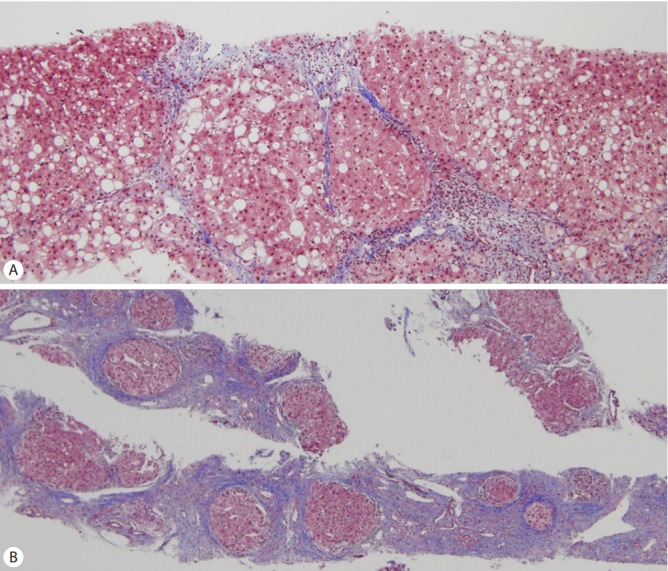
(A) Mixtures of thin and broad fibrous septa are seen in this case of cirrhosis with Laennec stage 4B. (B) A needle biopsy of Laennec stage 4C cirrhosis demonstrating very broad fibrous septa with micronodules (Masson’s trichrome stain, original magnification ×100 [A], ×40 [B]).

Histological features of fibrosis regression. (A) Presence of delicate perforated fibrous septa (arrowhead) and (B) clusters of hepatocytes within split septa (Masson’s trichrome stain, original magnification ×100).
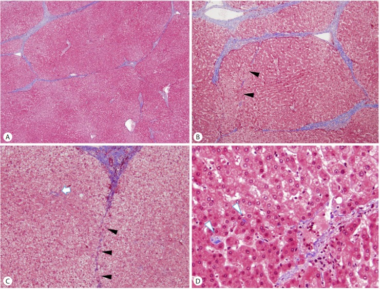
An example of a cirrhosis with predominantly regressive pattern. (A) Cirrhosis with thin fibrous septa, corresponding to Laennec stage 4A. (B, C, D) Features of cirrhosis regression are seen in the field adjacent to (A), including perforated delicate septa (B, C; arrowheads), remnant portal tracts (C; arrow) and isolated collagen fibers (D; arrowheads). (Masson’s trichrome stain, original magnification ×40 (A, B), ×100 (C), ×200 (D))
References
-
- Rappaport AM, MacPhee PJ, Fisher MM, Phillips MJ. The scarring of the liver acini (Cirrhosis). Tridimensional and microcirculatory considerations. Virchows Arch A Pathol Anat Histopathol. 1983;402:107–137. - PubMed
-
- Bouwens L, Baekeland M, Wisse E. Cytokinetic analysis of the expanding Kupffer-cell population in rat liver. Cell Tissue Kinet. 1986;19:217–226. - PubMed
-
- Tsuchida T, Friedman SL. Mechanisms of hepatic stellate cell activation. Nat Rev Gastroenterol Hepatol. 2017;14:397–411. - PubMed
-
- Brunt EM, Janney CG, Di Bisceglie AM, Neuschwander-Tetri BA, Bacon BR. Nonalcoholic steatohepatitis: a proposal for grading and staging the histological lesions. Am J Gastroenterol. 1999;94:2467–2474. - PubMed
Publication types
MeSH terms
LinkOut - more resources
Full Text Sources
Other Literature Sources
Medical
